SlideShare a Scribd company logo
1 of 10
Lungs 
Structure 
The lungs are the site of gas exchange 
in mammals. 
They are not locate outside the body 
because 
1) the air is not dense enough to 
support them 
2) they would dry out 
3) the lungs are very delicate 
the lungs are supported by the ribcage 
which can be moved by the use of the 
intercostal muscle. 
Trachea 
This is a flexible airways supported by 
rings of cartilage which prevent the trachea from collapsing when the air 
pressure inside falls. 
The walls are made up of muscle, lined with ciliated epithelium and goblet cells 
1) the goblet cells produce mucus which traps dirt and bacteria. 
2) The cilium moves the mucus up into the throat where it is swallowed. 
Bronchi: 
Bronchi have 2 divisions of the trachea. The larger bronchi are supported by 
cartilage. 
Bronchioles: 
These are branching subdivisions of the bronchi. The walls are made of the 
muscle lined with epithelial cells. The muscle enables them to control the air in 
and out. 
Alveoli: 
Very small air-sacs at the end of the bronchioles. It contains collagen and elastic 
fibres. Alveoli is the site of gas exchange where the CO2 goes into he alveoli and 
the O2 goes into the blood stream. 
Danger: 
The lungs increase and decrease in size continuously. When an organ increases 
in size in the body, it could touch other organs because there isn’t enough space. 
This can become a risk because when living tissue rubs against another, it could 
cause friction, which could cause tissue damage and kill the cells.
Mechanism of Breathing: 
The two breathing processes are INHALATION and EXHALATION. 
The breathing system does not have a foxed shape as it has the ability to move, 
whilst staying in a protective ribcage. This means that the rob cage too can 
moves positions. 
INHALATION 
1) When we inhale, our 
lungs fill with air. 
2) AS they fill with air, 
they become 
ENLARGED. 
3) Thus, the ribcage 
moves UPWARDS and 
OUTWARDS to make 
more room in the 
thorax. 
4) The overall effect is 
that our chest 
EXPANDS. 
The pressure changes in 
the lungs due to… 
1) The DIAPHRAGM - a sheet of muscle that separates the thorax and the 
abdomen. 
2) The INTERNAL INTERCOSTAL MUSCLES – leads to EXPIRATION 
3) The EXTERNAL INTERCOSTAL MUSCLES - leads to INSPIRATION 
DIAPHRAGM 
The diaphragm Is a dome shape when we exhale. However, when we inhale it 
CONTRACTS and FLATTENS. This results in a change in volume in the thorax as 
the volume increases when the diaphragm is flattened. 
As the volume increases, the internal air pressure drops meaning that the air 
pressure outside the lings is greater than the air pressure inside the lungs. 
PULMONARY VENTILATION 
This is the total volume of air that is moved into the lungfs in a minute. 
Tidal Volume- the volume of air normally taken in for each breathe when the 
body is at rest 
Ventilation Rate- the number of breathes taken in one minute (normally 12-20) 
Pulmonary Ventilation Rate= Tidal Volume x Ventilation 
(dm3min-1) (dm3) (min-1)
Exchange of Gases in the Lungs 
Alveoli 
The site of gas exchange in mammals is the 
epithelium of the alveoli. 
The air sacs are very small at 100mm- 
400mm each. 
There are about 300 alveoli in each lung. 
Each alveolus is lined with epithelial cells 
0.05mm-0.3mm thick. 
A network of pulmonary capillaries 
surrounds each alveolus. 
Oxygen passes into the alveolus and dissolves 
in s small moist lining of the epithelial. 
The moist lining also stops the alveolus from 
drying and cracking and keeps the insides 
lubricated. 
The oxygen molecules diffuse into the blood 
stream through the lining of the alveolus and 
the capillaries. The red blood cell pick up the 
oxygen and carries it to the heart by the 
pulmonary vein. 
Laws of Diffusion 
The movement of the oxygen from the blood 
to the cells also follows the laws of diffusion. 
1) There is a high concentration within 
the blood. 
2) There is a low concentration within the cell. 
3) Therefore the oxygen passes into the body cells. 
Alveoli Adaptations 
They are really good surface for gas exchange because: 
1) Large surface area to volume ratio- speeds up the rate of exchange. 
2) Thin exchange surface- SHORT diffusion pathway 
3) Partially Permeable- Allows selected materials to diffuse easily. 
4) Steep concentration gradient maintained by breathing and blood flow. 
5) The walls are moist- allows oxygen to dissolve into liquid before diffusing. 
6) Special liquid SURFACTANT- reduces surface tension and prevents air 
sacs collapsing.
Pulmonary Tuberculosis: 
Used to be called consumption. 
Cause 
Caused by a rod shaped bacterium: 
1) Mycobacterium Tuberculosis 
2) Mycobacterium Bovis 
It is a slow growing organism (only divides every 16-20 hours) 
Most commonly affects the lungs but can spread to other parts of the body. 
Transmission 
Spread through: 
1) Droplet Infection such as sneeze, cough, laughing and talking 
2) People with prolonged, frequent or intense contact are at highest risk. 
3) Cows to Human mycobacterium BOVIS) 
About 90% have asymptomatic latent TB that cannot be passed on. 
There is a 10% lifetime chance that the latent infection will become active TB. If 
untreated there is a more than 50% of dying. 
Mycobacterium Bovis affects cattle. However they are regularly checked and 
the milk is pasteurised which kills the bacterium. 
Some groups are at greater risk at contracting TB: 
1) Those in close contact with infected individuals 
2) Those from the countries where TB is common. 
3) Have reduced immunity such elderly people, AIDs sufferers, people with 
medical conditions, homeless, alcoholics, drug users, etc. 
Course of Infection 
Bacteria grow and divide in the upper regions of the lungs. 
Body’s immune system responds and white blood cells accumulate at the 
infected area. 
This leads to inflammation of the lungs and enlargement of the lymph nodes. 
In a healthy person there are few symptoms and infection is controlled within 
weeks. 
However some bacteria still remains, 
Many years later, the bacteria may re-emerge. 
Bacteria can destroy the lung tissue, this results in cavities and scar tissue. 
Suffers cough up damaged lung tissue and blood. 
The TB can spread to the rest of the body if it’s not treated. 
Treatment 
Latent TB- usually single antibiotics. 
Active TB- combinations of several antibiotics to reduce the bacterial growth. 
Prevention 
Children are vaccinated and those with TB are identified and treated. 
Increase in TB in Eastern Europe and Africa due to AIDs epidemic causing 
people to be more susceptible to TB.
Pulmonary Fibrosis 
This is when scars form on the epithelium of the lungs causing them to become 
irreversibly thickened. 
Oxygen uptake will be less efficient because 
1) The volume of air that the lung can contain reduces. 
2) There is a grater diffusion pathway. 
Fibrosis reduces the elasticity of the lungs so it is harder to breathe. 
Shortness of breath-especially when exercising due to the amount of fibrous 
tissues in the lungs. 
1) Less air is taken in by each breath 
2) Increased diffusion pathway due to the thickened epithelium 
3) Loss of elasticity makes ventilation difficult and thus makes it hard to 
maintain a diffusion gradient 
Chronic, dry cough because the fibrous tissue creates an obstruction in the 
airways of the lungs. The body naturally reacts to clear the obstruction by 
coughing but as the tissue is immovable the cough is ‘dry’. 
Pain and discomfort in the chest due to the pressure and damage from the 
fibrous tissue and coughing. 
Weakness and fatigue from reduced oxygen uptake. The energy released by 
cellular respiration is reduced, leading to tiredness. 
The exact cause is unclear, but evidence suggests that it is a reaction to 
microscopic lung injury, to which some individuals are genetically more 
susceptible. 
Emphysema 
Walls of the alveoli are broken down 
Symptoms 
1) Coughing, shortness of breath, and wheezing, developing into extreme 
difficulty in breathing, bluish skin colour 
2) Physical damage by repeated coughing and loss of elastin from walls of 
alveoli. Less surface area is available for the exchange of gases 
CAUSE 
Smoking and air pollution 
Treatment 
Emphysema is irreversible 
Asthma 
Affects up to 10% of the worlds population.
2000 deaths in UK each year. 
Asthma tends to run in families – genetic element 
Allergens include 
1) Pollen, animal fur, faeces of house dust mite. 
Can be triggered by 
2) air pollutants(sulphur dioxide, nitrogen oxides and ozone), exercise, cold 
air, infection, anxiety and stress. 
3) Number of cases rising due to 
4) Increase in air pollution 
5) Increase in stress 
6) Increase in the chemicals in our food 
7) Cleaner lifestyle 
The allergens cause the white blood cells lining the bronchi and bronchioles 
to release histamine. This has the following effects: 
a. Inflammation of the lining of the airways 
b. Cells of the epithelial lining secrete larger quantities of mucus 
c. Fluid leaves the capillaries and enters the airways 
d. The muscle surrounding the bronchioles contracts ands so 
constricts the airways 
Overall there is a much greater resistance to the flow of air in and out of the 
alveoli and therefore it is difficult to maintain the diffusion gradient. 
Symptoms 
Difficulty in breathing due to 
1) Constriction of bronchi and bronchioles 
2) Inflamed lining 
3) Presence of additional mucus and fluid 
Wheezing caused by air passing through constricted bronchi and bronchioles 
Tight feeling in chest, a consequence of not being able to ventilate the lungs 
properly 
Coughing, a reflex response to the obstructed airways 
Chronic Bronchitis 
Having a productive cough for at least 3 months during 2 successive years 
Symptoms: 
Productive cough, breathlessness 
Smoking and air pollution paralyse the cilia in the bronchial tubes so mucus 
builds up in clumps that are coughed up (productive cough). The lining of the 
bronchial tubes becomes irritated and inflamed. 
Cause 
Smoking and air pollution 
Treatment 
Drug treatment, oxygen therapy, and lung transplant 
June 2013
Q3) ai) Name the process by which oxygen passes from an alveolus in the 
lungs into the blood. 
1) Diffusion 
3aii) Describe 2 adaptions of the structure of alveoli for efficient gas 
exchange. 
1) Thin moist walls 
2) Large Surface area 
3b) People who have been fire-breathers for many years often find they 
cannot breathe out properly. Explain why. 
1) Loss of elasticity of the lungs 
2) Scar Tissue 
3) Less recoil 
January 2013 
1 (a) The diagram shows the structure of the human gas exchange system. 
1) Trachea 
2) Bronchus 
1 (b) Explain how downward movement of the diaphragm leads to air 
entering the lungs. 
1. Increases the volume in the thorax 
2. Lowers the pressure in the thorax 
3. Pressure outside the lungs is greater so air is pushed inside. 
1 c) Complete the equation. 
Pulmonary ventilation = ...................................... × ........................................ 
1. Tidal Volume 
2. Ventilation Rate 
June 2012
3a) Describe what happens to the diaphragm between times P and Q to 
bring about the change in its shape. 
1. Flattens 
2. Contracts 
3b) Air moves into the lungs between times P and Q. Explain how the 
diaphragm causes this. 
1. Diaphragm contracts and Flattens 
2. Increases the volume in the thorax 
3. Decreases the pressure inside the lungs. 
4. Air moves from high to lower pressure. 
3c) Describe how oxygen in air in the alveoli enters the blood in capillaries. 
1. Oxygen diffuses into the capillary 
2. Across as the alveoli’s’ epithelium and the capillary’s endothelium 
January 2012 
2a) The diaphragm helps to bring about the changes shown by the curve A. 
Explain how. 
1. Before his inhaler the Diaphragm was relaxed 
2. The diaphragm was dome shaped 
3. Which increased the pressure inside the lungs 
4. Decreased the volume in the thorax. 
2b) You could use curve A to find the total volume of air that this person 
could breathe out in one complete breath. Describe how. 
1. Extend the curve 
2. Until the curve flattens and has reached a peak 
3. You could use curve A to find the total volume of air that this person could 
breathe out in one complete breath. Describe how. 
2c) The inhaler which the person used contained a substance that dilates 
bronchioles. Use this information to explain why curve A is different from 
curve B. 
1. Before the inhaler, the bronchioles were narrower. 
2. The muscles around the bronchioles contracted. 
3. Which causes less air to pass through making it harder to breathe 
June 2011
4a) The two people were breathing out during the time shown. What 
evidence in the table supports this statement? 
1. The volume of air in the lungs decreases. 
4b) Calculate the rate at which person A breathed air out of his lungs 
between 0 and 3 seconds. Show your working. 
1. 6.5-2.3/3 secs = 4.2/3 = 1.4 
4c) Person B has emphysema. Give one piece of evidence from the table 
that shows this. 
1. Reduces flow rates / less air breathed out / more air left in the lungs 
4d) Emphysema reduces the efficiency of gas exchange in the lungs. Explain 
why. 
1. The alveoli breaks down which leaves fewer of them that are large 
2. This increases the diffusion pathway 
3. So less diffusion takes place 
4. There is less elasticity in the lungs 
5. Which reduced the flow rate 
6. So a small concentration gradient. 
January 2011 
7a) Pulmonary tuberculosis is a disease of the lungs. 
Describe the transmission and course of infection of pulmonary 
tuberculosis. 
1. Bacteria is transmitted by droplets from a sneeze or cough 
2. The bacteria destroys the alveoli 
3. Which leads to scar tissue and fibrosis 
4. Damage leads to less diffusion as there’s an increased pathway 
5. The bacteria could spread to other organs via bloodstream 
7b) Emphysema is another disease of the lungs. People with emphysema 
may feel weak and tired. Explain why. 
1. The alveoli breaks down 
2. Which increases the diffusion pathway so takes a longer time 
3. Decreases the diffusion gradient 
4. So less oxygen enters the blood 
5. Less respiration takes place, which leaves the person tired. 
June 2010
2a) i) Explain how the graph shows that the person was breathing in 
between times A and B. 
1. The volume of air was increasing 
ii) Describe and explain what happens to the shape of the diaphragm 
between times A and B. 
1. The diaphragm flattens 
2. The muscle contracts 
2b) The person’s pulmonary ventilation changed between times C and D. 
Describe how the graph shows that the pulmonary ventilation changed. 
1. Pulmonary ventilation= tidal volume x breathing rate 
2. The breathing rate increases per minute. 
3. The tidal volume increases between C and D as they are deeper breathes. 
January 2010 
2a) i) Describe the path by which oxygen goes from an alveolus to the 
blood. 
1. The oxygen diffuses across the epithelium of the alveolus 
2. And through the epithelium of the capillary 
2a) ii) Explain why people with miner’s lung have a lower concentration of 
oxygen in their blood. 
1. Longer Diffusion Pathway 
2b) i) Describe how ventilation helps to maintain this difference in oxygen 
concentration. 
1. Brings in air that has a higher oxygen concentration. 
2. Removes air with a lower oxygen concentration. 
2b) ii) Give one other way that helps to maintain the difference in oxygen 
concentration. 
1. Circulation of blood 
2c) Evaluate this conclusion. 
1. Correlation does not prove causation, there may be other causes 
2. Improved Diagnosis methods 
3. Not all cases are reported. We don’t know the number of cases.

More Related Content

What's hot (20)

Structure and function of the lungs
Structure and function of the lungsStructure and function of the lungs
Structure and function of the lungs
 
Physiology of hearing,smell and taste
Physiology of hearing,smell and tastePhysiology of hearing,smell and taste
Physiology of hearing,smell and taste
 
Respiratory system
Respiratory systemRespiratory system
Respiratory system
 
Epithelium, cells,tissues & histology
Epithelium, cells,tissues & histologyEpithelium, cells,tissues & histology
Epithelium, cells,tissues & histology
 
C tfinal iiii
C tfinal iiiiC tfinal iiii
C tfinal iiii
 
Asthma
AsthmaAsthma
Asthma
 
L1 introduction to respiratory medicine
L1 introduction to respiratory medicineL1 introduction to respiratory medicine
L1 introduction to respiratory medicine
 
Anatomy of bones
Anatomy of bones Anatomy of bones
Anatomy of bones
 
ear anatomy, structures, functions
ear anatomy, structures, functions ear anatomy, structures, functions
ear anatomy, structures, functions
 
Respiration
RespirationRespiration
Respiration
 
Respiratory system
 Respiratory system Respiratory system
Respiratory system
 
Cartilage & bones
Cartilage & bonesCartilage & bones
Cartilage & bones
 
Unit 1 introduction to physiology
Unit 1 introduction to physiologyUnit 1 introduction to physiology
Unit 1 introduction to physiology
 
Lab Exam #2 Histology (labeled)
Lab Exam #2 Histology (labeled)Lab Exam #2 Histology (labeled)
Lab Exam #2 Histology (labeled)
 
Respiratory system slide show
Respiratory system slide showRespiratory system slide show
Respiratory system slide show
 
Anatomy of blood vessels ppt
Anatomy of blood vessels pptAnatomy of blood vessels ppt
Anatomy of blood vessels ppt
 
Nervous system- SYMPATHETIC and PARASYMPATHETIC
Nervous system- SYMPATHETIC and PARASYMPATHETICNervous system- SYMPATHETIC and PARASYMPATHETIC
Nervous system- SYMPATHETIC and PARASYMPATHETIC
 
Unit 11 Lymphatic System
Unit 11 Lymphatic SystemUnit 11 Lymphatic System
Unit 11 Lymphatic System
 
respiratorysystem (16).ppt
respiratorysystem (16).pptrespiratorysystem (16).ppt
respiratorysystem (16).ppt
 
Epithelium cellstissues histology
Epithelium cellstissues histologyEpithelium cellstissues histology
Epithelium cellstissues histology
 

Similar to AQA Biology As level Lungs revision booklet

Introduction Yoga Therapy For The Management Of Respiratory Disorders
Introduction Yoga Therapy For The Management Of Respiratory DisordersIntroduction Yoga Therapy For The Management Of Respiratory Disorders
Introduction Yoga Therapy For The Management Of Respiratory DisordersShama
 
states of matter
states of matterstates of matter
states of matterSue Aris
 
GAS EXCHANGE pRESENTATION2.ppt
GAS EXCHANGE pRESENTATION2.pptGAS EXCHANGE pRESENTATION2.ppt
GAS EXCHANGE pRESENTATION2.pptCretileSlayer
 
CHRONIC OBSTRUCTIVE PULMONARY DISEASE
CHRONIC OBSTRUCTIVE PULMONARY DISEASECHRONIC OBSTRUCTIVE PULMONARY DISEASE
CHRONIC OBSTRUCTIVE PULMONARY DISEASETomcy Thankachan
 
respiratory system
respiratory systemrespiratory system
respiratory systemEspirituanna
 
Lecture 21 respiratory system
Lecture 21    respiratory systemLecture 21    respiratory system
Lecture 21 respiratory systemShakir Ellahi
 
Unit3NutritionalprocessRespiratory
Unit3NutritionalprocessRespiratoryUnit3NutritionalprocessRespiratory
Unit3NutritionalprocessRespiratoryaurorabiologia
 
Lecture 4 (Respiratory tract).pptx
Lecture 4 (Respiratory tract).pptxLecture 4 (Respiratory tract).pptx
Lecture 4 (Respiratory tract).pptxWaniaIzm
 
Gaseous exchange mahlangu n_218018380
Gaseous exchange mahlangu n_218018380Gaseous exchange mahlangu n_218018380
Gaseous exchange mahlangu n_218018380Ntokozo Mahlangu
 
Chap_8_Respiration_and_Circulation.pptx
Chap_8_Respiration_and_Circulation.pptxChap_8_Respiration_and_Circulation.pptx
Chap_8_Respiration_and_Circulation.pptxRahul Badve
 
Unit 14 Respiratory System
Unit 14 Respiratory SystemUnit 14 Respiratory System
Unit 14 Respiratory SystemIshtiaqAhmad114
 

Similar to AQA Biology As level Lungs revision booklet (20)

Introduction Yoga Therapy For The Management Of Respiratory Disorders
Introduction Yoga Therapy For The Management Of Respiratory DisordersIntroduction Yoga Therapy For The Management Of Respiratory Disorders
Introduction Yoga Therapy For The Management Of Respiratory Disorders
 
states of matter
states of matterstates of matter
states of matter
 
GAS EXCHANGE pRESENTATION2.ppt
GAS EXCHANGE pRESENTATION2.pptGAS EXCHANGE pRESENTATION2.ppt
GAS EXCHANGE pRESENTATION2.ppt
 
Tuberculosis
Tuberculosis Tuberculosis
Tuberculosis
 
Respiratory system
Respiratory systemRespiratory system
Respiratory system
 
Chapter1
Chapter1Chapter1
Chapter1
 
Gaseous Exchange
Gaseous ExchangeGaseous Exchange
Gaseous Exchange
 
RESPIRATION
RESPIRATIONRESPIRATION
RESPIRATION
 
Respiratory system
Respiratory systemRespiratory system
Respiratory system
 
Respiratory system
Respiratory systemRespiratory system
Respiratory system
 
CHRONIC OBSTRUCTIVE PULMONARY DISEASE
CHRONIC OBSTRUCTIVE PULMONARY DISEASECHRONIC OBSTRUCTIVE PULMONARY DISEASE
CHRONIC OBSTRUCTIVE PULMONARY DISEASE
 
respiratory system
respiratory systemrespiratory system
respiratory system
 
Lecture 21 respiratory system
Lecture 21    respiratory systemLecture 21    respiratory system
Lecture 21 respiratory system
 
Unit3NutritionalprocessRespiratory
Unit3NutritionalprocessRespiratoryUnit3NutritionalprocessRespiratory
Unit3NutritionalprocessRespiratory
 
Lecture 4 (Respiratory tract).pptx
Lecture 4 (Respiratory tract).pptxLecture 4 (Respiratory tract).pptx
Lecture 4 (Respiratory tract).pptx
 
Gaseous exchange mahlangu n_218018380
Gaseous exchange mahlangu n_218018380Gaseous exchange mahlangu n_218018380
Gaseous exchange mahlangu n_218018380
 
The Respiratory System
The Respiratory SystemThe Respiratory System
The Respiratory System
 
Chap_8_Respiration_and_Circulation.pptx
Chap_8_Respiration_and_Circulation.pptxChap_8_Respiration_and_Circulation.pptx
Chap_8_Respiration_and_Circulation.pptx
 
Unit 14 Respiratory System
Unit 14 Respiratory SystemUnit 14 Respiratory System
Unit 14 Respiratory System
 
Ch22respiratorysystem
Ch22respiratorysystemCh22respiratorysystem
Ch22respiratorysystem
 

Recently uploaded

G9 Science Q4- Week 1-2 Projectile Motion.ppt
G9 Science Q4- Week 1-2 Projectile Motion.pptG9 Science Q4- Week 1-2 Projectile Motion.ppt
G9 Science Q4- Week 1-2 Projectile Motion.pptMAESTRELLAMesa2
 
Spermiogenesis or Spermateleosis or metamorphosis of spermatid
Spermiogenesis or Spermateleosis or metamorphosis of spermatidSpermiogenesis or Spermateleosis or metamorphosis of spermatid
Spermiogenesis or Spermateleosis or metamorphosis of spermatidSarthak Sekhar Mondal
 
NAVSEA PEO USC - Unmanned & Small Combatants 26Oct23.pdf
NAVSEA PEO USC - Unmanned & Small Combatants 26Oct23.pdfNAVSEA PEO USC - Unmanned & Small Combatants 26Oct23.pdf
NAVSEA PEO USC - Unmanned & Small Combatants 26Oct23.pdfWadeK3
 
Call Girls in Mayapuri Delhi 💯Call Us 🔝9953322196🔝 💯Escort.
Call Girls in Mayapuri Delhi 💯Call Us 🔝9953322196🔝 💯Escort.Call Girls in Mayapuri Delhi 💯Call Us 🔝9953322196🔝 💯Escort.
Call Girls in Mayapuri Delhi 💯Call Us 🔝9953322196🔝 💯Escort.aasikanpl
 
Recombinant DNA technology (Immunological screening)
Recombinant DNA technology (Immunological screening)Recombinant DNA technology (Immunological screening)
Recombinant DNA technology (Immunological screening)PraveenaKalaiselvan1
 
Cultivation of KODO MILLET . made by Ghanshyam pptx
Cultivation of KODO MILLET . made by Ghanshyam pptxCultivation of KODO MILLET . made by Ghanshyam pptx
Cultivation of KODO MILLET . made by Ghanshyam pptxpradhanghanshyam7136
 
zoogeography of pakistan.pptx fauna of Pakistan
zoogeography of pakistan.pptx fauna of Pakistanzoogeography of pakistan.pptx fauna of Pakistan
zoogeography of pakistan.pptx fauna of Pakistanzohaibmir069
 
Natural Polymer Based Nanomaterials
Natural Polymer Based NanomaterialsNatural Polymer Based Nanomaterials
Natural Polymer Based NanomaterialsAArockiyaNisha
 
STERILITY TESTING OF PHARMACEUTICALS ppt by DR.C.P.PRINCE
STERILITY TESTING OF PHARMACEUTICALS ppt by DR.C.P.PRINCESTERILITY TESTING OF PHARMACEUTICALS ppt by DR.C.P.PRINCE
STERILITY TESTING OF PHARMACEUTICALS ppt by DR.C.P.PRINCEPRINCE C P
 
Orientation, design and principles of polyhouse
Orientation, design and principles of polyhouseOrientation, design and principles of polyhouse
Orientation, design and principles of polyhousejana861314
 
Nanoparticles synthesis and characterization​ ​
Nanoparticles synthesis and characterization​  ​Nanoparticles synthesis and characterization​  ​
Nanoparticles synthesis and characterization​ ​kaibalyasahoo82800
 
Bentham & Hooker's Classification. along with the merits and demerits of the ...
Bentham & Hooker's Classification. along with the merits and demerits of the ...Bentham & Hooker's Classification. along with the merits and demerits of the ...
Bentham & Hooker's Classification. along with the merits and demerits of the ...Nistarini College, Purulia (W.B) India
 
GFP in rDNA Technology (Biotechnology).pptx
GFP in rDNA Technology (Biotechnology).pptxGFP in rDNA Technology (Biotechnology).pptx
GFP in rDNA Technology (Biotechnology).pptxAleenaTreesaSaji
 
SOLUBLE PATTERN RECOGNITION RECEPTORS.pptx
SOLUBLE PATTERN RECOGNITION RECEPTORS.pptxSOLUBLE PATTERN RECOGNITION RECEPTORS.pptx
SOLUBLE PATTERN RECOGNITION RECEPTORS.pptxkessiyaTpeter
 
Disentangling the origin of chemical differences using GHOST
Disentangling the origin of chemical differences using GHOSTDisentangling the origin of chemical differences using GHOST
Disentangling the origin of chemical differences using GHOSTSérgio Sacani
 
VIRUSES structure and classification ppt by Dr.Prince C P
VIRUSES structure and classification ppt by Dr.Prince C PVIRUSES structure and classification ppt by Dr.Prince C P
VIRUSES structure and classification ppt by Dr.Prince C PPRINCE C P
 
Analytical Profile of Coleus Forskohlii | Forskolin .pptx
Analytical Profile of Coleus Forskohlii | Forskolin .pptxAnalytical Profile of Coleus Forskohlii | Forskolin .pptx
Analytical Profile of Coleus Forskohlii | Forskolin .pptxSwapnil Therkar
 
Behavioral Disorder: Schizophrenia & it's Case Study.pdf
Behavioral Disorder: Schizophrenia & it's Case Study.pdfBehavioral Disorder: Schizophrenia & it's Case Study.pdf
Behavioral Disorder: Schizophrenia & it's Case Study.pdfSELF-EXPLANATORY
 

Recently uploaded (20)

G9 Science Q4- Week 1-2 Projectile Motion.ppt
G9 Science Q4- Week 1-2 Projectile Motion.pptG9 Science Q4- Week 1-2 Projectile Motion.ppt
G9 Science Q4- Week 1-2 Projectile Motion.ppt
 
Spermiogenesis or Spermateleosis or metamorphosis of spermatid
Spermiogenesis or Spermateleosis or metamorphosis of spermatidSpermiogenesis or Spermateleosis or metamorphosis of spermatid
Spermiogenesis or Spermateleosis or metamorphosis of spermatid
 
NAVSEA PEO USC - Unmanned & Small Combatants 26Oct23.pdf
NAVSEA PEO USC - Unmanned & Small Combatants 26Oct23.pdfNAVSEA PEO USC - Unmanned & Small Combatants 26Oct23.pdf
NAVSEA PEO USC - Unmanned & Small Combatants 26Oct23.pdf
 
Call Girls in Mayapuri Delhi 💯Call Us 🔝9953322196🔝 💯Escort.
Call Girls in Mayapuri Delhi 💯Call Us 🔝9953322196🔝 💯Escort.Call Girls in Mayapuri Delhi 💯Call Us 🔝9953322196🔝 💯Escort.
Call Girls in Mayapuri Delhi 💯Call Us 🔝9953322196🔝 💯Escort.
 
Recombinant DNA technology (Immunological screening)
Recombinant DNA technology (Immunological screening)Recombinant DNA technology (Immunological screening)
Recombinant DNA technology (Immunological screening)
 
Cultivation of KODO MILLET . made by Ghanshyam pptx
Cultivation of KODO MILLET . made by Ghanshyam pptxCultivation of KODO MILLET . made by Ghanshyam pptx
Cultivation of KODO MILLET . made by Ghanshyam pptx
 
zoogeography of pakistan.pptx fauna of Pakistan
zoogeography of pakistan.pptx fauna of Pakistanzoogeography of pakistan.pptx fauna of Pakistan
zoogeography of pakistan.pptx fauna of Pakistan
 
Natural Polymer Based Nanomaterials
Natural Polymer Based NanomaterialsNatural Polymer Based Nanomaterials
Natural Polymer Based Nanomaterials
 
STERILITY TESTING OF PHARMACEUTICALS ppt by DR.C.P.PRINCE
STERILITY TESTING OF PHARMACEUTICALS ppt by DR.C.P.PRINCESTERILITY TESTING OF PHARMACEUTICALS ppt by DR.C.P.PRINCE
STERILITY TESTING OF PHARMACEUTICALS ppt by DR.C.P.PRINCE
 
Orientation, design and principles of polyhouse
Orientation, design and principles of polyhouseOrientation, design and principles of polyhouse
Orientation, design and principles of polyhouse
 
Nanoparticles synthesis and characterization​ ​
Nanoparticles synthesis and characterization​  ​Nanoparticles synthesis and characterization​  ​
Nanoparticles synthesis and characterization​ ​
 
The Philosophy of Science
The Philosophy of ScienceThe Philosophy of Science
The Philosophy of Science
 
Bentham & Hooker's Classification. along with the merits and demerits of the ...
Bentham & Hooker's Classification. along with the merits and demerits of the ...Bentham & Hooker's Classification. along with the merits and demerits of the ...
Bentham & Hooker's Classification. along with the merits and demerits of the ...
 
GFP in rDNA Technology (Biotechnology).pptx
GFP in rDNA Technology (Biotechnology).pptxGFP in rDNA Technology (Biotechnology).pptx
GFP in rDNA Technology (Biotechnology).pptx
 
SOLUBLE PATTERN RECOGNITION RECEPTORS.pptx
SOLUBLE PATTERN RECOGNITION RECEPTORS.pptxSOLUBLE PATTERN RECOGNITION RECEPTORS.pptx
SOLUBLE PATTERN RECOGNITION RECEPTORS.pptx
 
Engler and Prantl system of classification in plant taxonomy
Engler and Prantl system of classification in plant taxonomyEngler and Prantl system of classification in plant taxonomy
Engler and Prantl system of classification in plant taxonomy
 
Disentangling the origin of chemical differences using GHOST
Disentangling the origin of chemical differences using GHOSTDisentangling the origin of chemical differences using GHOST
Disentangling the origin of chemical differences using GHOST
 
VIRUSES structure and classification ppt by Dr.Prince C P
VIRUSES structure and classification ppt by Dr.Prince C PVIRUSES structure and classification ppt by Dr.Prince C P
VIRUSES structure and classification ppt by Dr.Prince C P
 
Analytical Profile of Coleus Forskohlii | Forskolin .pptx
Analytical Profile of Coleus Forskohlii | Forskolin .pptxAnalytical Profile of Coleus Forskohlii | Forskolin .pptx
Analytical Profile of Coleus Forskohlii | Forskolin .pptx
 
Behavioral Disorder: Schizophrenia & it's Case Study.pdf
Behavioral Disorder: Schizophrenia & it's Case Study.pdfBehavioral Disorder: Schizophrenia & it's Case Study.pdf
Behavioral Disorder: Schizophrenia & it's Case Study.pdf
 

AQA Biology As level Lungs revision booklet

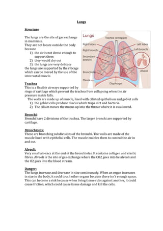
  • 1. Lungs Structure The lungs are the site of gas exchange in mammals. They are not locate outside the body because 1) the air is not dense enough to support them 2) they would dry out 3) the lungs are very delicate the lungs are supported by the ribcage which can be moved by the use of the intercostal muscle. Trachea This is a flexible airways supported by rings of cartilage which prevent the trachea from collapsing when the air pressure inside falls. The walls are made up of muscle, lined with ciliated epithelium and goblet cells 1) the goblet cells produce mucus which traps dirt and bacteria. 2) The cilium moves the mucus up into the throat where it is swallowed. Bronchi: Bronchi have 2 divisions of the trachea. The larger bronchi are supported by cartilage. Bronchioles: These are branching subdivisions of the bronchi. The walls are made of the muscle lined with epithelial cells. The muscle enables them to control the air in and out. Alveoli: Very small air-sacs at the end of the bronchioles. It contains collagen and elastic fibres. Alveoli is the site of gas exchange where the CO2 goes into he alveoli and the O2 goes into the blood stream. Danger: The lungs increase and decrease in size continuously. When an organ increases in size in the body, it could touch other organs because there isn’t enough space. This can become a risk because when living tissue rubs against another, it could cause friction, which could cause tissue damage and kill the cells.
  • 2. Mechanism of Breathing: The two breathing processes are INHALATION and EXHALATION. The breathing system does not have a foxed shape as it has the ability to move, whilst staying in a protective ribcage. This means that the rob cage too can moves positions. INHALATION 1) When we inhale, our lungs fill with air. 2) AS they fill with air, they become ENLARGED. 3) Thus, the ribcage moves UPWARDS and OUTWARDS to make more room in the thorax. 4) The overall effect is that our chest EXPANDS. The pressure changes in the lungs due to… 1) The DIAPHRAGM - a sheet of muscle that separates the thorax and the abdomen. 2) The INTERNAL INTERCOSTAL MUSCLES – leads to EXPIRATION 3) The EXTERNAL INTERCOSTAL MUSCLES - leads to INSPIRATION DIAPHRAGM The diaphragm Is a dome shape when we exhale. However, when we inhale it CONTRACTS and FLATTENS. This results in a change in volume in the thorax as the volume increases when the diaphragm is flattened. As the volume increases, the internal air pressure drops meaning that the air pressure outside the lings is greater than the air pressure inside the lungs. PULMONARY VENTILATION This is the total volume of air that is moved into the lungfs in a minute. Tidal Volume- the volume of air normally taken in for each breathe when the body is at rest Ventilation Rate- the number of breathes taken in one minute (normally 12-20) Pulmonary Ventilation Rate= Tidal Volume x Ventilation (dm3min-1) (dm3) (min-1)
  • 3. Exchange of Gases in the Lungs Alveoli The site of gas exchange in mammals is the epithelium of the alveoli. The air sacs are very small at 100mm- 400mm each. There are about 300 alveoli in each lung. Each alveolus is lined with epithelial cells 0.05mm-0.3mm thick. A network of pulmonary capillaries surrounds each alveolus. Oxygen passes into the alveolus and dissolves in s small moist lining of the epithelial. The moist lining also stops the alveolus from drying and cracking and keeps the insides lubricated. The oxygen molecules diffuse into the blood stream through the lining of the alveolus and the capillaries. The red blood cell pick up the oxygen and carries it to the heart by the pulmonary vein. Laws of Diffusion The movement of the oxygen from the blood to the cells also follows the laws of diffusion. 1) There is a high concentration within the blood. 2) There is a low concentration within the cell. 3) Therefore the oxygen passes into the body cells. Alveoli Adaptations They are really good surface for gas exchange because: 1) Large surface area to volume ratio- speeds up the rate of exchange. 2) Thin exchange surface- SHORT diffusion pathway 3) Partially Permeable- Allows selected materials to diffuse easily. 4) Steep concentration gradient maintained by breathing and blood flow. 5) The walls are moist- allows oxygen to dissolve into liquid before diffusing. 6) Special liquid SURFACTANT- reduces surface tension and prevents air sacs collapsing.
  • 4. Pulmonary Tuberculosis: Used to be called consumption. Cause Caused by a rod shaped bacterium: 1) Mycobacterium Tuberculosis 2) Mycobacterium Bovis It is a slow growing organism (only divides every 16-20 hours) Most commonly affects the lungs but can spread to other parts of the body. Transmission Spread through: 1) Droplet Infection such as sneeze, cough, laughing and talking 2) People with prolonged, frequent or intense contact are at highest risk. 3) Cows to Human mycobacterium BOVIS) About 90% have asymptomatic latent TB that cannot be passed on. There is a 10% lifetime chance that the latent infection will become active TB. If untreated there is a more than 50% of dying. Mycobacterium Bovis affects cattle. However they are regularly checked and the milk is pasteurised which kills the bacterium. Some groups are at greater risk at contracting TB: 1) Those in close contact with infected individuals 2) Those from the countries where TB is common. 3) Have reduced immunity such elderly people, AIDs sufferers, people with medical conditions, homeless, alcoholics, drug users, etc. Course of Infection Bacteria grow and divide in the upper regions of the lungs. Body’s immune system responds and white blood cells accumulate at the infected area. This leads to inflammation of the lungs and enlargement of the lymph nodes. In a healthy person there are few symptoms and infection is controlled within weeks. However some bacteria still remains, Many years later, the bacteria may re-emerge. Bacteria can destroy the lung tissue, this results in cavities and scar tissue. Suffers cough up damaged lung tissue and blood. The TB can spread to the rest of the body if it’s not treated. Treatment Latent TB- usually single antibiotics. Active TB- combinations of several antibiotics to reduce the bacterial growth. Prevention Children are vaccinated and those with TB are identified and treated. Increase in TB in Eastern Europe and Africa due to AIDs epidemic causing people to be more susceptible to TB.
  • 5. Pulmonary Fibrosis This is when scars form on the epithelium of the lungs causing them to become irreversibly thickened. Oxygen uptake will be less efficient because 1) The volume of air that the lung can contain reduces. 2) There is a grater diffusion pathway. Fibrosis reduces the elasticity of the lungs so it is harder to breathe. Shortness of breath-especially when exercising due to the amount of fibrous tissues in the lungs. 1) Less air is taken in by each breath 2) Increased diffusion pathway due to the thickened epithelium 3) Loss of elasticity makes ventilation difficult and thus makes it hard to maintain a diffusion gradient Chronic, dry cough because the fibrous tissue creates an obstruction in the airways of the lungs. The body naturally reacts to clear the obstruction by coughing but as the tissue is immovable the cough is ‘dry’. Pain and discomfort in the chest due to the pressure and damage from the fibrous tissue and coughing. Weakness and fatigue from reduced oxygen uptake. The energy released by cellular respiration is reduced, leading to tiredness. The exact cause is unclear, but evidence suggests that it is a reaction to microscopic lung injury, to which some individuals are genetically more susceptible. Emphysema Walls of the alveoli are broken down Symptoms 1) Coughing, shortness of breath, and wheezing, developing into extreme difficulty in breathing, bluish skin colour 2) Physical damage by repeated coughing and loss of elastin from walls of alveoli. Less surface area is available for the exchange of gases CAUSE Smoking and air pollution Treatment Emphysema is irreversible Asthma Affects up to 10% of the worlds population.
  • 6. 2000 deaths in UK each year. Asthma tends to run in families – genetic element Allergens include 1) Pollen, animal fur, faeces of house dust mite. Can be triggered by 2) air pollutants(sulphur dioxide, nitrogen oxides and ozone), exercise, cold air, infection, anxiety and stress. 3) Number of cases rising due to 4) Increase in air pollution 5) Increase in stress 6) Increase in the chemicals in our food 7) Cleaner lifestyle The allergens cause the white blood cells lining the bronchi and bronchioles to release histamine. This has the following effects: a. Inflammation of the lining of the airways b. Cells of the epithelial lining secrete larger quantities of mucus c. Fluid leaves the capillaries and enters the airways d. The muscle surrounding the bronchioles contracts ands so constricts the airways Overall there is a much greater resistance to the flow of air in and out of the alveoli and therefore it is difficult to maintain the diffusion gradient. Symptoms Difficulty in breathing due to 1) Constriction of bronchi and bronchioles 2) Inflamed lining 3) Presence of additional mucus and fluid Wheezing caused by air passing through constricted bronchi and bronchioles Tight feeling in chest, a consequence of not being able to ventilate the lungs properly Coughing, a reflex response to the obstructed airways Chronic Bronchitis Having a productive cough for at least 3 months during 2 successive years Symptoms: Productive cough, breathlessness Smoking and air pollution paralyse the cilia in the bronchial tubes so mucus builds up in clumps that are coughed up (productive cough). The lining of the bronchial tubes becomes irritated and inflamed. Cause Smoking and air pollution Treatment Drug treatment, oxygen therapy, and lung transplant June 2013
  • 7. Q3) ai) Name the process by which oxygen passes from an alveolus in the lungs into the blood. 1) Diffusion 3aii) Describe 2 adaptions of the structure of alveoli for efficient gas exchange. 1) Thin moist walls 2) Large Surface area 3b) People who have been fire-breathers for many years often find they cannot breathe out properly. Explain why. 1) Loss of elasticity of the lungs 2) Scar Tissue 3) Less recoil January 2013 1 (a) The diagram shows the structure of the human gas exchange system. 1) Trachea 2) Bronchus 1 (b) Explain how downward movement of the diaphragm leads to air entering the lungs. 1. Increases the volume in the thorax 2. Lowers the pressure in the thorax 3. Pressure outside the lungs is greater so air is pushed inside. 1 c) Complete the equation. Pulmonary ventilation = ...................................... × ........................................ 1. Tidal Volume 2. Ventilation Rate June 2012
  • 8. 3a) Describe what happens to the diaphragm between times P and Q to bring about the change in its shape. 1. Flattens 2. Contracts 3b) Air moves into the lungs between times P and Q. Explain how the diaphragm causes this. 1. Diaphragm contracts and Flattens 2. Increases the volume in the thorax 3. Decreases the pressure inside the lungs. 4. Air moves from high to lower pressure. 3c) Describe how oxygen in air in the alveoli enters the blood in capillaries. 1. Oxygen diffuses into the capillary 2. Across as the alveoli’s’ epithelium and the capillary’s endothelium January 2012 2a) The diaphragm helps to bring about the changes shown by the curve A. Explain how. 1. Before his inhaler the Diaphragm was relaxed 2. The diaphragm was dome shaped 3. Which increased the pressure inside the lungs 4. Decreased the volume in the thorax. 2b) You could use curve A to find the total volume of air that this person could breathe out in one complete breath. Describe how. 1. Extend the curve 2. Until the curve flattens and has reached a peak 3. You could use curve A to find the total volume of air that this person could breathe out in one complete breath. Describe how. 2c) The inhaler which the person used contained a substance that dilates bronchioles. Use this information to explain why curve A is different from curve B. 1. Before the inhaler, the bronchioles were narrower. 2. The muscles around the bronchioles contracted. 3. Which causes less air to pass through making it harder to breathe June 2011
  • 9. 4a) The two people were breathing out during the time shown. What evidence in the table supports this statement? 1. The volume of air in the lungs decreases. 4b) Calculate the rate at which person A breathed air out of his lungs between 0 and 3 seconds. Show your working. 1. 6.5-2.3/3 secs = 4.2/3 = 1.4 4c) Person B has emphysema. Give one piece of evidence from the table that shows this. 1. Reduces flow rates / less air breathed out / more air left in the lungs 4d) Emphysema reduces the efficiency of gas exchange in the lungs. Explain why. 1. The alveoli breaks down which leaves fewer of them that are large 2. This increases the diffusion pathway 3. So less diffusion takes place 4. There is less elasticity in the lungs 5. Which reduced the flow rate 6. So a small concentration gradient. January 2011 7a) Pulmonary tuberculosis is a disease of the lungs. Describe the transmission and course of infection of pulmonary tuberculosis. 1. Bacteria is transmitted by droplets from a sneeze or cough 2. The bacteria destroys the alveoli 3. Which leads to scar tissue and fibrosis 4. Damage leads to less diffusion as there’s an increased pathway 5. The bacteria could spread to other organs via bloodstream 7b) Emphysema is another disease of the lungs. People with emphysema may feel weak and tired. Explain why. 1. The alveoli breaks down 2. Which increases the diffusion pathway so takes a longer time 3. Decreases the diffusion gradient 4. So less oxygen enters the blood 5. Less respiration takes place, which leaves the person tired. June 2010
  • 10. 2a) i) Explain how the graph shows that the person was breathing in between times A and B. 1. The volume of air was increasing ii) Describe and explain what happens to the shape of the diaphragm between times A and B. 1. The diaphragm flattens 2. The muscle contracts 2b) The person’s pulmonary ventilation changed between times C and D. Describe how the graph shows that the pulmonary ventilation changed. 1. Pulmonary ventilation= tidal volume x breathing rate 2. The breathing rate increases per minute. 3. The tidal volume increases between C and D as they are deeper breathes. January 2010 2a) i) Describe the path by which oxygen goes from an alveolus to the blood. 1. The oxygen diffuses across the epithelium of the alveolus 2. And through the epithelium of the capillary 2a) ii) Explain why people with miner’s lung have a lower concentration of oxygen in their blood. 1. Longer Diffusion Pathway 2b) i) Describe how ventilation helps to maintain this difference in oxygen concentration. 1. Brings in air that has a higher oxygen concentration. 2. Removes air with a lower oxygen concentration. 2b) ii) Give one other way that helps to maintain the difference in oxygen concentration. 1. Circulation of blood 2c) Evaluate this conclusion. 1. Correlation does not prove causation, there may be other causes 2. Improved Diagnosis methods 3. Not all cases are reported. We don’t know the number of cases.