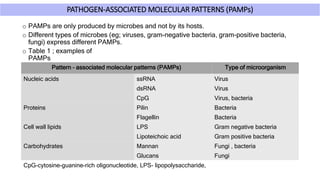
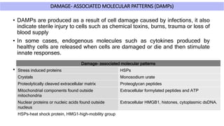
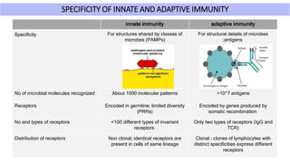
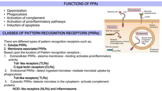
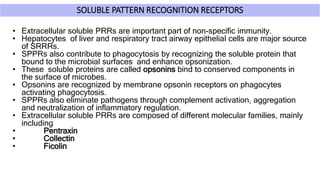
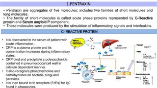
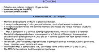
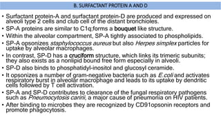
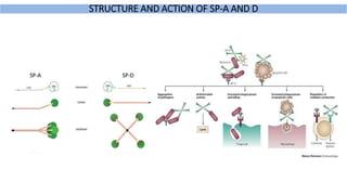
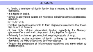
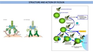
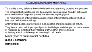
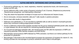
Pattern recognition receptors are type of receptors that plays a major role in innate immunity by recognizing conserved molecular components of the pathogen called pathogens- associated molecular patterns (PAMPs).There are different kinds of PRRS such as soluble pattern recognition receptors and membrane associated PRRs that recognises different kinds of PAMPs such as Carbohydrates,Proteins, lipids and nucleic acids and thereby eliminating the pathogen through different mechanisms.
2. INTRODUCTION
• Vertebrates are protected by both innate and adaptive immunity.
• Innate immunity consists of the defenses against infection that are
ready for immediate action when a host is attacked by a
pathogen.
• The major function of innate immunity is the recognition of foreign
substances or organisms that have penetrated our outer defenses
such as skin epithelium.
• The elimination of such agents are carried out
by different cells and molecules of immune system.
• Thus a critical role of immune system is to determine what is
foreign or non self from what is normally present in body.
3. INNATE IMMUNITY AND PATTERN RECOGNITION RECEPTORS (PRRs)
o The cells that participate in innate immunity recognize conserved molecular components of
pathogen through cell surface or intracellular receptors called pattern recognition
receptors (PRRs).
o The innate immune system uses these receptors to recognize conserved molecular structures
that are produced by microbial pathogens called Pathogen- associated molecular patterns
(PAMPs) and also endogenous molecules that are produced and released by damaged and
dying cells called Damaged –associated molecular patterns (DAMPs).
o Pattern recognition receptors are expressed mainly by immune cells such as dendritic cells,
macrophages, monocytes, neutrophiles and non-immune cells such as epithelial cells, they also
expressed on the surface, in phagocytic vesicles and in the cytosol of various cell types.
o When PAMPs and DAMPs bind to the pattern recognition receptors they activate signal
transduction pathways that promote the antimicrobial and proinflammatory functions of
the cells in which they are expressed.
o In addition ; many proteins present in the blood and extracellular fluids recognize PAMPs and
facilitate the clearance of microbes from blood.
4. PATHOGEN-ASSOCIATED MOLECULAR PATTERNS (PAMPs)
o PAMPs are only produced by microbes and not by its hosts.
o Different types of microbes (eg; viruses, gram-negative bacteria, gram-positive bacteria,
fungi) express different PAMPs.
o Table 1 ; examples of
PAMPs
Pattern – associated molecular patterns (PAMPs) Type of microorganism
Nucleic acids ssRNA Virus
dsRNA Virus
CpG Virus, bacteria
Proteins Pilin Bacteria
Flagellin Bacteria
Cell wall lipids LPS Gram negative bacteria
Lipoteichoic acid Gram positive bacteria
Carbohydrates Mannan Fungi , bacteria
Glucans Fungi
CpG-cytosine-guanine-rich oligonucleotide, LPS- lipopolysaccharide,
5. DAMAGE- ASSOCIATED MOLECULAR PATTERNS (DAMPs)
• DAMPs are produced as a result of cell damage caused by infections, it also
indicate sterile injury to cells such as chemical toxins, burns, trauma or loss of
blood supply
• In some cases, endogenous molecules such as cytokines produced by
healthy cells are released when cells are damaged or die and then stimulate
innate responses.
• These molecules are subset of DAMPs and are called alarmins.
Damage- associated molecular patterns
Stress induced proteins HSPs
Crystals Monosodium urate
Proteolytically cleaved extracellular matrix Proteoglycan peptides
Mitochondrial components found outside
mitochondria
Extracellular formylated peptides and ATP
Nuclear proteins or nucleic acids found outside
nucleus
Extracellular HMGB1, histones, cytoplasmic dsDNA.
HSPs-heat shock protein, HMG1-high-mobility group
6. SPECIFICITY OF INNATE AND ADAPTIVE IMMUNITY
innate immunity adaptive immunity
Specificity For structures shared by classes of
microbes (PAMPs)
For structural details of microbes
;antigens
No of microbial molecules recognized About 1000 molecular patterns >10^7 antigens
Receptors Encoded in germline; limited diversity
(PRRs)
Encoded by genes produced by
somatic recombination
No and types of receptors <100 different types of invariant
receptors
Only two types of receptors (IgG and
TCR)
Distribution of receptors Non clonal; identical receptors are
present in cells of same lineage
Clonal ; clones of lymphocytes with
distinct specificities express different
receptors
7. FUNCTIONS OF PPRs
• Opsonization
• Phagocytosis
• Activation of complement
• Activation of proinflammatory pathways
• Induction of apoptosis
CLASSES OF PATTERN RECOGNITION RECEPTORS (PRRs)
There are different types of pattern recognition receptors such as;
1. Soluble PRRs
2. Membrane associated PRRs
Based upon the location of Pattern recognition receptors ;
1. Extracellular PRRs – plasma membrane – binding activates proinflammatory
activity
Toll- like receptors (TLRs)
C-type lectin receptors (CLRs)
2 . Endosomal PRRs – detect ingested microbes- mediate microbial uptake by
phagocytosis
Toll-like receptors( TLRs)
3. Cytosolic PRRs- detects microbes in the cytoplasm- activate complement
proteins
NOD- like receptors (NLRs) and Inflammasome
8. SOLUBLE PATTERN RECOGNITION RECEPTORS
• Extracellular soluble PRRs are important part of non-specific immunity.
• Hepatocytes of liver and respiratory tract airway epithelial cells are major source
of SRRRs.
• SPPRs also contribute to phagocytosis by recognizing the soluble protein that
bound to the microbial surfaces and enhance opsonization.
• These soluble proteins are called opsonins bind to conserved components in
the surface of microbes.
• Opsonins are recognized by membrane opsonin receptors on phagocytes
activating phagocytosis.
• SPPRs also eliminate pathogens through complement activation, aggregation
and neutralization of inflammatory regulation.
• Extracellular soluble PRRs are composed of different molecular families, mainly
including
• Pentraxin
• Collectin
• Ficolin
9. 1.PENTRAXIN
• Pentraxin are aggregates of five molecules; includes two families of short molecules and
long molecules.
• The family of short molecules is called acute phase proteins represented by C-Reactive
protein and Serum amyloid P component.
• These molecules were produced by the stimulation of inflammatory signals and interleukins.
C- REACTIVE PROTEIN
• It is discovered in the serum of patient with
acute inflammation .
• CRP is a plasma protein and its
concentration increases during inflammatory
states.
• CRP bind and precipitate c polysaccharide
contained in pneumococcal cell wall in
calcium dependent manner.
• It also recognize phosphocholine and
carbohydrates on bacteria, fungi and
parasites.
• It is then bound to fc receptors (FcRs) for IgG
found in phagocytes.
10. 2.COLLECTINS
• Collectins are collagen containing C-type lectins.
• Mannose-binding lectins (MBL)
• Surfactant protein A and D
A.MANNOSE-BINDING LECTINS (MBL)
• Mannose binding lectins are found in plasma and alveoli.
• It recognizes large array of pathogens and activates classical pathway of complement.
• It recognizes carbohydrates with terminal mannose and fucose and various microbial structures.
• STRUCTURE
• MBL is composed of 3 identical 32kDa polypeptide chains, which associate to a hexamer.
• The individual polypeptide chains are composed of a C- terminal fibrinogen like recognition
or carbohydrate binding domain (CDRs) and an N-terminal cysteine rich neck domain which is
followed by a collagen domain.
• The end of CDRs can identify sugar structures of pathogens such as mannose, fucose, glucose etc.
Yeast , parasites, gram bacteria and so on.
• In circulation MBL is complexed to MBL –associated serine protease MASP-2 and MASP-3.
• The MASPs then activate the C1 complement pathways .
12. B. SURFACTANT PROTEIN A AND D
• Surfactant protein-A and surfactant protein-D are produced and expressed on
alveoli type 2 cells and club cell of the distant bronchioles.
• SP-A proteins are similar to C1q;forms a bouquet like structure.
• Within the alveolar compartment, SP-A tightly associated to phospholipids.
• SP-A opsonizes staphylococcus aureus but also Herpes simplex particles for
uptake by alveolar macrophages.
• In contrast, SP-D has a cruciform structure, which links its trimeric subunits;
they also exists as a nonlipid bound free form especially in alveoli.
• SP-D also binds to phosphatidyl-inositol and glucosyl ceramide.
• It opsonizes a number of gram-negative bacteria such as E.coli and activates
respiratory burst in alveolar macrophage and leads to its uptake by dendritic
cells followed by T cell activation.
• SP-A and SP-D contributes to clearance of the fungal respiratory pathogens
such as Pneumocytosis caririi, a major cause of pneumonia on HIV patients.
• After binding to microbes they are recognized by CD91opsonin receptors and
promote phagocytosis.
14. 3.FICOLINS
• L ficolin, a member of ficolin family that is related to MBL and other
collectins.
• It is found in blood.
• Binds to acetylated sugars on microbes including some streptococcal
species.
• STRUCTURE
• Ficolins are lectins assemble to form oligomeric structures that looks
like 'bunch of flowers'.
• It has high calcium dependent binding specificity for N-acetyl
glucosamine, a cell wall component of Aspergillus fumigatus.
• Primarily function as opsonins, induce phagocytosis of fungi.
• Contribute to the activation of lectin pathway by interacting and
activating the MBL-associated serine proteases.
• Trigger the production of inflammatory cytokines and nitric oxide by
macrophages.
16. 4. ANTIMICROBIAL PROTEINS AND PEPTIDES
• To provide strong defense the epithelial cells secrete many proteins and peptides
• The antimicrobial substances can be enzymes such as saliva found in saliva and
tears and fluids of respiratory tracts that cleaves peptidoglycan.
• The major class of antimicrobial components is antimicrobial peptides which is
less than 100 amino acid long.
• Antimicrobial peptides are cysteine- rich, cationic and amphipathic in nature.
• They interact with acidic phospholipids in lipid bilayer and disrupts the membranes
of microbes by inhibiting the synthesis of DNA, RNA or proteins and
activating antimicrobial enzymes resulting in cell death.
• Major types of antimicrobial peptides
• α and β defensins
• cathelicidin
17. ΑLPHA AND BETA DEFENSINS AND CATHELICIDINS
• Produced by epithelia (eg; oro, nasal, respiratory, intestinal, reproductive tracts, skin keratinocytes,
kidney) and NK cells.
• Human defensins kills a wide variety of bacteria including E.coli, S.aureus, Streptococcus pneumonia,
Pseudomonas aeruginosa and Haemophilus influenzae.
• They also attack the lipoprotein envelope of viruses such as influenzae and herpes viruses.
• Act on monocytes ,immature dendritic cells and T cells results in cytokine activation.
• Act on mast cells results in degranulation.
• Defensins and cathelicidin LL-37 are secreted by epithelial cells as well as stored in neutrophil granules
where they contribute to killing phagocytosed microbes.
• Human α-defensins antimicrobial peptides secreted into the gut by intestinal epithelial paneth cells, located
in crypts between the villi are required for maintaining beneficial bacterial flora.
• On mucosal and glandular epithelial cells, skin keratinocytes and NK cells the expression of AMPs are
induced by particular PRRs (toll-like receptors and NOD NLRs).
• Macrophages cannot produce AMPs followed by PRRs activation, but there is an indirect pathway for the
production of cathelicidin.
• Macrophages TLRs binds to the microbial ligand increases the expression of receptors for vitamin D ; this
binding activates the macrophages to produce cathelicidin which helps to kill pathogen.