SlideShare a Scribd company logo
1 of 32
Running Head: ASTHMA
ANATOMY AND PHYSIOLOGY OF THE RESPIRATORY SYSTEM
The respiratory system is situated in the thorax, and is responsible for gaseous
exchange which involves transfer of oxygen and carbon dioxide between the
atmosphere and the blood.
The respiratory system assists in gas exchange and performs other functions as well.
1. Gas exchange. The respiratory system allows oxygen from the air to enter the
blood and carbon dioxide to leave the blood and enter the air. The cardiovascular
system transports oxygen from the lungs to the cells of the body and carbon
dioxide from the cells of the body to the lungs. Thus the respiratory and
cardiovascular systems work together to supply oxygen to all cells and to remove
carbon dioxide.
2. Regulation of blood pH. The respiratory system can alter blood pH by changing
blood carbon dioxide levels.
ASTHMA
3. Voice production. Air movement past the vocal cords makes sound and speech
possible.
4. Olfaction. The sensation of smell occurs when airborne molecules are drawn
into the nasal cavity.
5. Innate immunity. The respiratory system provides protection against some
microorganisms by preventing their entry into the body and by removing them
from respiratory surfaces.
The respiratory system is divided into two parts: The upper respiratory tract and
lower respiratory tract. The upper respiratory tract includes the nose, pharynx,
adenoids, tonsils, epiglottis, larynx and trachea. The lower respiratory tract consists of
the bronchi, bronchioles alveolar ducts and alveoli. With the exception of the right and
left main-stem bronchi, all lower airway structures are contained within lungs. The right
lung is divided into three lobes (upper, middle and lower) and the left lung into two lobes
(upper and lower). The structures of the chest wall (ribs, pleura, muscles of respiration)
are also essential to respiration.
Nose - is the primary upper respiratory organ in which air enters into and exits
from the body. Cilia and mucus line the nasal cavity and traps bacteria and foreign
particles that enter in through the nose. In addition, air that passes through the nasal
cavity is humidified and moistened.
The nasal septum divides the nose into two narrow nasal cavities: one area is
responsible for smell and the other area is responsible for respiration. Within the walls
ASTHMA
of the nasal cavity there are frontal, nasal, ethmoid, maxillary, and sphenoid bones.
Cartilage helps form the shape of the nose.
Pharynx - besides the nose, air can enter into the lungs through the mouth. The
pharynx is a tubular structure, positioned behind the oral and nasal cavities, that allows
air to pass from the mouth to the lungs. The pharynx contains three parts: The
nasopharynx, which connects the upper part of the throat with the nasal cavity; the
oropharynx, positioned between the top of the epiglottis and the soft palate; and the
laryngopharynx, located below the epiglottis.
Larynx - from the pharynx, air enters into the larynx, commonly called the voice
box. The larynx is part of the upper respiratory tract that has two main functions: a
passageway for air to enter into the lungs, and a source of vocalization. The larynx is
made up of the hyoid bone and cartilage, which helps regulate the flow of air. The
epiglottis is a flap-like cartilage structure contained in the larynx that protects the
trachea against food aspiration.
Trachea - is sometimes called the windpipe. The trachea filters the air we
breathe and branches into the bronchi.
Bronchi - the bronchi allow the passage of air to the lungs. The trachea is made
of c-shaped ringed cartilage that divides into the right and left bronchus. The right main
bronchus is shorter and wider than the left main bronchus. The right bronchus is
subdivided into three lobar bronchi, while the left one is divided into two lobar bronchi.
Lungs - the lungs are the main organs of the respiratory system. In the lungs
oxygen is taken into the body and carbon dioxide is breathed out. The red blood cells
ASTHMA
are responsible for picking up the oxygen in the lungs and carrying the oxygen to all the
body cells that need it. The red blood cells drop off the oxygen to the body cells, then
pick up the carbon dioxide which is a waste gas product produced by our cells.
Diaphragm - breathing starts with a dome-shaped muscle at the bottom of the
lungs called the diaphragm. When you breathe in, the diaphragm contracts. When it
contracts it flattens out and pulls downward. When you breathe out, the diaphragm
expands reducing the amount of space for the lungs and forcing air out. The diaphragm
is the main muscle used in breathing.
Expiration is mainly due to the natural elasticity of the lungs, which tend to collapse if
they are not held against the thoracic wall. This is the mechanism behind lung collapse
if there is air in the pleural space (pneumothorax).
• Tidal volume - this is the volume of air moved in and out of the lungs with each
respiratory movement. The average tidal volume is 500 ml.
• Inspiratory reserve – this is the volume of air, in excess of the tidal, that can be
inhaled by the deepest possible inspiration. The average inspiratory reserve is
3,000 ml.
• Expiratory reserve – this is the volume of air, in excess of the tidal, which can be
exhaled by the deepest possible expiration. It is about 1,200 ml.
PHYSIOLOGY OF GAS EXCHANGE
Each branch of the bronchial tree eventually sub-divides to form very narrow
terminal bronchioles, which terminate in the alveoli. There are many millions of alveloi in
ASTHMA
each lung, and these are the areas responsible for gaseous exchange, presenting a
massive surface area for exchange to occur over.
Each alveolus is very closely associated with a network of capillaries containing
deoxygenated blood from the pulmonary artery. The capillary and alveolar walls are
very thin, allowing rapid exchange of gases by passive diffusion along concentration
gradients. CO2 moves into the alveolus as the concentration is much lower in the
alveolus than in the blood, and O2 moves out of the alveolus as the continuous flow of
blood through the capillaries prevents saturation of the blood with O2 and allows
maximal transfer across the membrane.
The lung can be conceptualized as a collection of 300
million bubbles (alveoli), each 0.3 mm in diameter. The
alveolar surface is composed of two kinds of cells: type
I and type II. Type I cells provide structure and type II
cells secrete surfactant.
Surfactant lowers surface tension in the alveoli, thereby
reducing the amount of pressure needed to inflate the
alveoli and decreasing the tendency of the alveoli
collapse. This sigh stretches the alveoli and causes
surfactant to be secreted by type II cells.
References: Lewis Heitkemper Dirksen,. Medical Surgical Nursing: Assessment and
Management of Clinical Problems,. 5th
edition.
The Virtual Autopsy of Different Systems. Retrieved: (November 07, 2014)
http://www.le.ac.uk/pa/teach/va/titlpag1.html
ASTHMA
OVERVIEW OF DISEASE
Over 10% of people have some history of asthma. It often runs in families. The
heritable nature of asthma is not well understood, however, and geneticists cannot
define the precise manner in which it is passed from parents to children.
Asthma affects the airways, which begin just below the throat as a single tube
called the trachea. The trachea is situated immediately in front of the esophagus, the
passageway that connects the throat with the stomach. The trachea divides into two
slightly narrower tubes called the main bronchi (each one is called a bronchus). Each
main bronchus then divides into progressively smaller tubes - the smallest are called
bronchioles - to carry air to and from microscopic air spaces called alveoli. It is in the
alveoli that the important work of the lung occurs, exchanging oxygen in the air for
carbon dioxide in the blood..
The airways are lined with a mucus membrane that secretes a fine layer of
mucus and fluid. This mucus washes the airways to remove any bacteria, dirt, or other
ASTHMA
foreign material that might get into our lungs. The overreaction or hyper-responsiveness
of the airways results in bronchospasm, which is excessive contraction or spasm of the
bronchial smooth muscle. The airways also become inflamed with swelling of the
bronchial mucous membrane (mucosa) and secretion of excessive thick mucus that is
difficult to expel. The airway hyper-responsiveness leading to obstruction of the airways
occurs from one or more of various stimuli that vary with the individual patient. These
include:
• Viral (but not bacterial) respiratory infections (common colds)
• Inhaled irritants (cigarette smoke, wood burning stoves and fireplaces, strong
odors, chemical fumes)
• Inhaled allergens (pollens, dusts, molds, animal danders)
• Cold air
• Exercise
Occasional ingested substances (aspirin, sulfite preservatives, specific foods).
Sometimes these exposures just act as triggers of brief symptoms with rapid relief once
exposure ends. Sensitivity of the airway may be increased, however, following even
brief exposure to one of these.
The obstruction of the airways decreases the rate at which air can flow. This is
felt as tightness in the chest and labored breathing (dyspnea). The obstruction and
inflammation causes coughing. Obstruction to air flow can be measured with pulmonary
ASTHMA
function tests, which can detect even degrees of airway obstruction not yet causing
symptoms. Pulmonary function measurements can be an extremely valuable tool for
your physician to make decisions regarding treatment.
Narrowing of the airway causes noises when air passes through them with
sufficient speed. This typical high-pitched noise is called wheezing. Mucus in the airway
causes a rattling sound called coarse crackles. Complete obstruction of some airways
can cause absorption of air from the alveoli (air sacks at the end of the airways in the
lungs). This causes portions of the lung to appear more dense and cast more of a
shadow on a chest x-ray (this is called atelectasis). The rattling sounds or increased
shadows on the x-ray are often misinterpreted as indicating pneumonia.
There are different patterns of asthma. Some people have only an intermittent
pattern of disease. They have self-limited episodes of varying severity followed by
extended symptom-free periods. The individual episodes are frequently triggered by
viral respiratory infections (causes of the common cold). This is particularly common in
young children in whom viral respiratory infections are frequent (as many as 8 to 12 per
year during the toddler and preschool age group). Others have these intermittent
symptomatic periods brought on by vigorous exertion, cold air, or specific environmental
exposures. This pattern is intermittent asthma.
Some patients have daily or very frequently recurring symptoms. Although
variable in severity, these patients do not have extended periods free of chest tightness,
labored breathing, exertional intolerance, or cough. They may additionally have acute
exacerbations triggered by the same factors that cause symptoms with an intermittent
ASTHMA
or seasonal allergic pattern of disease. Thus, viral respiratory infections (common colds)
and specific environmental exposures may further increase the severity of symptoms in
these patients. This pattern is chronic asthma (sometimes called persistent asthma). All
patterns of disease are associated with varying degrees of severity ranging from mild to
severe.
The airway obstruction of asthma is generally completely reversible and usually
does not cause permanent damage to the lungs, heart, or other organs. However,
severe acute episodes of asthma can be associated with life threatening events and
even fatalities. Survival of severe life threatening events can be associated with damage
from lack of oxygen during the severe exacerbation, and lack of oxygen to the brain can
cause loss of consciousness and brain damage.
Other factors can also worsen asthma on occasion. Hyperventilation, excessively
rapid and deep breathing can worsen asthma. This occurs from anxiety in some
patients, particularly when asthma symptoms have begun for some other reason. A
vicious cycle then occurs of asthma causing anxiety, which then worsens asthma,
thereby causing more anxiety, etc. Ingested substances, such as aspirin, sulfite
preservatives, and specific foods can cause acute attacks of asthma in sensitive
patients.
References: Miles Weinberger, MD Professor of Pediatrics, Allergy,
Immunology, and Pulmonary., Overview of Asthma. Retrieved: (November 07, 2014)
http://www.uichildrens.org/childrens-content.aspx?id=228741
American Academy of Allergy Asthma and Immunology,. Overview of asthma.
Retrieved: (November 07, 2014) http://www.aaaai.org/conditions-and-
treatments/asthma.aspx
ASTHMA
ASSESSMENT
MILD
exertional dyspnea/cough
+/- nocturnal symptoms
increased use of β –agonists
good response to β –agonists
MODERATE
dyspnea at rest
cough
nocturnal symptoms
partial relief from β –agonists
β –agonists needed >8 puffs/ day
chest tightness
SEVERE
labored respiration
agitated, diaphoretic
difficulty of speaking
tachycardia
no relief with β –agonists
NEAR DEATH
exhausted, confused
diaphoretic, cyanotic
silent chest, decreased respiratory effort
falling heart rate
ASTHMA
During the physical assessment the primary symptom will be shortness of breath,
other symptoms may include:
• Anxiety
• Cough
• Chest tightness
• Diaphoresis.
• You may also find signs such as
• Barrel chest
• Diffuse or local wheezes
• Pallor
• Pulse paradoxus (pulse becomes weaker with inspiration and stronger with
expiration)
• Accessory muscle use.
In mild cases of asthma wheezing may only be heard at the end of expiration. In
more severe cases, wheezing will be present throughout expiration. As the attack
worsens, wheezing will be present during both inspiration and expiration. During the
most severe attacks wheezing may be absent. This indicates that the smaller airways
ASTHMA
have become obstructed by mucus or that the patient is so fatigued that they are not
moving enough air to make the sound.
Wheezing is not sign that is exclusive to asthma. It is important to gather a good
history and consider other possibilities prior to deciding to treat the patient for asthma.
Laryngeal abnormalities, growths and foreign body obstructions can all cause wheezing.
Asthma attacks can be placed into three severity categories mild,
moderate and severe.
• In a mild attack, the patient will be able to speak in sentences and will be willing to lie
down. They may be anxious but are easily calmed. The patient’s respiratory rate will be
increased and accessory muscles will not be used. End expiatory wheezes may be
present. Oxygen saturations will be above 95% on room air.
• During a moderate attack the patient will be breathless while talking and may only
use short sentences. Respiratory rates will be increased and accessory muscles will be
used. The heart rate will be increased and a pulse paradox may be present. The patient
will have loud expiatory wheezes. In infants, trouble feeding and a short soft cry will be
noted. Oxygen saturations will be between 91% and 95% on room air.
• In severe attacks the patient will be breathless even while resting. They will be very
anxious, sitting upright and will be unwilling to lie down. They will only be able to speak
in single words. Respiratory rate will be 30 bpm or higher. Accessory muscles will be
used. The patients 9 pulse rate will be as high as 120 bpm and the patient may have a
low blood pressure as the hyper inflated lungs are affecting the pre-load of the heart.
ASTHMA
Both inspiratory and expiratory wheezes can be heard. Pulse paradox is often present.
The patients Oxygen saturations will be below 90%.
While assessing an asthma patient consideration should be given to the amount
of respiratory effort being made. A patient who fits the description of one having a
moderate attack but is working hard to breath will soon become tired and quickly
progress into respiratory arrest.
Reference: Lindell Forbes EMT-P.,Assessing the Asthmatic Airway.
Retrieved: (November 08, 2014)
http://www.umchealthsystem.com/downloads/ems/Assessing_the_Asthmatic_Airway.pd
f
ASTHMA
PATHOPHYSIOLOGY
HEREDITARY FACTORS
-Genetic predisposition
-Atopy
-Airway hyper responsiveness
-Male sex (< 10 years old)
-Female sex (adult)
ACQUIRED FACTORS
-Indoor allergens (domestic mites, animal
allergens/ danders, cockroach allergens,
fungi)
-Outdoor allergens (pollen, fungi)
-Irritants (tobacco smoke, air pollutants)
-Respiratory infections (rhinoviruses,
coronaviruses, influenza, para influenza,
respiratory syncytial virus (RSV),
adenoviruses)
-High socio-economic status
-Small family size
-Higher Body Mass Index
TRIGGERS
-Allergens
-Air pollutants including tobacco smoke
-Respiratory infections
-Exercise and Hyper ventilation
-Weather changes
-Extreme Emotional Expressions
-Drugs (acetylsalicylic acid, beta blockers,
contrast agents, nebulized drugs like
beclomethasone)
-Foods and additives
Release of mediators from mast cells, eosinophils, macrophages, lymphocytes
IgE- mast cell mediated response
Infiltration with monocytes and lymphocytes
LATE PHASE RESPONSE
(Peaks in 5-6 hour)
EARLY PHASE RESPONSE
(Peaks in 30-60 min)
Bronchial smooth muscle constriction
Mucus secretion
Vascular leakage
Mucosal edema
Obstruction of large and small airways
Air trapping
Respiratory acidosis
Hypoxemia
Infiltration with eosinophils and neutrophils
Inflammation
Bronchial hyper reactivity
(Within 1-2 days)
Reference: Lewis Heitkemper Dirksen., Medical-Surgical Nursing: Assessment & Management of Clinical Problems., 5th
Edition.
ASTHMA
MEDICAL MANAGEMENT
Asthma is not so much "treated" as it is "controlled". As a chronic, long-term
disease, there is no cure. However, there are tools and medicines to help you control
asthma as well as benchmarks to gauge your progress.
The Peak Flow Meter – peak flow meter is a simple, small, hand-held tool that can
help you maintain control of asthma by providing a measurement of how well air moves
out of the lungs.If peak flow tests begin to decline - even before other symptoms are
present - it may indicate a looming asthma attack. After taking asthma medication, the
peak flow meter can be used to test the effectiveness of drug therapy.
Inhaler – medication may also be administered using a nebulizer, providing a
larger, continuous dose. Nebulizers vaporize a dose of medication in a saline solution
into a steady stream of foggy vapor that is inhaled by the patient.
SMART (Single Inhaler Maintenance and Reliever Therapy), is better for the
relief and preventive treatment of asthma symptoms in adults compared to standard
therapy, researchers reported in The Lancet Respiratory Medicine (March 2013 issue).
SMART refers to using ICS (corticosteroid) plus LABA (long-acting β2 agonist) in one
inhaler. Medical guidelines advise doctors to prescribe corticosteroids (ICS) plus rapid-
onset long-acting β2 agonist (LABA) combination inhaler to achieve control, together
with a second short-acting β2 agonist (SABA) inhaler for rescue usage, for the
treatment of symptoms. SMART, on the other hand, uses only a single ICS/LABA
inhaler for both relief and preventive treatment.
ASTHMA
Long-term control medicines are taken every day and are designed to prevent
asthma symptom such as airway inflammation. Inhaled corticosteroids are the most
effective long-term control medicine - the best at relieving airway inflammation and
swelling. They are usually taken daily to greatly reduce the inflammation that initiates
the chain reaction of the asthma attack.
There are other long-term control medicines available that doctors may
prescribe. Most of them are taken by mouth and are designed to open the airways and
prevent airway inflammation. Examples include inhaled long-acting B2-agonists (used
with low-dose inhaled corticosteroids), leukotriene modifiers, cromolyn and nedocromil,
and theophylline.
Quick-relief medicines relieve asthma symptoms when they occur. The most
common of these are inhaled short-acting B2-agonists - bronchodilators that quickly
relax tight muscles around the airways, allowing air to flow through them. The quick-
relief inhaler should be used when asthma symptoms are first noticed, but should not be
used more than 2 days a week. Most people carry the quick-relief inhaler with them at
all times. Quick-relief medicines usually do not reduce inflammation and therefore
should not be used as a replacement for long-term control medicines.
Emergency Care – lifesaving treatments at the hospital will consist of direct
oxygen (to alleviate hypoxia) and higher doses of medicines. Emergency personnel will
likely administer a cocktail of short-acting B-2 agonists, systemic oral or intravenous
steroids, other bronchodilators; nonspecific injected or inhaled B-2 agonists,
anticholinergics, inhalation anesthetics, the dissociative anesthetic ketamine, and
ASTHMA
intravenous magnesium sulfate. Intubation (a breathing tube down one's throat) and
mechanical ventilation may also be used in patients undergoing respiratory arrest.
Vitamin D MayReduce Asthma Symptoms
Researchers from King’s College London have discovered how vitamin D can
reduce asthma symptoms. Catherine Hawrylowicz and team explained in the Journal of
Allergy and Clinical Immunology (May 2013 issue) that their findings may offer a new
way of treating the debilitating and usually chronic condition.
Asthma patients are currently prescribed steroid tablets, which may have harmful
side effects. There is a type of asthma, however, that is resistant to steroid therapy.
Patients with this type are susceptible to severe and often life-threatening asthma
attacks.The scientists found that people with asthma have higher levels of IL-17A
(interleukin-17A). IL-17A is part of the immune system that protects the body against
infection. However, this natural compound also worsens asthma symptoms. Large
amounts of IL-17A can reduce the clinical effects of steroids.
The team found that asthma patients who were on steroids had the highest levels
of IL-17A. They also found that vitamin D significantly lowers IL-17A production in cells.
Hawrylowicz believes vitamin D could be a safe and useful add-on treatment.
Reference: This asthma information section was written by Peter Crosta. It was
first published in September 2007 and last updated on 5 March 2013. The
information may not be re-produced in any way without the permission of Medical
News Today. Retrieved: (November 08, 2014)
http://www.medicalnewstoday.com/info/asthma/treatment-for-asthma.php
ASTHMA
SURGICAL MANAGEMENT
Methods of surgical treatment of bronchial asthma (BA) are:
GLOMECTOMY for ASTHMA: The carotid body is a chemoreceptor, sensitive
to: hypoxemia, decreased pH, and CO2retention. Stimulation causes hyperpnoea. This
is beneficial if airways are clear. In the asthmatic, such a reflex initiates a vicious cycle.
An already diminished functional capacity, due to bronchospasm. Mucosal swelling, and
secretion accumulation, is further reduced by hyperpnoea. Removal of one carotid body
apparently prevents such an unwanted sequence, as well as partially interrupts
pathways of communication to the pulmonary plexus which contains bronchodilator
fibers.
Reference: RICHARD H. OVERHOLT., December 1961, Vol 40, No. 6., Glomectomy
for Asthma. Retrieved: (November 08, 2014)
http://journal.publications.chestnet.org/article.aspx?articleid=1055280
GANGLIECTOMY: Excision of a ganglion; surgical removal of a mass of tissue
in lungs.
VAGOTOMY: Unilateral application of histamine in one segmental bronchus
potentiated the airway resistance increase caused by ACH challenge of the bronchial
tree. Unilateral or contralateral blockade of the N. vagus reduces the severity of the
reaction by about 70% of the values before the blockade. The arterial blood gases were
not influenced by the unilateral blockade of the N. vagus. The decrease of the arterial
oxygen pressure following the ACH induced bronchoconstriction was not changed by
the unilateral vagotomy.
ASTHMA
Reference: M. S. Islam, I. Zimmermann, W. T. Ulmer., The Role of Unilateral
Vagotomy on Reflex Broncho constriction. 14. II. 1975, Volume 152, Issue 4, pp
281-289. Retrieved: (November 08, 2014)
http://link.springer.com/article/10.1007%2FBF02094942
IMPLANTATION OF NEUROSTIMULATORS OF SINOCAROTID also called
an implanted pulse generator (IPG) is a battery powered device designed to
deliver electrical stimulation to the brain, central and peripheral nervous system.
RADIOFREQUENCY ELECTRO STIMULATION OF SYMPATHETIC NERVE
BOUNDARY TRUNK IN PATIENTS WITH BRONCHIAL ASTHMA. The boundary trunk
of the sympathic nerve (BTSN) has been implanted in the neck can produce both
bronchial dilatation and bronchial spasm resulted in a decrease of bronchial hyper
reactivity, frequency of asthmatic attacks
Reference: Surgical treatment of bronchial asthma. Retrieved: (November 08, 2014)
http://www.ncbi.nlm.nih.gov/pubmed/12162078
ASTHMA
NURSING DIAGNOSIS AND MANAGEMENT
Nursing Care Plans
1. Ineffective Airway Clearance— the presence of a foreign microorganism
triggers the B lymphocyte to produce antibodies that are specific to that antigen.
These antibodies then attach to mast cells in the lungs. The mast cells with the
antibody attach to the antigen and begin to degranulate. This degranulation
causes the release of certain chemical mediators, namely, histamine, bradykinin,
prostaglandin, and leukotriene. These chemical mediators cause bronchospasm
leading to bronchoconstriction, increased vascular permeability leading to fluid
leakage from the lung vasculature and increased mucus production. These lead
to swelling of the bronchi, mucus buildup that plugs the airway and decreased
bronchial diameter. This causes an increased airway resistance and a
constricted pathway for air. Air cannot pass effectively and this manifests as a
whistling sound. Coughing is a way to expel the obstruction (mucus plug) while
dyspnea is a manifestation of the increased airway resistance.
Patient may manifest
• Difficulty breathing
• Changes in depth and rate of respiration
• Use of respiratory accessory muscles
• Persistent ineffective cough with or without sputum production
• Wheezing upon inspiration and expiration
ASTHMA
• Dyspnea
• Coughing
• Tachypnea, prolonged expiration
• Tachycardia
• Chest tightness
• Suprasternal retraction
• Restlessness
• Anxiety
• Cyanosis
• Loss of consciousness
NURSING DIAGNOSIS
Ineffective airway clearance RT bronchoconstriction, increased mucus production, and
respiratory infection AEB wheezing, dyspnea, and cough
May be related to:
• Increased production or retainment of pulmonary secretions
• Bronchospasms
• Decreased energy
• Fatigue
ASTHMA
Planning – patient will maintain/improve airway clearance AEB absence of signs of
respiratory distress.
- Patient will verbalize understanding that allergens like dust, fumes, animal
dander, pollen, and extremes of temperature and humidity are irritants or factors
that can contribute to ineffective airway clearance and should be avoided.
- Patient will demonstrate behaviors that would prevent the recurrence of the
problem.
Nursing Interventions Rationale
Keep the patient adequately hydrated. Systemic hydration keeps secretion moist
and easier to expectorate.
Teach and encourage the use of
diaphragmatic breathing and coughing
exercises.
These techniques help to improve
ventilation and mobilize secretions without
causing breathlessness and fatigue.
Instruct patient to avoid bronchial
irritants such as cigarette smoke,
aerosols, extremes of temperature, and
fumes.
Bronchial irritants cause
bronchoconstriction and increased mucus
production, which then interfere with airway
clearance.
Teach early signs of infection that are to
be reported to the clinician immediately.
Minor respiratory infections that are of no
consequence to the person with normal
lungs can produce fatal disturbances in the
ASTHMA
Nursing Interventions Rationale
lungs of an asthmatic person. Early
recognition is crucial.
Assist and prepare patient for postural
drainage.
Uses gravity to help raise secretions so
they can be more easily expectorated.
Administer nebulization as ordered. This ensures adequate delivery of
medications to the airways.
Administer medications as ordered. Antibiotics may be prescribed to treat the
infection.
2. Ineffective Breathing Pattern— presence of secretions in the bronchi will result
into a blockage of air that will enter the body and thus producing insufficient air
needed by the body and inability to maintain clear airway. This obstruction is
further heightened by bronchospasm due to the contraction of the smooth
muscles in the bronchi. This is caused by parasympathetic stimulation of the
muscarinic m2 receptors as well as by chemical mediators released in response
to the presence of allergens.
NURSING DIAGNOSIS
ASTHMA
Ineffective breathing pattern r/t presence of secretions AEB productive cough and
dyspnea
Planning – patient will demonstrate pursed-lip breathing and diaphragmatic breathing.
- Patient will manifest signs of decreased respiratory effort AEB absence of
dyspnea
- Patient will verbalize understanding of causative factors and demonstrate
behaviors that would improve breathing pattern
Nursing Interventions Rationale
Assess patient’s respiratory rate, depth,
and rhythm. Obtain pulse oximetry.
To obtain baseline data
Monitor and record vital signs. Increase in respiratory rate could mean
worsening condition.
Auscultate breath sounds and assess
airway pattern
to check for the presence of adventitious
breath sounds
Elevate head of the bed and change
position of the pt. every 2 hours.
To minimize difficulty in breathing
Encourage deep breathing and
coughing exercises.
To maximize effort for expectoration.
Demonstrate diaphragmatic and To decrease air trapping and for efficient
ASTHMA
Nursing Interventions Rationale
pursed-lip breathing. breathing.
Encourage increase in fluid intake To prevent fatigue.
Encourage opportunities for rest and
limit physical activities.
To prevent situations that will aggravate
the condition
Reinforce low salt, low fat diet as
ordered.
To mobilize secretions.
3. Impaired Gas Exchange— bronchial asthma is a condition wherein the airway
diameter is highly reduced. This is due to severe bronchospasm, mucosal edema
and mucus plug formation. There is a rise in airway resistance which leads to
decreased amount of air that enters upon inspiration as well as expiration. Thus,
ventilation is impaired. In bronchial asthma, perfusion is not directly affected.
However, the balance between ventilation and perfusion (V/Q ratio) is lost
because despite the adequate perfusion (capillary circulation), not much gas is
available to diffuse from the alveoli to the capillaries. Conversely, the gases in
the capillaries do diffuse to the alveoli but since expiration is impaired, such
gases fail to be ventilated out. Thus, gas exchange is impaired.
NURSING DIAGNOSIS
ASTHMA
Impaired gas exchange RT ventilation perfusion imbalance AEB dyspnea, tachypnea,
and tachycardia
May be related to:
• altered delivery of inspired O2 or air trapping
Planning –patient will improve gas exchange AEB absence of respiratory distress
- Patient will demonstrate improved ventilation and adequate oxygenation of
tissues by ABG’s within client’s normal limits and absence of symptoms of
respiratory distress.
- Patient will verbalize understand of causative factors and appropriate
interventions (deep breathing, cough exercises, etc)
Nursing Interventions Rationale
Assess vital signs, noting respiratory rate,
depth, and rhythm.
To obtain baseline data
VS monitor and record Serve to track important changes
Auscultate breath sounds and assess
airway pattern
to check for the presence of adventitious
breath sounds
Elevate head of the bed and change
position of the pt. every 2 hours.
To minimize difficulty in breathing and
promote maximum lung expansion.
ASTHMA
Nursing Interventions Rationale
Encourage deep breathing and coughing
exercises.
To maximize effort for expectoration.
Demonstrate diaphragmatic and pursed-
lip breathing.
To decrease air trapping and for efficient
breathing.
Encourage increase in fluid intake To prevent fatigue.
Encourage opportunities for rest and limit
physical activities.
To prevent situations that will aggravate the
condition
Reinforce low salt, low fat diet as ordered. To mobilize secretions.
4. Fatigue— fluid accumulation in the lungs makes it difficult to breathe. The fluid
inside prohibits the lungs to expand thus it is harder to breathe. The client, to
have adequate ventilation makes use of his accessory muscles to breathe to
have sufficient air. With too much use of the accessory muscles, feeling of
tiredness may be present resulting to fatigue which is experienced by the client.
ASTHMA
NURSING DIAGNOSIS
Fatigue r/t physical exertion to maintain adequate ventilation AEB use of accessory
muscles to breathe
Patient may manifest:
• Generalized weakness
• Verbalization of overwhelming lack of energy
• Inability to maintain usual routines
• Tired
• Lethargic
• Compromised concentration
• Decreased performance
ASTHMA
Planning – patient will verbalize understand on health teachings given and report
improved sense of energy.
- Patient will perform ADL’s within client’s ability and participates in desired
activities.
- Patient will be able to identify basis of fatigue and be able to cope up with the
problem.
Nursing Interventions Rationale
Establish rapport To gain patient’s trust
Monitor and record vital signs. For baseline data.
Provide environment conducive to relief of
fatigue.
Temperature and level of humidity are known
to affect exhaustion.
Assist client to identify appropriate coping
behaviors.
Promotes sense of control and improves self-
esteem.
Encourage patient to restrict activity and
rest in bed as much as possible.
Helps counteract effects of increased
metabolism.
Avoid topics that irritate or upset patient.
Discuss ways to respond to these feelings.
Increased irritability of the CNS may cause
patient to be easily excited, agitated and
prone to emotional outbursts.
ASTHMA
Nursing Interventions Rationale
Discuss with the patient the need for
activity. Plan schedule with patient and
identify activities that lead to fatigue.
Education may provide motivation to increase
activity level even though patient may feel too
weak initially.
Alternate activity with rest periods. Prevents excessive fatigue.
Monitor VS before and after activity. Indicates physiological levels of tolerance.
Increase patient participation in ADL’s as
tolerated.
Increases confidence level and/or self-
esteem and tolerance level
5. Risk for Activity Intolerance— inadequate oxygen in the circulation can
develop weakness in our muscles. Muscles need oxygen to move and to do its
function. If the patient cannot tolerate any activities because of the low
oxygenation caused by the ventilation-perfusion imbalance caused by the
pathological minimized lung expansion.
NURSING DIAGNOSIS
Risk for Activity Intolerance r/t decrease oxygenation
Planning – patient will participate willingly in necessary/ desired activities such as deep
breathing exercises.
- Patient will perform ADL’s within client’s ability and participates in desired
activities.
ASTHMA
- Patient will be able to increase activity tolerance AEB attendance of self-care
needs.
- Patient will be able to gradually increase activity within level of ability
Nursing Interventions Rationale
Monitor VS. For baseline data.
Assess motor function. To identify causative factors.
Note contributing factors to fatigue. To identify precipitating factors.
Evaluate degree of deficit. To identify severity.
Ascertain ability to stand and move about. To identify necessity of assistive devices.
Assess emotional or psychological factors Stress and/or depression may increase the
effects of illness.
Plan care with rest periods between
activities
To reduce fatigue
Increase activity/exercise gradually such as
assisting the patient in doing PROM to
active or full range of motions.
Minimizes muscle atrophy, promotes
circulation, helps to prevent contractures
Provide adequate rest periods. To replenish energy.
Assist client in doing self-care needs To promote independence and increase
ASTHMA
Nursing Interventions Rationale
activity tolerance
Elevate arm and hand Promotes venous
Place knees and hips in extended position Maintains functional
Other Possible Nursing Care Plans
• Anxiety—may be related to perceived threat of death, possibly evidenced by
apprehension, fearful expression, and extraneous movements.
• Risk for contamination—risk factors may include presence of atmospheric
pollutants, environmental contaminants in the home.
Reference: 5 Bronchial Asthma Nursing Care Plans., February 11, 2012.
Retrieved: (November 08, 2014) http://nurseslabs.com/bronchial-asthma-nursing-care-
plans/

More Related Content

What's hot

Tracheostomy care
Tracheostomy careTracheostomy care
Tracheostomy careTHANUJA MATHEW
 
Ot nursing
Ot nursingOt nursing
Ot nursingRam Prasad
 
CARDIO-PULMONARY RESUSCITATION
CARDIO-PULMONARY RESUSCITATIONCARDIO-PULMONARY RESUSCITATION
CARDIO-PULMONARY RESUSCITATIONresmigs
 
Nursing care plan bronchial asthma part 2
Nursing care plan bronchial asthma part 2Nursing care plan bronchial asthma part 2
Nursing care plan bronchial asthma part 2NUMED SCIENCE
 
Urinary Tract Infection with Nursing Management
Urinary Tract Infection with Nursing ManagementUrinary Tract Infection with Nursing Management
Urinary Tract Infection with Nursing ManagementSwatilekha Das
 
Upper respiratory disorders and nursing mangement
Upper respiratory disorders and nursing mangementUpper respiratory disorders and nursing mangement
Upper respiratory disorders and nursing mangementANILKUMAR BR
 
Urinary Catheterization Nursing Procedure & Management ppt.pptx
Urinary Catheterization Nursing Procedure & Management ppt.pptxUrinary Catheterization Nursing Procedure & Management ppt.pptx
Urinary Catheterization Nursing Procedure & Management ppt.pptxanjalatchi
 
Subcutaneous injection
Subcutaneous injectionSubcutaneous injection
Subcutaneous injectionSusmitaBarman2
 
Tuberculosis-Medical and Nursing Managements
Tuberculosis-Medical and Nursing ManagementsTuberculosis-Medical and Nursing Managements
Tuberculosis-Medical and Nursing ManagementsReynel Dan
 
Breathing and coughing exercises
Breathing and coughing exercisesBreathing and coughing exercises
Breathing and coughing exercisesABHIJIT BHOYAR
 
Lesson plan on surgical wound dressing
Lesson plan on surgical wound dressingLesson plan on surgical wound dressing
Lesson plan on surgical wound dressingAnvin Thomas
 
suctioning procedure ppt.pptx
suctioning procedure  ppt.pptxsuctioning procedure  ppt.pptx
suctioning procedure ppt.pptxanjalatchi
 
Nursing management of critically ill patient in intensive care units
Nursing management of critically   ill patient in intensive care unitsNursing management of critically   ill patient in intensive care units
Nursing management of critically ill patient in intensive care unitsANILKUMAR BR
 
oxygen administration procedure
oxygen administration procedureoxygen administration procedure
oxygen administration procedureSachin Chauhan
 
Endotracheal intubation nursing procedure
Endotracheal intubation nursing procedureEndotracheal intubation nursing procedure
Endotracheal intubation nursing procedureABHIJIT BHOYAR
 

What's hot (20)

Tracheostomy care
Tracheostomy careTracheostomy care
Tracheostomy care
 
Ot nursing
Ot nursingOt nursing
Ot nursing
 
CARDIO-PULMONARY RESUSCITATION
CARDIO-PULMONARY RESUSCITATIONCARDIO-PULMONARY RESUSCITATION
CARDIO-PULMONARY RESUSCITATION
 
Emphysema
EmphysemaEmphysema
Emphysema
 
Copd ppt
Copd pptCopd ppt
Copd ppt
 
Nursing care plan bronchial asthma part 2
Nursing care plan bronchial asthma part 2Nursing care plan bronchial asthma part 2
Nursing care plan bronchial asthma part 2
 
Urinary Tract Infection with Nursing Management
Urinary Tract Infection with Nursing ManagementUrinary Tract Infection with Nursing Management
Urinary Tract Infection with Nursing Management
 
Upper respiratory disorders and nursing mangement
Upper respiratory disorders and nursing mangementUpper respiratory disorders and nursing mangement
Upper respiratory disorders and nursing mangement
 
Urinary Catheterization Nursing Procedure & Management ppt.pptx
Urinary Catheterization Nursing Procedure & Management ppt.pptxUrinary Catheterization Nursing Procedure & Management ppt.pptx
Urinary Catheterization Nursing Procedure & Management ppt.pptx
 
Subcutaneous injection
Subcutaneous injectionSubcutaneous injection
Subcutaneous injection
 
Tuberculosis-Medical and Nursing Managements
Tuberculosis-Medical and Nursing ManagementsTuberculosis-Medical and Nursing Managements
Tuberculosis-Medical and Nursing Managements
 
Breathing and coughing exercises
Breathing and coughing exercisesBreathing and coughing exercises
Breathing and coughing exercises
 
Lesson plan on surgical wound dressing
Lesson plan on surgical wound dressingLesson plan on surgical wound dressing
Lesson plan on surgical wound dressing
 
suctioning procedure ppt.pptx
suctioning procedure  ppt.pptxsuctioning procedure  ppt.pptx
suctioning procedure ppt.pptx
 
Colostomy care
Colostomy careColostomy care
Colostomy care
 
Nursing management of critically ill patient in intensive care units
Nursing management of critically   ill patient in intensive care unitsNursing management of critically   ill patient in intensive care units
Nursing management of critically ill patient in intensive care units
 
oxygen administration procedure
oxygen administration procedureoxygen administration procedure
oxygen administration procedure
 
Endotracheal intubation nursing procedure
Endotracheal intubation nursing procedureEndotracheal intubation nursing procedure
Endotracheal intubation nursing procedure
 
Abdominal paracentesis
Abdominal paracentesis Abdominal paracentesis
Abdominal paracentesis
 
Drug administration
Drug administrationDrug administration
Drug administration
 

Similar to Respiratory System Anatomy Guide

The lungs presentation
The lungs presentationThe lungs presentation
The lungs presentationglenda
 
The respiratory system
The respiratory systemThe respiratory system
The respiratory systemitutor
 
Respiratory system by A.H..pptx
Respiratory system by A.H..pptxRespiratory system by A.H..pptx
Respiratory system by A.H..pptxSadiyaAbubakar7
 
Human Body - Respiratory System
Human Body - Respiratory SystemHuman Body - Respiratory System
Human Body - Respiratory Systemvzt00
 
Resp-System.ppt human anatomy and physiology
Resp-System.ppt human anatomy and physiologyResp-System.ppt human anatomy and physiology
Resp-System.ppt human anatomy and physiologyRubikhan18
 
Introduction Yoga Therapy For The Management Of Respiratory Disorders
Introduction Yoga Therapy For The Management Of Respiratory DisordersIntroduction Yoga Therapy For The Management Of Respiratory Disorders
Introduction Yoga Therapy For The Management Of Respiratory DisordersShama
 
The Respiratory System
The Respiratory SystemThe Respiratory System
The Respiratory SystemAbhay Rajpoot
 
Report in Respiratory System
Report in Respiratory SystemReport in Respiratory System
Report in Respiratory SystemAmy Saguin
 
Respiratory system
Respiratory systemRespiratory system
Respiratory systemShaik Afsar
 
Respiratory System
 Respiratory System Respiratory System
Respiratory SystemFatimaAhmed148
 
respiratory%20system.pptx
respiratory%20system.pptxrespiratory%20system.pptx
respiratory%20system.pptxRohitSharma67482
 
therespiratorysystem-130524022043-phpapp01.pptx
therespiratorysystem-130524022043-phpapp01.pptxtherespiratorysystem-130524022043-phpapp01.pptx
therespiratorysystem-130524022043-phpapp01.pptxPawanKanherkar1
 
EVFQLeOYfTDgnNQm679.pptx
EVFQLeOYfTDgnNQm679.pptxEVFQLeOYfTDgnNQm679.pptx
EVFQLeOYfTDgnNQm679.pptxShivamsharma15812
 
Respiratory system(M.NAGAPRADHEESH).pptx
Respiratory system(M.NAGAPRADHEESH).pptxRespiratory system(M.NAGAPRADHEESH).pptx
Respiratory system(M.NAGAPRADHEESH).pptxMNAGAPRADHEESH
 
03 respiratory system2
03   respiratory system203   respiratory system2
03 respiratory system2mrtangextrahelp
 

Similar to Respiratory System Anatomy Guide (20)

Respiratory syst
Respiratory systRespiratory syst
Respiratory syst
 
Respiration
RespirationRespiration
Respiration
 
The lungs presentation
The lungs presentationThe lungs presentation
The lungs presentation
 
The respiratory system
The respiratory systemThe respiratory system
The respiratory system
 
Respiratory system by A.H..pptx
Respiratory system by A.H..pptxRespiratory system by A.H..pptx
Respiratory system by A.H..pptx
 
Human Body - Respiratory System
Human Body - Respiratory SystemHuman Body - Respiratory System
Human Body - Respiratory System
 
Resp-System.ppt human anatomy and physiology
Resp-System.ppt human anatomy and physiologyResp-System.ppt human anatomy and physiology
Resp-System.ppt human anatomy and physiology
 
Introduction Yoga Therapy For The Management Of Respiratory Disorders
Introduction Yoga Therapy For The Management Of Respiratory DisordersIntroduction Yoga Therapy For The Management Of Respiratory Disorders
Introduction Yoga Therapy For The Management Of Respiratory Disorders
 
the lungs
the lungsthe lungs
the lungs
 
The Respiratory System
The Respiratory SystemThe Respiratory System
The Respiratory System
 
Report in Respiratory System
Report in Respiratory SystemReport in Respiratory System
Report in Respiratory System
 
Respiratory system
Respiratory systemRespiratory system
Respiratory system
 
Respiratory System
 Respiratory System Respiratory System
Respiratory System
 
respiratory%20system.pptx
respiratory%20system.pptxrespiratory%20system.pptx
respiratory%20system.pptx
 
Lungs Grade 4
Lungs Grade 4Lungs Grade 4
Lungs Grade 4
 
therespiratorysystem-130524022043-phpapp01.pptx
therespiratorysystem-130524022043-phpapp01.pptxtherespiratorysystem-130524022043-phpapp01.pptx
therespiratorysystem-130524022043-phpapp01.pptx
 
EVFQLeOYfTDgnNQm679.pptx
EVFQLeOYfTDgnNQm679.pptxEVFQLeOYfTDgnNQm679.pptx
EVFQLeOYfTDgnNQm679.pptx
 
Respiratory system(M.NAGAPRADHEESH).pptx
Respiratory system(M.NAGAPRADHEESH).pptxRespiratory system(M.NAGAPRADHEESH).pptx
Respiratory system(M.NAGAPRADHEESH).pptx
 
Respiratory system of goat
Respiratory system of goatRespiratory system of goat
Respiratory system of goat
 
03 respiratory system2
03   respiratory system203   respiratory system2
03 respiratory system2
 

More from RoxanneMae Birador (20)

Typhoid fever
Typhoid feverTyphoid fever
Typhoid fever
 
Herniated Nucleus Pulposus
Herniated Nucleus PulposusHerniated Nucleus Pulposus
Herniated Nucleus Pulposus
 
Intractable Pain
Intractable PainIntractable Pain
Intractable Pain
 
ELECTROENCEPHALOGRAM/ ELECTROENCEPHALOGRAPHY
ELECTROENCEPHALOGRAM/ ELECTROENCEPHALOGRAPHYELECTROENCEPHALOGRAM/ ELECTROENCEPHALOGRAPHY
ELECTROENCEPHALOGRAM/ ELECTROENCEPHALOGRAPHY
 
Heart failure
Heart failureHeart failure
Heart failure
 
X-RAY , SPECIAL ORTHOPEDIC BEDS
X-RAY , SPECIAL ORTHOPEDIC BEDSX-RAY , SPECIAL ORTHOPEDIC BEDS
X-RAY , SPECIAL ORTHOPEDIC BEDS
 
PHEOCHROMOCYTOMA
PHEOCHROMOCYTOMAPHEOCHROMOCYTOMA
PHEOCHROMOCYTOMA
 
ALDOSTERONISM
ALDOSTERONISM ALDOSTERONISM
ALDOSTERONISM
 
OBESITY
OBESITYOBESITY
OBESITY
 
DENTAL PLAQUE & CARIES
DENTAL PLAQUE & CARIESDENTAL PLAQUE & CARIES
DENTAL PLAQUE & CARIES
 
Philosophy of Man
Philosophy of ManPhilosophy of Man
Philosophy of Man
 
Microsope
MicrosopeMicrosope
Microsope
 
Suicide report
Suicide report Suicide report
Suicide report
 
Community Health Nursing
Community Health NursingCommunity Health Nursing
Community Health Nursing
 
Hygiene Practice
Hygiene PracticeHygiene Practice
Hygiene Practice
 
Urinary Tract Infection
Urinary Tract InfectionUrinary Tract Infection
Urinary Tract Infection
 
Breastfeed
BreastfeedBreastfeed
Breastfeed
 
Non-Communicable Disease
Non-Communicable DiseaseNon-Communicable Disease
Non-Communicable Disease
 
Control of Communicable Diseases
Control of Communicable Diseases Control of Communicable Diseases
Control of Communicable Diseases
 
Biochemistry
BiochemistryBiochemistry
Biochemistry
 

Recently uploaded

Call Girls Gurgaon Parul 9711199012 Independent Escort Service Gurgaon
Call Girls Gurgaon Parul 9711199012 Independent Escort Service GurgaonCall Girls Gurgaon Parul 9711199012 Independent Escort Service Gurgaon
Call Girls Gurgaon Parul 9711199012 Independent Escort Service GurgaonCall Girls Service Gurgaon
 
College Call Girls Hyderabad Sakshi 9907093804 Independent Escort Service Hyd...
College Call Girls Hyderabad Sakshi 9907093804 Independent Escort Service Hyd...College Call Girls Hyderabad Sakshi 9907093804 Independent Escort Service Hyd...
College Call Girls Hyderabad Sakshi 9907093804 Independent Escort Service Hyd...delhimodelshub1
 
VIP Call Girls Sector 67 Gurgaon Just Call Me 9711199012
VIP Call Girls Sector 67 Gurgaon Just Call Me 9711199012VIP Call Girls Sector 67 Gurgaon Just Call Me 9711199012
VIP Call Girls Sector 67 Gurgaon Just Call Me 9711199012Call Girls Service Gurgaon
 
Gurgaon Sector 68 Call Girls ( 9873940964 ) Book Hot And Sexy Girls In A Few ...
Gurgaon Sector 68 Call Girls ( 9873940964 ) Book Hot And Sexy Girls In A Few ...Gurgaon Sector 68 Call Girls ( 9873940964 ) Book Hot And Sexy Girls In A Few ...
Gurgaon Sector 68 Call Girls ( 9873940964 ) Book Hot And Sexy Girls In A Few ...ggsonu500
 
College Call Girls Mumbai Alia 9910780858 Independent Escort Service Mumbai
College Call Girls Mumbai Alia 9910780858 Independent Escort Service MumbaiCollege Call Girls Mumbai Alia 9910780858 Independent Escort Service Mumbai
College Call Girls Mumbai Alia 9910780858 Independent Escort Service Mumbaisonalikaur4
 
Russian Call Girls Hyderabad Saloni 9907093804 Independent Escort Service Hyd...
Russian Call Girls Hyderabad Saloni 9907093804 Independent Escort Service Hyd...Russian Call Girls Hyderabad Saloni 9907093804 Independent Escort Service Hyd...
Russian Call Girls Hyderabad Saloni 9907093804 Independent Escort Service Hyd...delhimodelshub1
 
hyderabad call girl.pdfRussian Call Girls in Hyderabad Amrita 9907093804 Inde...
hyderabad call girl.pdfRussian Call Girls in Hyderabad Amrita 9907093804 Inde...hyderabad call girl.pdfRussian Call Girls in Hyderabad Amrita 9907093804 Inde...
hyderabad call girl.pdfRussian Call Girls in Hyderabad Amrita 9907093804 Inde...delhimodelshub1
 
Gurgaon Sector 90 Call Girls ( 9873940964 ) Book Hot And Sexy Girls In A Few ...
Gurgaon Sector 90 Call Girls ( 9873940964 ) Book Hot And Sexy Girls In A Few ...Gurgaon Sector 90 Call Girls ( 9873940964 ) Book Hot And Sexy Girls In A Few ...
Gurgaon Sector 90 Call Girls ( 9873940964 ) Book Hot And Sexy Girls In A Few ...ggsonu500
 
Call Girl Chandigarh Mallika ❤️🍑 9907093804 👄🫦 Independent Escort Service Cha...
Call Girl Chandigarh Mallika ❤️🍑 9907093804 👄🫦 Independent Escort Service Cha...Call Girl Chandigarh Mallika ❤️🍑 9907093804 👄🫦 Independent Escort Service Cha...
Call Girl Chandigarh Mallika ❤️🍑 9907093804 👄🫦 Independent Escort Service Cha...High Profile Call Girls Chandigarh Aarushi
 
Call Girls LB Nagar 7001305949 all area service COD available Any Time
Call Girls LB Nagar 7001305949 all area service COD available Any TimeCall Girls LB Nagar 7001305949 all area service COD available Any Time
Call Girls LB Nagar 7001305949 all area service COD available Any Timedelhimodelshub1
 
indian Call Girl Panchkula ❤️🍑 9907093804 Low Rate Call Girls Ludhiana Tulsi
indian Call Girl Panchkula ❤️🍑 9907093804 Low Rate Call Girls Ludhiana Tulsiindian Call Girl Panchkula ❤️🍑 9907093804 Low Rate Call Girls Ludhiana Tulsi
indian Call Girl Panchkula ❤️🍑 9907093804 Low Rate Call Girls Ludhiana TulsiHigh Profile Call Girls Chandigarh Aarushi
 
Russian Call Girls Hyderabad Indira 9907093804 Independent Escort Service Hyd...
Russian Call Girls Hyderabad Indira 9907093804 Independent Escort Service Hyd...Russian Call Girls Hyderabad Indira 9907093804 Independent Escort Service Hyd...
Russian Call Girls Hyderabad Indira 9907093804 Independent Escort Service Hyd...delhimodelshub1
 
VIP Call Girls Hyderabad Megha 9907093804 Independent Escort Service Hyderabad
VIP Call Girls Hyderabad Megha 9907093804 Independent Escort Service HyderabadVIP Call Girls Hyderabad Megha 9907093804 Independent Escort Service Hyderabad
VIP Call Girls Hyderabad Megha 9907093804 Independent Escort Service Hyderabaddelhimodelshub1
 
Call Girls Hyderabad Kirti 9907093804 Independent Escort Service Hyderabad
Call Girls Hyderabad Kirti 9907093804 Independent Escort Service HyderabadCall Girls Hyderabad Kirti 9907093804 Independent Escort Service Hyderabad
Call Girls Hyderabad Kirti 9907093804 Independent Escort Service Hyderabaddelhimodelshub1
 
Call Girl Gurgaon Saloni 9711199012 Independent Escort Service Gurgaon
Call Girl Gurgaon Saloni 9711199012 Independent Escort Service GurgaonCall Girl Gurgaon Saloni 9711199012 Independent Escort Service Gurgaon
Call Girl Gurgaon Saloni 9711199012 Independent Escort Service GurgaonCall Girls Service Gurgaon
 
Gurgaon iffco chowk 🔝 Call Girls Service 🔝 ( 8264348440 ) unlimited hard sex ...
Gurgaon iffco chowk 🔝 Call Girls Service 🔝 ( 8264348440 ) unlimited hard sex ...Gurgaon iffco chowk 🔝 Call Girls Service 🔝 ( 8264348440 ) unlimited hard sex ...
Gurgaon iffco chowk 🔝 Call Girls Service 🔝 ( 8264348440 ) unlimited hard sex ...soniya singh
 

Recently uploaded (20)

Call Girls Gurgaon Parul 9711199012 Independent Escort Service Gurgaon
Call Girls Gurgaon Parul 9711199012 Independent Escort Service GurgaonCall Girls Gurgaon Parul 9711199012 Independent Escort Service Gurgaon
Call Girls Gurgaon Parul 9711199012 Independent Escort Service Gurgaon
 
College Call Girls Hyderabad Sakshi 9907093804 Independent Escort Service Hyd...
College Call Girls Hyderabad Sakshi 9907093804 Independent Escort Service Hyd...College Call Girls Hyderabad Sakshi 9907093804 Independent Escort Service Hyd...
College Call Girls Hyderabad Sakshi 9907093804 Independent Escort Service Hyd...
 
VIP Call Girls Sector 67 Gurgaon Just Call Me 9711199012
VIP Call Girls Sector 67 Gurgaon Just Call Me 9711199012VIP Call Girls Sector 67 Gurgaon Just Call Me 9711199012
VIP Call Girls Sector 67 Gurgaon Just Call Me 9711199012
 
Gurgaon Sector 68 Call Girls ( 9873940964 ) Book Hot And Sexy Girls In A Few ...
Gurgaon Sector 68 Call Girls ( 9873940964 ) Book Hot And Sexy Girls In A Few ...Gurgaon Sector 68 Call Girls ( 9873940964 ) Book Hot And Sexy Girls In A Few ...
Gurgaon Sector 68 Call Girls ( 9873940964 ) Book Hot And Sexy Girls In A Few ...
 
VIP Call Girls Lucknow Isha 🔝 9719455033 🔝 🎶 Independent Escort Service Lucknow
VIP Call Girls Lucknow Isha 🔝 9719455033 🔝 🎶 Independent Escort Service LucknowVIP Call Girls Lucknow Isha 🔝 9719455033 🔝 🎶 Independent Escort Service Lucknow
VIP Call Girls Lucknow Isha 🔝 9719455033 🔝 🎶 Independent Escort Service Lucknow
 
Model Call Girl in Subhash Nagar Delhi reach out to us at 🔝9953056974🔝
Model Call Girl in Subhash Nagar Delhi reach out to us at 🔝9953056974🔝Model Call Girl in Subhash Nagar Delhi reach out to us at 🔝9953056974🔝
Model Call Girl in Subhash Nagar Delhi reach out to us at 🔝9953056974🔝
 
Call Girl Lucknow Gauri 🔝 8923113531 🔝 🎶 Independent Escort Service Lucknow
Call Girl Lucknow Gauri 🔝 8923113531  🔝 🎶 Independent Escort Service LucknowCall Girl Lucknow Gauri 🔝 8923113531  🔝 🎶 Independent Escort Service Lucknow
Call Girl Lucknow Gauri 🔝 8923113531 🔝 🎶 Independent Escort Service Lucknow
 
College Call Girls Mumbai Alia 9910780858 Independent Escort Service Mumbai
College Call Girls Mumbai Alia 9910780858 Independent Escort Service MumbaiCollege Call Girls Mumbai Alia 9910780858 Independent Escort Service Mumbai
College Call Girls Mumbai Alia 9910780858 Independent Escort Service Mumbai
 
Russian Call Girls Hyderabad Saloni 9907093804 Independent Escort Service Hyd...
Russian Call Girls Hyderabad Saloni 9907093804 Independent Escort Service Hyd...Russian Call Girls Hyderabad Saloni 9907093804 Independent Escort Service Hyd...
Russian Call Girls Hyderabad Saloni 9907093804 Independent Escort Service Hyd...
 
hyderabad call girl.pdfRussian Call Girls in Hyderabad Amrita 9907093804 Inde...
hyderabad call girl.pdfRussian Call Girls in Hyderabad Amrita 9907093804 Inde...hyderabad call girl.pdfRussian Call Girls in Hyderabad Amrita 9907093804 Inde...
hyderabad call girl.pdfRussian Call Girls in Hyderabad Amrita 9907093804 Inde...
 
Gurgaon Sector 90 Call Girls ( 9873940964 ) Book Hot And Sexy Girls In A Few ...
Gurgaon Sector 90 Call Girls ( 9873940964 ) Book Hot And Sexy Girls In A Few ...Gurgaon Sector 90 Call Girls ( 9873940964 ) Book Hot And Sexy Girls In A Few ...
Gurgaon Sector 90 Call Girls ( 9873940964 ) Book Hot And Sexy Girls In A Few ...
 
Call Girl Chandigarh Mallika ❤️🍑 9907093804 👄🫦 Independent Escort Service Cha...
Call Girl Chandigarh Mallika ❤️🍑 9907093804 👄🫦 Independent Escort Service Cha...Call Girl Chandigarh Mallika ❤️🍑 9907093804 👄🫦 Independent Escort Service Cha...
Call Girl Chandigarh Mallika ❤️🍑 9907093804 👄🫦 Independent Escort Service Cha...
 
Call Girls LB Nagar 7001305949 all area service COD available Any Time
Call Girls LB Nagar 7001305949 all area service COD available Any TimeCall Girls LB Nagar 7001305949 all area service COD available Any Time
Call Girls LB Nagar 7001305949 all area service COD available Any Time
 
indian Call Girl Panchkula ❤️🍑 9907093804 Low Rate Call Girls Ludhiana Tulsi
indian Call Girl Panchkula ❤️🍑 9907093804 Low Rate Call Girls Ludhiana Tulsiindian Call Girl Panchkula ❤️🍑 9907093804 Low Rate Call Girls Ludhiana Tulsi
indian Call Girl Panchkula ❤️🍑 9907093804 Low Rate Call Girls Ludhiana Tulsi
 
Russian Call Girls Hyderabad Indira 9907093804 Independent Escort Service Hyd...
Russian Call Girls Hyderabad Indira 9907093804 Independent Escort Service Hyd...Russian Call Girls Hyderabad Indira 9907093804 Independent Escort Service Hyd...
Russian Call Girls Hyderabad Indira 9907093804 Independent Escort Service Hyd...
 
VIP Call Girls Hyderabad Megha 9907093804 Independent Escort Service Hyderabad
VIP Call Girls Hyderabad Megha 9907093804 Independent Escort Service HyderabadVIP Call Girls Hyderabad Megha 9907093804 Independent Escort Service Hyderabad
VIP Call Girls Hyderabad Megha 9907093804 Independent Escort Service Hyderabad
 
Call Girls Hyderabad Kirti 9907093804 Independent Escort Service Hyderabad
Call Girls Hyderabad Kirti 9907093804 Independent Escort Service HyderabadCall Girls Hyderabad Kirti 9907093804 Independent Escort Service Hyderabad
Call Girls Hyderabad Kirti 9907093804 Independent Escort Service Hyderabad
 
Call Girls in Lucknow Esha 🔝 8923113531 🔝 🎶 Independent Escort Service Lucknow
Call Girls in Lucknow Esha 🔝 8923113531  🔝 🎶 Independent Escort Service LucknowCall Girls in Lucknow Esha 🔝 8923113531  🔝 🎶 Independent Escort Service Lucknow
Call Girls in Lucknow Esha 🔝 8923113531 🔝 🎶 Independent Escort Service Lucknow
 
Call Girl Gurgaon Saloni 9711199012 Independent Escort Service Gurgaon
Call Girl Gurgaon Saloni 9711199012 Independent Escort Service GurgaonCall Girl Gurgaon Saloni 9711199012 Independent Escort Service Gurgaon
Call Girl Gurgaon Saloni 9711199012 Independent Escort Service Gurgaon
 
Gurgaon iffco chowk 🔝 Call Girls Service 🔝 ( 8264348440 ) unlimited hard sex ...
Gurgaon iffco chowk 🔝 Call Girls Service 🔝 ( 8264348440 ) unlimited hard sex ...Gurgaon iffco chowk 🔝 Call Girls Service 🔝 ( 8264348440 ) unlimited hard sex ...
Gurgaon iffco chowk 🔝 Call Girls Service 🔝 ( 8264348440 ) unlimited hard sex ...
 

Respiratory System Anatomy Guide

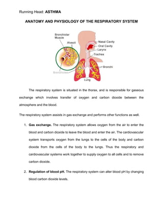
  • 1. Running Head: ASTHMA ANATOMY AND PHYSIOLOGY OF THE RESPIRATORY SYSTEM The respiratory system is situated in the thorax, and is responsible for gaseous exchange which involves transfer of oxygen and carbon dioxide between the atmosphere and the blood. The respiratory system assists in gas exchange and performs other functions as well. 1. Gas exchange. The respiratory system allows oxygen from the air to enter the blood and carbon dioxide to leave the blood and enter the air. The cardiovascular system transports oxygen from the lungs to the cells of the body and carbon dioxide from the cells of the body to the lungs. Thus the respiratory and cardiovascular systems work together to supply oxygen to all cells and to remove carbon dioxide. 2. Regulation of blood pH. The respiratory system can alter blood pH by changing blood carbon dioxide levels.
  • 2. ASTHMA 3. Voice production. Air movement past the vocal cords makes sound and speech possible. 4. Olfaction. The sensation of smell occurs when airborne molecules are drawn into the nasal cavity. 5. Innate immunity. The respiratory system provides protection against some microorganisms by preventing their entry into the body and by removing them from respiratory surfaces. The respiratory system is divided into two parts: The upper respiratory tract and lower respiratory tract. The upper respiratory tract includes the nose, pharynx, adenoids, tonsils, epiglottis, larynx and trachea. The lower respiratory tract consists of the bronchi, bronchioles alveolar ducts and alveoli. With the exception of the right and left main-stem bronchi, all lower airway structures are contained within lungs. The right lung is divided into three lobes (upper, middle and lower) and the left lung into two lobes (upper and lower). The structures of the chest wall (ribs, pleura, muscles of respiration) are also essential to respiration. Nose - is the primary upper respiratory organ in which air enters into and exits from the body. Cilia and mucus line the nasal cavity and traps bacteria and foreign particles that enter in through the nose. In addition, air that passes through the nasal cavity is humidified and moistened. The nasal septum divides the nose into two narrow nasal cavities: one area is responsible for smell and the other area is responsible for respiration. Within the walls
  • 3. ASTHMA of the nasal cavity there are frontal, nasal, ethmoid, maxillary, and sphenoid bones. Cartilage helps form the shape of the nose. Pharynx - besides the nose, air can enter into the lungs through the mouth. The pharynx is a tubular structure, positioned behind the oral and nasal cavities, that allows air to pass from the mouth to the lungs. The pharynx contains three parts: The nasopharynx, which connects the upper part of the throat with the nasal cavity; the oropharynx, positioned between the top of the epiglottis and the soft palate; and the laryngopharynx, located below the epiglottis. Larynx - from the pharynx, air enters into the larynx, commonly called the voice box. The larynx is part of the upper respiratory tract that has two main functions: a passageway for air to enter into the lungs, and a source of vocalization. The larynx is made up of the hyoid bone and cartilage, which helps regulate the flow of air. The epiglottis is a flap-like cartilage structure contained in the larynx that protects the trachea against food aspiration. Trachea - is sometimes called the windpipe. The trachea filters the air we breathe and branches into the bronchi. Bronchi - the bronchi allow the passage of air to the lungs. The trachea is made of c-shaped ringed cartilage that divides into the right and left bronchus. The right main bronchus is shorter and wider than the left main bronchus. The right bronchus is subdivided into three lobar bronchi, while the left one is divided into two lobar bronchi. Lungs - the lungs are the main organs of the respiratory system. In the lungs oxygen is taken into the body and carbon dioxide is breathed out. The red blood cells
  • 4. ASTHMA are responsible for picking up the oxygen in the lungs and carrying the oxygen to all the body cells that need it. The red blood cells drop off the oxygen to the body cells, then pick up the carbon dioxide which is a waste gas product produced by our cells. Diaphragm - breathing starts with a dome-shaped muscle at the bottom of the lungs called the diaphragm. When you breathe in, the diaphragm contracts. When it contracts it flattens out and pulls downward. When you breathe out, the diaphragm expands reducing the amount of space for the lungs and forcing air out. The diaphragm is the main muscle used in breathing. Expiration is mainly due to the natural elasticity of the lungs, which tend to collapse if they are not held against the thoracic wall. This is the mechanism behind lung collapse if there is air in the pleural space (pneumothorax). • Tidal volume - this is the volume of air moved in and out of the lungs with each respiratory movement. The average tidal volume is 500 ml. • Inspiratory reserve – this is the volume of air, in excess of the tidal, that can be inhaled by the deepest possible inspiration. The average inspiratory reserve is 3,000 ml. • Expiratory reserve – this is the volume of air, in excess of the tidal, which can be exhaled by the deepest possible expiration. It is about 1,200 ml. PHYSIOLOGY OF GAS EXCHANGE Each branch of the bronchial tree eventually sub-divides to form very narrow terminal bronchioles, which terminate in the alveoli. There are many millions of alveloi in
  • 5. ASTHMA each lung, and these are the areas responsible for gaseous exchange, presenting a massive surface area for exchange to occur over. Each alveolus is very closely associated with a network of capillaries containing deoxygenated blood from the pulmonary artery. The capillary and alveolar walls are very thin, allowing rapid exchange of gases by passive diffusion along concentration gradients. CO2 moves into the alveolus as the concentration is much lower in the alveolus than in the blood, and O2 moves out of the alveolus as the continuous flow of blood through the capillaries prevents saturation of the blood with O2 and allows maximal transfer across the membrane. The lung can be conceptualized as a collection of 300 million bubbles (alveoli), each 0.3 mm in diameter. The alveolar surface is composed of two kinds of cells: type I and type II. Type I cells provide structure and type II cells secrete surfactant. Surfactant lowers surface tension in the alveoli, thereby reducing the amount of pressure needed to inflate the alveoli and decreasing the tendency of the alveoli collapse. This sigh stretches the alveoli and causes surfactant to be secreted by type II cells. References: Lewis Heitkemper Dirksen,. Medical Surgical Nursing: Assessment and Management of Clinical Problems,. 5th edition. The Virtual Autopsy of Different Systems. Retrieved: (November 07, 2014) http://www.le.ac.uk/pa/teach/va/titlpag1.html
  • 6. ASTHMA OVERVIEW OF DISEASE Over 10% of people have some history of asthma. It often runs in families. The heritable nature of asthma is not well understood, however, and geneticists cannot define the precise manner in which it is passed from parents to children. Asthma affects the airways, which begin just below the throat as a single tube called the trachea. The trachea is situated immediately in front of the esophagus, the passageway that connects the throat with the stomach. The trachea divides into two slightly narrower tubes called the main bronchi (each one is called a bronchus). Each main bronchus then divides into progressively smaller tubes - the smallest are called bronchioles - to carry air to and from microscopic air spaces called alveoli. It is in the alveoli that the important work of the lung occurs, exchanging oxygen in the air for carbon dioxide in the blood.. The airways are lined with a mucus membrane that secretes a fine layer of mucus and fluid. This mucus washes the airways to remove any bacteria, dirt, or other
  • 7. ASTHMA foreign material that might get into our lungs. The overreaction or hyper-responsiveness of the airways results in bronchospasm, which is excessive contraction or spasm of the bronchial smooth muscle. The airways also become inflamed with swelling of the bronchial mucous membrane (mucosa) and secretion of excessive thick mucus that is difficult to expel. The airway hyper-responsiveness leading to obstruction of the airways occurs from one or more of various stimuli that vary with the individual patient. These include: • Viral (but not bacterial) respiratory infections (common colds) • Inhaled irritants (cigarette smoke, wood burning stoves and fireplaces, strong odors, chemical fumes) • Inhaled allergens (pollens, dusts, molds, animal danders) • Cold air • Exercise Occasional ingested substances (aspirin, sulfite preservatives, specific foods). Sometimes these exposures just act as triggers of brief symptoms with rapid relief once exposure ends. Sensitivity of the airway may be increased, however, following even brief exposure to one of these. The obstruction of the airways decreases the rate at which air can flow. This is felt as tightness in the chest and labored breathing (dyspnea). The obstruction and inflammation causes coughing. Obstruction to air flow can be measured with pulmonary
  • 8. ASTHMA function tests, which can detect even degrees of airway obstruction not yet causing symptoms. Pulmonary function measurements can be an extremely valuable tool for your physician to make decisions regarding treatment. Narrowing of the airway causes noises when air passes through them with sufficient speed. This typical high-pitched noise is called wheezing. Mucus in the airway causes a rattling sound called coarse crackles. Complete obstruction of some airways can cause absorption of air from the alveoli (air sacks at the end of the airways in the lungs). This causes portions of the lung to appear more dense and cast more of a shadow on a chest x-ray (this is called atelectasis). The rattling sounds or increased shadows on the x-ray are often misinterpreted as indicating pneumonia. There are different patterns of asthma. Some people have only an intermittent pattern of disease. They have self-limited episodes of varying severity followed by extended symptom-free periods. The individual episodes are frequently triggered by viral respiratory infections (causes of the common cold). This is particularly common in young children in whom viral respiratory infections are frequent (as many as 8 to 12 per year during the toddler and preschool age group). Others have these intermittent symptomatic periods brought on by vigorous exertion, cold air, or specific environmental exposures. This pattern is intermittent asthma. Some patients have daily or very frequently recurring symptoms. Although variable in severity, these patients do not have extended periods free of chest tightness, labored breathing, exertional intolerance, or cough. They may additionally have acute exacerbations triggered by the same factors that cause symptoms with an intermittent
  • 9. ASTHMA or seasonal allergic pattern of disease. Thus, viral respiratory infections (common colds) and specific environmental exposures may further increase the severity of symptoms in these patients. This pattern is chronic asthma (sometimes called persistent asthma). All patterns of disease are associated with varying degrees of severity ranging from mild to severe. The airway obstruction of asthma is generally completely reversible and usually does not cause permanent damage to the lungs, heart, or other organs. However, severe acute episodes of asthma can be associated with life threatening events and even fatalities. Survival of severe life threatening events can be associated with damage from lack of oxygen during the severe exacerbation, and lack of oxygen to the brain can cause loss of consciousness and brain damage. Other factors can also worsen asthma on occasion. Hyperventilation, excessively rapid and deep breathing can worsen asthma. This occurs from anxiety in some patients, particularly when asthma symptoms have begun for some other reason. A vicious cycle then occurs of asthma causing anxiety, which then worsens asthma, thereby causing more anxiety, etc. Ingested substances, such as aspirin, sulfite preservatives, and specific foods can cause acute attacks of asthma in sensitive patients. References: Miles Weinberger, MD Professor of Pediatrics, Allergy, Immunology, and Pulmonary., Overview of Asthma. Retrieved: (November 07, 2014) http://www.uichildrens.org/childrens-content.aspx?id=228741 American Academy of Allergy Asthma and Immunology,. Overview of asthma. Retrieved: (November 07, 2014) http://www.aaaai.org/conditions-and- treatments/asthma.aspx
  • 10. ASTHMA ASSESSMENT MILD exertional dyspnea/cough +/- nocturnal symptoms increased use of β –agonists good response to β –agonists MODERATE dyspnea at rest cough nocturnal symptoms partial relief from β –agonists β –agonists needed >8 puffs/ day chest tightness SEVERE labored respiration agitated, diaphoretic difficulty of speaking tachycardia no relief with β –agonists NEAR DEATH exhausted, confused diaphoretic, cyanotic silent chest, decreased respiratory effort falling heart rate
  • 11. ASTHMA During the physical assessment the primary symptom will be shortness of breath, other symptoms may include: • Anxiety • Cough • Chest tightness • Diaphoresis. • You may also find signs such as • Barrel chest • Diffuse or local wheezes • Pallor • Pulse paradoxus (pulse becomes weaker with inspiration and stronger with expiration) • Accessory muscle use. In mild cases of asthma wheezing may only be heard at the end of expiration. In more severe cases, wheezing will be present throughout expiration. As the attack worsens, wheezing will be present during both inspiration and expiration. During the most severe attacks wheezing may be absent. This indicates that the smaller airways
  • 12. ASTHMA have become obstructed by mucus or that the patient is so fatigued that they are not moving enough air to make the sound. Wheezing is not sign that is exclusive to asthma. It is important to gather a good history and consider other possibilities prior to deciding to treat the patient for asthma. Laryngeal abnormalities, growths and foreign body obstructions can all cause wheezing. Asthma attacks can be placed into three severity categories mild, moderate and severe. • In a mild attack, the patient will be able to speak in sentences and will be willing to lie down. They may be anxious but are easily calmed. The patient’s respiratory rate will be increased and accessory muscles will not be used. End expiatory wheezes may be present. Oxygen saturations will be above 95% on room air. • During a moderate attack the patient will be breathless while talking and may only use short sentences. Respiratory rates will be increased and accessory muscles will be used. The heart rate will be increased and a pulse paradox may be present. The patient will have loud expiatory wheezes. In infants, trouble feeding and a short soft cry will be noted. Oxygen saturations will be between 91% and 95% on room air. • In severe attacks the patient will be breathless even while resting. They will be very anxious, sitting upright and will be unwilling to lie down. They will only be able to speak in single words. Respiratory rate will be 30 bpm or higher. Accessory muscles will be used. The patients 9 pulse rate will be as high as 120 bpm and the patient may have a low blood pressure as the hyper inflated lungs are affecting the pre-load of the heart.
  • 13. ASTHMA Both inspiratory and expiratory wheezes can be heard. Pulse paradox is often present. The patients Oxygen saturations will be below 90%. While assessing an asthma patient consideration should be given to the amount of respiratory effort being made. A patient who fits the description of one having a moderate attack but is working hard to breath will soon become tired and quickly progress into respiratory arrest. Reference: Lindell Forbes EMT-P.,Assessing the Asthmatic Airway. Retrieved: (November 08, 2014) http://www.umchealthsystem.com/downloads/ems/Assessing_the_Asthmatic_Airway.pd f
  • 14. ASTHMA PATHOPHYSIOLOGY HEREDITARY FACTORS -Genetic predisposition -Atopy -Airway hyper responsiveness -Male sex (< 10 years old) -Female sex (adult) ACQUIRED FACTORS -Indoor allergens (domestic mites, animal allergens/ danders, cockroach allergens, fungi) -Outdoor allergens (pollen, fungi) -Irritants (tobacco smoke, air pollutants) -Respiratory infections (rhinoviruses, coronaviruses, influenza, para influenza, respiratory syncytial virus (RSV), adenoviruses) -High socio-economic status -Small family size -Higher Body Mass Index TRIGGERS -Allergens -Air pollutants including tobacco smoke -Respiratory infections -Exercise and Hyper ventilation -Weather changes -Extreme Emotional Expressions -Drugs (acetylsalicylic acid, beta blockers, contrast agents, nebulized drugs like beclomethasone) -Foods and additives Release of mediators from mast cells, eosinophils, macrophages, lymphocytes IgE- mast cell mediated response Infiltration with monocytes and lymphocytes LATE PHASE RESPONSE (Peaks in 5-6 hour) EARLY PHASE RESPONSE (Peaks in 30-60 min) Bronchial smooth muscle constriction Mucus secretion Vascular leakage Mucosal edema Obstruction of large and small airways Air trapping Respiratory acidosis Hypoxemia Infiltration with eosinophils and neutrophils Inflammation Bronchial hyper reactivity (Within 1-2 days) Reference: Lewis Heitkemper Dirksen., Medical-Surgical Nursing: Assessment & Management of Clinical Problems., 5th Edition.
  • 15. ASTHMA MEDICAL MANAGEMENT Asthma is not so much "treated" as it is "controlled". As a chronic, long-term disease, there is no cure. However, there are tools and medicines to help you control asthma as well as benchmarks to gauge your progress. The Peak Flow Meter – peak flow meter is a simple, small, hand-held tool that can help you maintain control of asthma by providing a measurement of how well air moves out of the lungs.If peak flow tests begin to decline - even before other symptoms are present - it may indicate a looming asthma attack. After taking asthma medication, the peak flow meter can be used to test the effectiveness of drug therapy. Inhaler – medication may also be administered using a nebulizer, providing a larger, continuous dose. Nebulizers vaporize a dose of medication in a saline solution into a steady stream of foggy vapor that is inhaled by the patient. SMART (Single Inhaler Maintenance and Reliever Therapy), is better for the relief and preventive treatment of asthma symptoms in adults compared to standard therapy, researchers reported in The Lancet Respiratory Medicine (March 2013 issue). SMART refers to using ICS (corticosteroid) plus LABA (long-acting β2 agonist) in one inhaler. Medical guidelines advise doctors to prescribe corticosteroids (ICS) plus rapid- onset long-acting β2 agonist (LABA) combination inhaler to achieve control, together with a second short-acting β2 agonist (SABA) inhaler for rescue usage, for the treatment of symptoms. SMART, on the other hand, uses only a single ICS/LABA inhaler for both relief and preventive treatment.
  • 16. ASTHMA Long-term control medicines are taken every day and are designed to prevent asthma symptom such as airway inflammation. Inhaled corticosteroids are the most effective long-term control medicine - the best at relieving airway inflammation and swelling. They are usually taken daily to greatly reduce the inflammation that initiates the chain reaction of the asthma attack. There are other long-term control medicines available that doctors may prescribe. Most of them are taken by mouth and are designed to open the airways and prevent airway inflammation. Examples include inhaled long-acting B2-agonists (used with low-dose inhaled corticosteroids), leukotriene modifiers, cromolyn and nedocromil, and theophylline. Quick-relief medicines relieve asthma symptoms when they occur. The most common of these are inhaled short-acting B2-agonists - bronchodilators that quickly relax tight muscles around the airways, allowing air to flow through them. The quick- relief inhaler should be used when asthma symptoms are first noticed, but should not be used more than 2 days a week. Most people carry the quick-relief inhaler with them at all times. Quick-relief medicines usually do not reduce inflammation and therefore should not be used as a replacement for long-term control medicines. Emergency Care – lifesaving treatments at the hospital will consist of direct oxygen (to alleviate hypoxia) and higher doses of medicines. Emergency personnel will likely administer a cocktail of short-acting B-2 agonists, systemic oral or intravenous steroids, other bronchodilators; nonspecific injected or inhaled B-2 agonists, anticholinergics, inhalation anesthetics, the dissociative anesthetic ketamine, and
  • 17. ASTHMA intravenous magnesium sulfate. Intubation (a breathing tube down one's throat) and mechanical ventilation may also be used in patients undergoing respiratory arrest. Vitamin D MayReduce Asthma Symptoms Researchers from King’s College London have discovered how vitamin D can reduce asthma symptoms. Catherine Hawrylowicz and team explained in the Journal of Allergy and Clinical Immunology (May 2013 issue) that their findings may offer a new way of treating the debilitating and usually chronic condition. Asthma patients are currently prescribed steroid tablets, which may have harmful side effects. There is a type of asthma, however, that is resistant to steroid therapy. Patients with this type are susceptible to severe and often life-threatening asthma attacks.The scientists found that people with asthma have higher levels of IL-17A (interleukin-17A). IL-17A is part of the immune system that protects the body against infection. However, this natural compound also worsens asthma symptoms. Large amounts of IL-17A can reduce the clinical effects of steroids. The team found that asthma patients who were on steroids had the highest levels of IL-17A. They also found that vitamin D significantly lowers IL-17A production in cells. Hawrylowicz believes vitamin D could be a safe and useful add-on treatment. Reference: This asthma information section was written by Peter Crosta. It was first published in September 2007 and last updated on 5 March 2013. The information may not be re-produced in any way without the permission of Medical News Today. Retrieved: (November 08, 2014) http://www.medicalnewstoday.com/info/asthma/treatment-for-asthma.php
  • 18. ASTHMA SURGICAL MANAGEMENT Methods of surgical treatment of bronchial asthma (BA) are: GLOMECTOMY for ASTHMA: The carotid body is a chemoreceptor, sensitive to: hypoxemia, decreased pH, and CO2retention. Stimulation causes hyperpnoea. This is beneficial if airways are clear. In the asthmatic, such a reflex initiates a vicious cycle. An already diminished functional capacity, due to bronchospasm. Mucosal swelling, and secretion accumulation, is further reduced by hyperpnoea. Removal of one carotid body apparently prevents such an unwanted sequence, as well as partially interrupts pathways of communication to the pulmonary plexus which contains bronchodilator fibers. Reference: RICHARD H. OVERHOLT., December 1961, Vol 40, No. 6., Glomectomy for Asthma. Retrieved: (November 08, 2014) http://journal.publications.chestnet.org/article.aspx?articleid=1055280 GANGLIECTOMY: Excision of a ganglion; surgical removal of a mass of tissue in lungs. VAGOTOMY: Unilateral application of histamine in one segmental bronchus potentiated the airway resistance increase caused by ACH challenge of the bronchial tree. Unilateral or contralateral blockade of the N. vagus reduces the severity of the reaction by about 70% of the values before the blockade. The arterial blood gases were not influenced by the unilateral blockade of the N. vagus. The decrease of the arterial oxygen pressure following the ACH induced bronchoconstriction was not changed by the unilateral vagotomy.
  • 19. ASTHMA Reference: M. S. Islam, I. Zimmermann, W. T. Ulmer., The Role of Unilateral Vagotomy on Reflex Broncho constriction. 14. II. 1975, Volume 152, Issue 4, pp 281-289. Retrieved: (November 08, 2014) http://link.springer.com/article/10.1007%2FBF02094942 IMPLANTATION OF NEUROSTIMULATORS OF SINOCAROTID also called an implanted pulse generator (IPG) is a battery powered device designed to deliver electrical stimulation to the brain, central and peripheral nervous system. RADIOFREQUENCY ELECTRO STIMULATION OF SYMPATHETIC NERVE BOUNDARY TRUNK IN PATIENTS WITH BRONCHIAL ASTHMA. The boundary trunk of the sympathic nerve (BTSN) has been implanted in the neck can produce both bronchial dilatation and bronchial spasm resulted in a decrease of bronchial hyper reactivity, frequency of asthmatic attacks Reference: Surgical treatment of bronchial asthma. Retrieved: (November 08, 2014) http://www.ncbi.nlm.nih.gov/pubmed/12162078
  • 20. ASTHMA NURSING DIAGNOSIS AND MANAGEMENT Nursing Care Plans 1. Ineffective Airway Clearance— the presence of a foreign microorganism triggers the B lymphocyte to produce antibodies that are specific to that antigen. These antibodies then attach to mast cells in the lungs. The mast cells with the antibody attach to the antigen and begin to degranulate. This degranulation causes the release of certain chemical mediators, namely, histamine, bradykinin, prostaglandin, and leukotriene. These chemical mediators cause bronchospasm leading to bronchoconstriction, increased vascular permeability leading to fluid leakage from the lung vasculature and increased mucus production. These lead to swelling of the bronchi, mucus buildup that plugs the airway and decreased bronchial diameter. This causes an increased airway resistance and a constricted pathway for air. Air cannot pass effectively and this manifests as a whistling sound. Coughing is a way to expel the obstruction (mucus plug) while dyspnea is a manifestation of the increased airway resistance. Patient may manifest • Difficulty breathing • Changes in depth and rate of respiration • Use of respiratory accessory muscles • Persistent ineffective cough with or without sputum production • Wheezing upon inspiration and expiration
  • 21. ASTHMA • Dyspnea • Coughing • Tachypnea, prolonged expiration • Tachycardia • Chest tightness • Suprasternal retraction • Restlessness • Anxiety • Cyanosis • Loss of consciousness NURSING DIAGNOSIS Ineffective airway clearance RT bronchoconstriction, increased mucus production, and respiratory infection AEB wheezing, dyspnea, and cough May be related to: • Increased production or retainment of pulmonary secretions • Bronchospasms • Decreased energy • Fatigue
  • 22. ASTHMA Planning – patient will maintain/improve airway clearance AEB absence of signs of respiratory distress. - Patient will verbalize understanding that allergens like dust, fumes, animal dander, pollen, and extremes of temperature and humidity are irritants or factors that can contribute to ineffective airway clearance and should be avoided. - Patient will demonstrate behaviors that would prevent the recurrence of the problem. Nursing Interventions Rationale Keep the patient adequately hydrated. Systemic hydration keeps secretion moist and easier to expectorate. Teach and encourage the use of diaphragmatic breathing and coughing exercises. These techniques help to improve ventilation and mobilize secretions without causing breathlessness and fatigue. Instruct patient to avoid bronchial irritants such as cigarette smoke, aerosols, extremes of temperature, and fumes. Bronchial irritants cause bronchoconstriction and increased mucus production, which then interfere with airway clearance. Teach early signs of infection that are to be reported to the clinician immediately. Minor respiratory infections that are of no consequence to the person with normal lungs can produce fatal disturbances in the
  • 23. ASTHMA Nursing Interventions Rationale lungs of an asthmatic person. Early recognition is crucial. Assist and prepare patient for postural drainage. Uses gravity to help raise secretions so they can be more easily expectorated. Administer nebulization as ordered. This ensures adequate delivery of medications to the airways. Administer medications as ordered. Antibiotics may be prescribed to treat the infection. 2. Ineffective Breathing Pattern— presence of secretions in the bronchi will result into a blockage of air that will enter the body and thus producing insufficient air needed by the body and inability to maintain clear airway. This obstruction is further heightened by bronchospasm due to the contraction of the smooth muscles in the bronchi. This is caused by parasympathetic stimulation of the muscarinic m2 receptors as well as by chemical mediators released in response to the presence of allergens. NURSING DIAGNOSIS
  • 24. ASTHMA Ineffective breathing pattern r/t presence of secretions AEB productive cough and dyspnea Planning – patient will demonstrate pursed-lip breathing and diaphragmatic breathing. - Patient will manifest signs of decreased respiratory effort AEB absence of dyspnea - Patient will verbalize understanding of causative factors and demonstrate behaviors that would improve breathing pattern Nursing Interventions Rationale Assess patient’s respiratory rate, depth, and rhythm. Obtain pulse oximetry. To obtain baseline data Monitor and record vital signs. Increase in respiratory rate could mean worsening condition. Auscultate breath sounds and assess airway pattern to check for the presence of adventitious breath sounds Elevate head of the bed and change position of the pt. every 2 hours. To minimize difficulty in breathing Encourage deep breathing and coughing exercises. To maximize effort for expectoration. Demonstrate diaphragmatic and To decrease air trapping and for efficient
  • 25. ASTHMA Nursing Interventions Rationale pursed-lip breathing. breathing. Encourage increase in fluid intake To prevent fatigue. Encourage opportunities for rest and limit physical activities. To prevent situations that will aggravate the condition Reinforce low salt, low fat diet as ordered. To mobilize secretions. 3. Impaired Gas Exchange— bronchial asthma is a condition wherein the airway diameter is highly reduced. This is due to severe bronchospasm, mucosal edema and mucus plug formation. There is a rise in airway resistance which leads to decreased amount of air that enters upon inspiration as well as expiration. Thus, ventilation is impaired. In bronchial asthma, perfusion is not directly affected. However, the balance between ventilation and perfusion (V/Q ratio) is lost because despite the adequate perfusion (capillary circulation), not much gas is available to diffuse from the alveoli to the capillaries. Conversely, the gases in the capillaries do diffuse to the alveoli but since expiration is impaired, such gases fail to be ventilated out. Thus, gas exchange is impaired. NURSING DIAGNOSIS
  • 26. ASTHMA Impaired gas exchange RT ventilation perfusion imbalance AEB dyspnea, tachypnea, and tachycardia May be related to: • altered delivery of inspired O2 or air trapping Planning –patient will improve gas exchange AEB absence of respiratory distress - Patient will demonstrate improved ventilation and adequate oxygenation of tissues by ABG’s within client’s normal limits and absence of symptoms of respiratory distress. - Patient will verbalize understand of causative factors and appropriate interventions (deep breathing, cough exercises, etc) Nursing Interventions Rationale Assess vital signs, noting respiratory rate, depth, and rhythm. To obtain baseline data VS monitor and record Serve to track important changes Auscultate breath sounds and assess airway pattern to check for the presence of adventitious breath sounds Elevate head of the bed and change position of the pt. every 2 hours. To minimize difficulty in breathing and promote maximum lung expansion.
  • 27. ASTHMA Nursing Interventions Rationale Encourage deep breathing and coughing exercises. To maximize effort for expectoration. Demonstrate diaphragmatic and pursed- lip breathing. To decrease air trapping and for efficient breathing. Encourage increase in fluid intake To prevent fatigue. Encourage opportunities for rest and limit physical activities. To prevent situations that will aggravate the condition Reinforce low salt, low fat diet as ordered. To mobilize secretions. 4. Fatigue— fluid accumulation in the lungs makes it difficult to breathe. The fluid inside prohibits the lungs to expand thus it is harder to breathe. The client, to have adequate ventilation makes use of his accessory muscles to breathe to have sufficient air. With too much use of the accessory muscles, feeling of tiredness may be present resulting to fatigue which is experienced by the client.
  • 28. ASTHMA NURSING DIAGNOSIS Fatigue r/t physical exertion to maintain adequate ventilation AEB use of accessory muscles to breathe Patient may manifest: • Generalized weakness • Verbalization of overwhelming lack of energy • Inability to maintain usual routines • Tired • Lethargic • Compromised concentration • Decreased performance
  • 29. ASTHMA Planning – patient will verbalize understand on health teachings given and report improved sense of energy. - Patient will perform ADL’s within client’s ability and participates in desired activities. - Patient will be able to identify basis of fatigue and be able to cope up with the problem. Nursing Interventions Rationale Establish rapport To gain patient’s trust Monitor and record vital signs. For baseline data. Provide environment conducive to relief of fatigue. Temperature and level of humidity are known to affect exhaustion. Assist client to identify appropriate coping behaviors. Promotes sense of control and improves self- esteem. Encourage patient to restrict activity and rest in bed as much as possible. Helps counteract effects of increased metabolism. Avoid topics that irritate or upset patient. Discuss ways to respond to these feelings. Increased irritability of the CNS may cause patient to be easily excited, agitated and prone to emotional outbursts.
  • 30. ASTHMA Nursing Interventions Rationale Discuss with the patient the need for activity. Plan schedule with patient and identify activities that lead to fatigue. Education may provide motivation to increase activity level even though patient may feel too weak initially. Alternate activity with rest periods. Prevents excessive fatigue. Monitor VS before and after activity. Indicates physiological levels of tolerance. Increase patient participation in ADL’s as tolerated. Increases confidence level and/or self- esteem and tolerance level 5. Risk for Activity Intolerance— inadequate oxygen in the circulation can develop weakness in our muscles. Muscles need oxygen to move and to do its function. If the patient cannot tolerate any activities because of the low oxygenation caused by the ventilation-perfusion imbalance caused by the pathological minimized lung expansion. NURSING DIAGNOSIS Risk for Activity Intolerance r/t decrease oxygenation Planning – patient will participate willingly in necessary/ desired activities such as deep breathing exercises. - Patient will perform ADL’s within client’s ability and participates in desired activities.
  • 31. ASTHMA - Patient will be able to increase activity tolerance AEB attendance of self-care needs. - Patient will be able to gradually increase activity within level of ability Nursing Interventions Rationale Monitor VS. For baseline data. Assess motor function. To identify causative factors. Note contributing factors to fatigue. To identify precipitating factors. Evaluate degree of deficit. To identify severity. Ascertain ability to stand and move about. To identify necessity of assistive devices. Assess emotional or psychological factors Stress and/or depression may increase the effects of illness. Plan care with rest periods between activities To reduce fatigue Increase activity/exercise gradually such as assisting the patient in doing PROM to active or full range of motions. Minimizes muscle atrophy, promotes circulation, helps to prevent contractures Provide adequate rest periods. To replenish energy. Assist client in doing self-care needs To promote independence and increase
  • 32. ASTHMA Nursing Interventions Rationale activity tolerance Elevate arm and hand Promotes venous Place knees and hips in extended position Maintains functional Other Possible Nursing Care Plans • Anxiety—may be related to perceived threat of death, possibly evidenced by apprehension, fearful expression, and extraneous movements. • Risk for contamination—risk factors may include presence of atmospheric pollutants, environmental contaminants in the home. Reference: 5 Bronchial Asthma Nursing Care Plans., February 11, 2012. Retrieved: (November 08, 2014) http://nurseslabs.com/bronchial-asthma-nursing-care- plans/