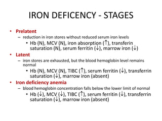
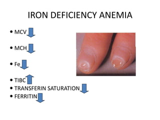
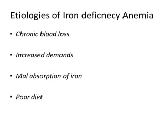
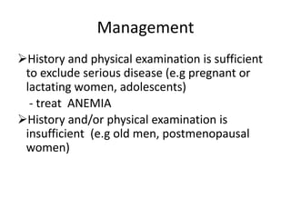
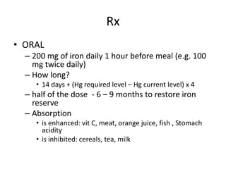
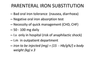
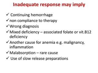
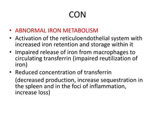
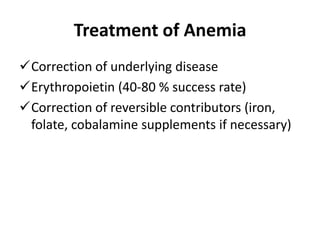
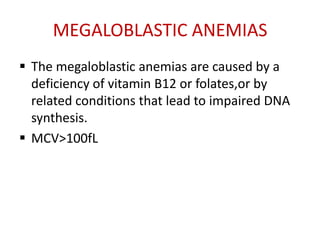
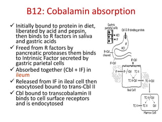
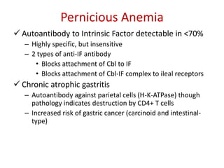
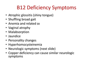
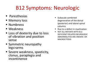
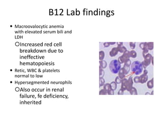
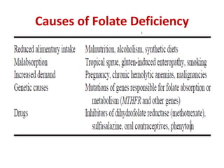
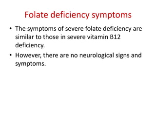
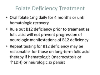
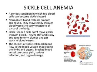
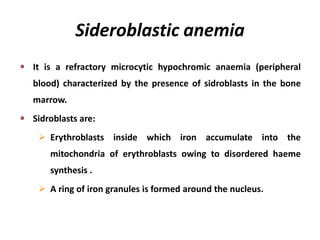
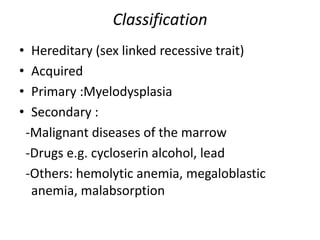
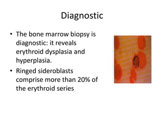
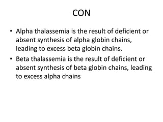
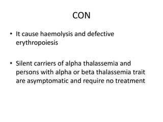
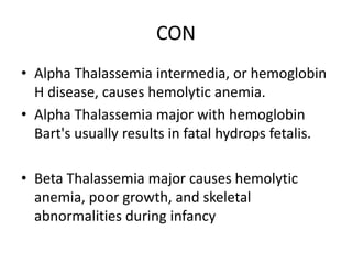
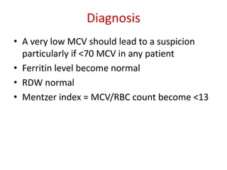
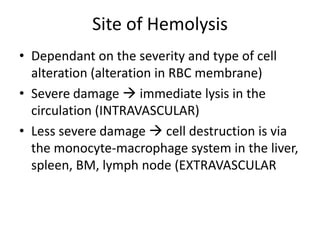
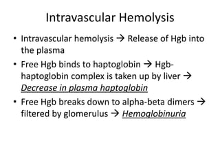
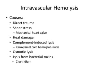
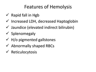
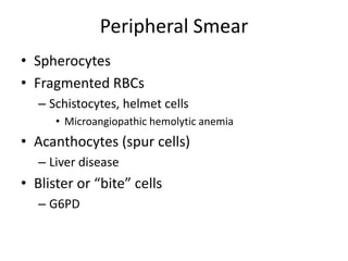
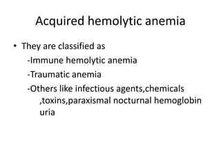
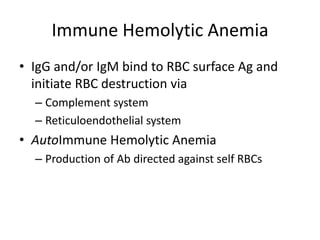
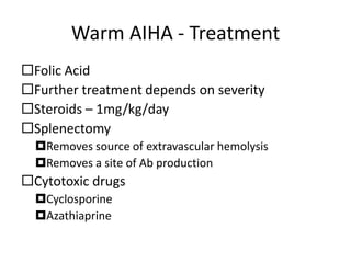
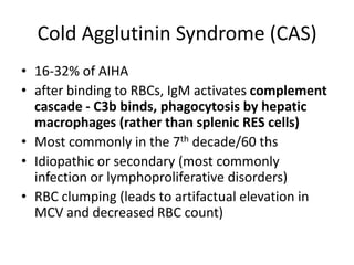
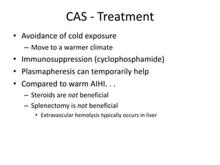
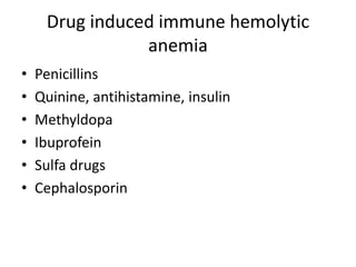
SEMINAR PRESENTATION ON APROACH
OF A PATIENT WITH ANEMIA
Niguse Desta, clerkship1
Yirgalem, MC 2014
OUTLINE
Objective
Introduction
Definition and major classification of anemia
Approach of a patient with anemia
Common types of anemia
:diagnosis,management and complications of
anemia
OBIECTIVE
To Discuss Definition,Etiology,Manifestations
,Diagnosis And Management Of Common
Types Of Anemia
IRON DEFICIENCY ANEMIA
Introduction
Iron metabolism:
Daily 10-30mg iron ingested, 5-10% absorbed
to balance precisely the amount lost (1mg)
under physiologic condition
 Amount absorbed can increases up to five
fold if body iron store are depleted or
erythropoiesis is accelerated.
 Absorbed as hem and non hem iron in the
duodenum and proximal jejunum
IRON DEFICIENCY ANEMIA
 Definition: - Iron deficiency anemia occurs
when body iron stores become inadequate for
the needs of normal RBC production
 On a worldwide basis, iron deficiency is
among the most frequent causes of anemia.
IRON DEFICENCY - STAGES
• Prelatent
– reduction in iron stores without reduced serum iron levels
• Hb (N), MCV (N), iron absorption ( ), transferin
saturation (N), serum ferritin ( ), marrow iron ( )
• Latent
– iron stores are exhausted, but the blood hemoglobin level remains
normal
• Hb (N), MCV (N), TIBC ( ), serum ferritin ( ), transferrin
saturation ( ), marrow iron (absent)
• Iron deficiency anemia
– blood hemoglobin concentration falls below the lower limit of normal
• Hb ( ), MCV ( ), TIBC ( ), serum ferritin ( ), transferrin
saturation ( ), marrow iron (absent)
IRON DEFICIENCY ANEMIA
 MCV
 MCH
 Fe
 TIBC
 TRANSFERIN SATURATION
 FERRITIN
Etiologies of Iron deficnecy Anemia
• Chronic blood loss
• Increased demands
• Mal absorption of iron
• Poor diet
Management
History and physical examination is sufficient
to exclude serious disease (e.g pregnant or
lactating women, adolescents)
- treat ANEMIA
History and/or physical examination is
insufficient (e.g old men, postmenopausal
women)
Rx
• ORAL
– 200 mg of iron daily 1 hour before meal (e.g. 100
mg twice daily)
– How long?
• 14 days + (Hg required level – Hg current level) x 4
– half of the dose - 6 – 9 months to restore iron
reserve
– Absorption
• is enhanced: vit C, meat, orange juice, fish , Stomach
acidity
• is inhibited: cereals, tea, milk
PARENTERAL IRON SUBSTITUTION
– Bad oral iron tolerance (nausea, diarrhoea)
– Negative oral iron absorption test
– Necessity of quick management (CHD, CHF)
– 50 - 100 mg daily
– I.v only in hospital (risk of anaphilactic shock)
– I.m in outpatient department
– iron to be injected (mg) = (15 - Hb/g%/) x body
weight (kg) x 3
Inadequate response may imply
 Continuing hemorrhage
non compliance to therapy
Wrong diagnosis
Mixed deficiency – associated folate or vit.B12
deficiency
Another cause for anemia e.g. malignancy,
inflammation
Malabsorption – rare cause
 Use of slow release preparations
Anemia of chronic disease
• ACD is a common type of anemia that occurs
in patients with infectious, inflammatory, or
neoplastic diseases that persist for more than
1 or 2 months.
• It does not include anemias caused by
marrow replacement, blood loss, hemolysis,
renal insufficiency, hepatic disease, or
endocrinopathy, even when these disirders
are chronic.
Etiologies
Chronic inflammatory diseases
- Infections e.g. pulmonary abscess, TB,
Osteomyelitis, pneumonia, bacterial
endocarditis
- Non infectious, e.g. Rheumatoid arthritis, SLE
and other connective tissue
diseases,sarcoidosis, Crohn’s disease
 Malignant diseases
E.g. carcinoma, lymphoma, sarcoma
Characteristic features are:
NCNC or mildly hypochromic indices and red
cell morphology
 Mild and non progressive anemia (Hgb rarely
< 9.0g/dl) : Severity being related to severity
of underlying disease
Both serum iron and TIBC are reduced
 Serum ferritine is normal or raised and
 Bone marrow storage iron is normal but
erythroblast iron is reduced
pathogenesis
• Shortened red cell life span, moderately 20-30%
(from 120 to 60-90 days)
• Relative bone marrow(erythropoiesis) failure
- Cytokines released from inflammatory cells (TNF- ,
IL-1, IFN- ) affects erythropoiesis by inhibiting the
growth of erythroid progenitors
- Serum erythropoietin levels in patiens with ACD are
normal when compared to healthy subjects but much
lower than levels in non-ACD anemic patients
CON
• ABNORMAL IRON METABOLISM
• Activation of the reticuloendothelial system with
increased iron retention and storage within it
• Impaired release of iron from macrophages to
circulating transferrin (impaired reutilization of
iron)
• Reduced concentration of transferrin
(decreased production, increase sequestration in
the spleen and in the foci of inflammation,
increase loss)
Treatment of Anemia
Correction of underlying disease
Erythropoietin (40-80 % success rate)
Correction of reversible contributors (iron,
folate, cobalamine supplements if necessary)
MEGALOBLASTIC ANEMIAS
 The megaloblastic anemias are caused by a
deficiency of vitamin B12 or folates,or by
related conditions that lead to impaired DNA
synthesis.
 MCV>100fL
Etiologies
Vitamin B12 deficiency
Folate deficiency
Abnormalities of vitamin B12 or folate
metabolism,transcobalamine
deficiency,antifolate-drugs
Other defects of DNA synthesis: congenital
enzyme deficiency, alcohol, treatment with
hydroxyurea
B12: Cobalamin absorption
 Initially bound to protein in diet,
liberated by acid and pepsin,
then binds to R factors in saliva
and gastric acids
 Freed from R factors by
pancreatic proteases them binds
to Intrinsic Factor secreted by
gastric parietal cells
 Absorbed together (Cbl + IF) in
ileum
 Released from IF in ileal cell then
exocytosed bound to trans-Cbl II
 Cbl bound to transcobalamin II
binds to cell surface receptors
and is endocytosed
Causes of Vit. B12 deficiency
 Nutritional: especially in vegans
 Malabsorption
Gastric causes
-pernicious anemia
-Congenital lack or abnormality of intrinsic factor
-Total or partial gastrectomy
Intestinal causes
-Ileal resection and Crohn’s disease
-Congenital selective malabsorption
Pernicious Anemia
 Autoantibody to Intrinsic Factor detectable in <70%
– Highly specific, but insensitive
– 2 types of anti-IF antibody
• Blocks attachment of Cbl to IF
• Blocks attachment of Cbl-IF complex to ileal receptors
 Chronic atrophic gastritis
– Autoantibody against parietal cells (H-K-ATPase) though
pathology indicates destruction by CD4+ T cells
– Increased risk of gastric cancer (carcinoid and intestinal-
type)
B12 Deficiency Symptoms
• Atrophic glossitis (shiny tongue)
• Shuffling broad gait
• Anemia and related sx
• Vaginal atrophy
• Malabsorption
• Jaundice
• Personality changes
• Hyperhomocysteinemia
• Neurologic symptoms (next slide)
• Copper deficiency can cause similar neurologic
symptoms
B12 Symptoms: Neurologic
 Paresthesias
 Memory loss
 Numbness
 Weakness
 Loss of dexterity due to loss
of vibration and position
sense
 Symmetric neuropathy
legs>arms
 Severe weakness, spasticity,
clonus, paraplegia and
incontinence
 Subacute combined
degeneration of the dorsal
(posterior) and lateral spinal
columns
 Due to a defect in myelination
 NOT ALL PATIENTS WITH B12
DEFICIENCY RELATED NEUROLOGIC
ABNORMALITIES ARE ANEMIA OR
MACROCYTOSIS
B12 Lab findings
 Macroovalocytic anemia
with elevated serum bili and
LDH
Increased red cell
breakdown due to
ineffective
hematopoiesis
 Retic, WBC & platelets
normal to low
 Hypersegmented neurophils
Also occur in renal
failure, fe deficiency,
inherited
Bone Marrow
Hypercellular marrow
Megaloblastic erythroid
hyperplasia
Giant metamyelocytes
Due to slowing of DNA synthesis
and delayed nuclear maturation
Methionine deficiency may play a
central role
Shilling Test
1. PART 1: Oral labeled B12 and
IM unlabeled B12 at the
same time to saturate tissue
stores
2. 24h urine to assess absorption
>5% normal
<5% impaired
3. PART 2: Repeat w/oral IF
if now normal =PA
if abnormal = malabsorption
4. Can continue with antibiotics
to look for bacterial
overgrowth, pancreatic
enzymes for exocrine
insufficiency
Part 1 test result Part 2 test result Diagnosis
Normal -
Normal or vitamin
B12 deficiency
Low Normal Pernicious anemia
Low Low Malabsorption
B12 Deficiency: Treatment
IM B12 1000mcg daily x 1 wk
– then 1000mcg weekly x 1 month
– Then 1000mcg monthly for life for PA
Oral high dose 1-2 mg daily
– As effective but less reliable than IM
– Currently only recommended after
full parenteral repletion
Sublingual, nasal spray and gel formulations
available
Folate
• Animal products (liver), yeast and leafy
vegetables
• Normal requirement 400mcg/day
• Pregnancy/Lactation: 500-800mcg/day
• Increased requirement in hemolytic anemia
and exfoliateive skin disease
• Body stores: 5-10mg
Causes of Folate Deficiency
Folate deficiency symptoms
• The symptoms of severe folate deficiency are
similar to those in severe vitamin B12
deficiency.
• However, there are no neurological signs and
symptoms.
Folate Deficiency Treatment
• Oral folate 1mg daily for 4 months or until
hematologic recovery
• Rule out B12 deficiency prior to treament as
folic acid will not prevent progression of
neurologic manifestations of B12 deficiency
• Repeat testing for B12 deficiency may be
reasonable for those on long-term folic acid
therapy if hematologic (macrocytosis or
↑LDH) or neurologic sx persist
SICKLE CELL ANEMIA
 A serious condition in which red blood
cells can become sickle-shaped
 Normal red blood cells are smooth
and round. They move easily through
blood vessels to carry oxygen to all
parts of the body.
 Sickle-shaped cells don’t move easily
through blood. They’re stiff and sticky
and tend to form clumps and get
stuck in blood vessels.
 The clumps of sickle cell block blood
flow in the blood vessels that lead to
the limbs and organs. Blocked blood
vessel can cause pain, serious
infection, and organ damage.
Normal and Sickled Red Blood Cells
in Blood Vessels
Mechanism
• Underlying mechanism of disease: sickle cell
anemia is a Glu->Val substitution in the sixth
amino acid of the β-globin gene
Sickle Cell Anemia vs. Sickle Cell Trait
• People who have sickle cell anemia are born with
it; means inherited, lifelong condition.
• They inherit two copies of sickle cell gene, one
from each parent.
• Sickle cell trait is different from sickle cell anemia.
People with sickle cell trait don’t have the
condition, but they have one of the genes that
cause the condition.
• People with sickle cell anemia and sickle cell trait
can pass the gene on when they have children
Inheritance of Sickle Cell Anemia
Why Anemia?
• In sickle cell anemia, a lower-than-normal
number of red blood cells occurs because
sickle cells don’t last very long.
• Sickle cells die faster than normal red blood
cells, usually after only about 10 to 20 days.
• The bone marrow can’t make new red blood
cells fast enough to replace the dying ones.
The result is anemia.
Signs and Symptoms
Complication of Sickle Cell Anemia
• Splenic Crisis
• Infections
• Acute Chest Syndrome
• Delayed growth and
puberty in children
• Stroke
• Eye problem
• Priapism
•Gallstone
•Ulcers on the
legs
•Pulmonary
Arterial
•Hypertension
• Organ Failure
Treatments
 Effective treatments are available to help relieve the
symptoms and complications of sickle cell anemia, but
in most cases there’s no cure.
 The goal is to relieve the pain; prevent infections, eye
damage, strokes and control complications if they
occur.
 Pain medicine: acetaminophen, nonsteroidal anti-
inflammatory drugs (NSAIDs), and narcotics such as
meperidine, morphine, oxycodone, and etc.
 Heating pads
 Hydroxyurea, Folic Acid
 Blood Transfusions
Prevention
 Identify what can trigger the “Crisis” such as
stress, avoid extremes of heat and cold weather
 Maintain healthy lifestyle habits
Eating healthy
Avoid dehydration
Exercise regularly
Get enough sleep and rest
Avoid alcohol and don’t smoke
 Regular medical checkups and treatment are
important
Sideroblastic anemia
It is a refractory microcytic hypochromic anaemia (peripheral
blood) characterized by the presence of sidroblasts in the bone
marrow.
Sidroblasts are:
 Erythroblasts inside which iron accumulate into the
mitochondria of erythroblasts owing to disordered haeme
synthesis .
 A ring of iron granules is formed around the nucleus.
Classification
• Hereditary (sex linked recessive trait)
• Acquired
• Primary :Myelodysplasia
• Secondary :
-Malignant diseases of the marrow
-Drugs e.g. cycloserin alcohol, lead
-Others: hemolytic anemia, megaloblastic
anemia, malabsorption
Diagnostic
• The bone marrow biopsy is
diagnostic: it reveals
erythroid dysplasia and
hyperplasia.
• Ringed sideroblasts
comprise more than 20% of
the erythroid series
Thalassemia
• Inherited hematologic disorders caused by
defects in the synthesis of one or more of the
haemoglobin chains
• Two type
- alpha Thalassemia
- beta Thalassemia
CON
• Alpha thalassemia is the result of deficient or
absent synthesis of alpha globin chains,
leading to excess beta globin chains.
• Beta thalassemia is the result of deficient or
absent synthesis of beta globin chains, leading
to excess alpha chains
CON
• It cause haemolysis and defective
erythropoiesis
• Silent carriers of alpha thalassemia and
persons with alpha or beta thalassemia trait
are asymptomatic and require no treatment
CON
• Alpha Thalassemia intermedia, or hemoglobin
H disease, causes hemolytic anemia.
• Alpha Thalassemia major with hemoglobin
Bart's usually results in fatal hydrops fetalis.
• Beta Thalassemia major causes hemolytic
anemia, poor growth, and skeletal
abnormalities during infancy
Diagnosis
• A very low MCV should lead to a suspicion
particularly if <70 MCV in any patient
• Ferritin level become normal
• RDW normal
• Mentzer index = MCV/RBC count become <13
Treatment
• Treatment include -life long transfution
-Bone marrow transplant
-Preconception councelling
• Prognosis -Persons with beta thalassemia
major often die from cardiac complications of
iron overload by 30 years of age.
Hemolytic anemia
 Anemia due to shortened survival of
circulating RBCs (Normal: 110-120 days)
– Hemolysis <100 days
 With intact bone marrow:
• Anemia  Compensatory increase in Epo
secretion  Enhances RBC production
(reticulocytosis)  Reduces degree of anemia
• This is most commonly seen with hemolytic
anemia, but not specific to hemolysis (can also be
seen with acute blood loss)
Causes of Hemolysis - Intrinsic
• Generally, a hereditary disorder
• Remember, the mature RBC has lost its nucleus,
mitochondria, and RNA, leaving Hgb, RBC
membrane
• Intrinsic hemolysis is caused by defects in Hgb,
RBC membrane or metabolic factors needed to
generate ATP
• Examples
• Thalassemia (defect in alpha or beta globin chains)
• Spherocytosis (missing RBC membrane proteins)
• G6PD deficiency (abnormality in reducing power (NADPH))
Causes of Hemolysis - Extrinsic
• Acquired disorder
• Causes include:
• Ab directed against RBC membrane components
-AIHA, delayed transfusion reaction
• Stasis/trapping/destruction in spleen (hypersplenism)
• Trauma
-Prosthetic heart valve
• Exposure to compounds with oxidant potential
-Sulfonamide in those with G6PD
• Destruction of RBC by pathogens
-Malaria, babesiosis
Site of Hemolysis
• Dependant on the severity and type of cell
alteration (alteration in RBC membrane)
• Severe damage  immediate lysis in the
circulation (INTRAVASCULAR)
• Less severe damage  cell destruction is via
the monocyte-macrophage system in the liver,
spleen, BM, lymph node (EXTRAVASCULAR
Intravascular Hemolysis
• Intravascular hemolysis  Release of Hgb into
the plasma
• Free Hgb binds to haptoglobin  Hgb-
haptoglobin complex is taken up by liver 
Decrease in plasma haptoglobin
• Free Hgb breaks down to alpha-beta dimers 
filtered by glomerulus  Hemoglobinuria
Intravascular Hemolysis
• Causes:
• Direct trauma
• Shear stress
– Mechanical heart valve
• Heat damage
• Complement-induced lysis
– Paroxysmal cold hemoglobinuria
• Osmotic lysis
• Lysis from bacterial toxins
• Clostridium
Extravascular Hemolysis
• Damaged RBCs are destroyed by liver (receives a larger
portion of the cardiac output) and spleen
• Spleen contains cords of Billroth, which end blindly (unlike
other vascular channels in body)
• RBCs must deform to pass through 2-3 micron slits in the wall
of the cords in order to get back into circulation
• RBCs unable to pass are phagocytosed by the monocyte-
macrophage system
– Degraded into biliverdin, iron, carbon monoxide
Features of Hemolysis
 Rapid fall in Hgb
 Increased LDH, decreased Haptoglobin
 Jaundice (elevated indirect bilirubin)
 Splenomegaly
 H/o pigmented gallstones
 Abnormally shaped RBCs
 Reticulocytosis
Peripheral Smear
• Spherocytes
• Fragmented RBCs
– Schistocytes, helmet cells
• Microangiopathic hemolytic anemia
• Acanthocytes (spur cells)
– Liver disease
• Blister or “bite” cells
– G6PD
con
• RBCs with inclusions
• Teardrop RBCs
• Red Cell “ghosts”
– Hemolyzed RBCs that reform, but have lost all
internal components, leaving only the membrane
– Indicates Intravascular Hemolysis
Labs
 LDH: elevated
 Indirect bilirubin: elevated (due to catabolism of Hgb)
 Haptoglobin: decreased
 Binds to Hgb and taken up by liver
 In a series of reports:
 Elevated LDH, low Haptoglobin was 90% specific
 Normal LDH, Haptoglobin >25 was 92% sensitive for ruling out
hemolysis
 Reticulocyte Count: elevated
 Normal is 0.5-1.5%
 Anemia leads to increase Epo production leading to a
reticulocytosis (4-5% increase above baseline)
 Positive Direct Antiglobulin Test (Coombs)
Acquired hemolytic anemia
• They are classified as
-Immune hemolytic anemia
-Traumatic anemia
-Others like infectious agents,chemicals
,toxins,paraxismal nocturnal hemoglobin
uria
Immune Hemolytic Anemia
• IgG and/or IgM bind to RBC surface Ag and
initiate RBC destruction via
– Complement system
– Reticuloendothelial system
• AutoImmune Hemolytic Anemia
– Production of Ab directed against self RBCs
Autoimmune Hemolytic Anemia
• Types
– Warm AIHA
– Cold Agglutinin Syndrome
– Paroxysmal Cold
Hemoglobinuria
– Mixed Type AIHA
– Drug Induced AIHA
Causes
-Idiopathic
Secondary causes
-Lymphoproliferative
disorders
-Autoimmune
disorders
-Viral Infection
-Immunodeficiency
-Drugs
Warm AIHA
48-79% of AIHA
Incidence increased at 40yrs
2:1 Female to Male ratio
No racial predilection
Idiopathic vs Secondary Causes
CLL, Hodgkins, non-Hodgkins,
Autoimmune, non-lymphoid neoplasms, immunodeficiency, viral
illnesses
Typically present with anemic signs and symptoms
These symptoms may precede and underlying illness by
months to years
Warm AIHA - Labs
• Elevated MCV (reflecting reticulocytosis)
• Mild leukocytosis with neutrophilia
• Blood smear:
– Polychromasia, macrocytosis, spherocytes, nucleated RBCs
• Elevated LDH and indirect bilirubin
• Haptoglobin typically low
– Because it is an acute phase reactant, it can also be normal
or elevated in mild disease
Warm AIHA – DAT (direct Coombs)
Detects Ab on the RBC surface
Pt’s RBCs are washed then reacted with
monoclonal Ab against various Ig (IgG) and C3
Warm autoantibodies react at warmer
temperatures (~37° C)
95% of Warm AIHA will have a positive DAT
Negative tests due to:
IgG quantity too low for detectable threshold
IgA autoantibodies
IgM autoantibodies
Warm AIHA - Treatment
Folic Acid
Further treatment depends on severity
Steroids – 1mg/kg/day
Splenectomy
Removes source of extravascular hemolysis
Removes a site of Ab production
Cytotoxic drugs
Cyclosporine
Azathiaprine
Cold Agglutinin Syndrome (CAS)
• 16-32% of AIHA
• after binding to RBCs, IgM activates complement
cascade - C3b binds, phagocytosis by hepatic
macrophages (rather than splenic RES cells)
• Most commonly in the 7th decade/60 ths
• Idiopathic or secondary (most commonly
infection or lymphoproliferative disorders)
• RBC clumping (leads to artifactual elevation in
MCV and decreased RBC count)
CAS - Treatment
• Avoidance of cold exposure
– Move to a warmer climate
• Immunosuppression (cyclophosphamide)
• Plasmapheresis can temporarily help
• Compared to warm AIHI. . .
– Steroids are not beneficial
– Splenectomy is not beneficial
• Extravascular hemolysis typically occurs in liver
Drug induced immune hemolytic
anemia
• Penicillins
• Quinine, antihistamine, insulin
• Methyldopa
• Ibuprofein
• Sulfa drugs
• Cephalosporin
Non immune Hemolytic anemia
• Fragmentation Hemolysis (Microangiopathy) –
result of mechanical shearing of RBCs from
damaged microvasculature, cardiac
abnormalities, AV shunts, turbulent flow, drugs
(cyclosporine, cocaine)
• Hypersplenism – functionally hyperactive spleen
too much sequestration of all blood cells
• Infection – can be several mechanisms of
hemolysis (direct attack, hypersplenism
induction, immune, toxin release, altered RBC
surface)
• Burn ,liver disease
Lead poisoning
• Inhibits both hem and globin synthesis
• Interferes with breakdown of RNA by inhibiting the
enzyme pyrimidine- nucleotidase ⇒ accumulation of
denatured RNA in red cells giving rise to Basophilic
stippling.
• Hypo chromic/Hemolytic anemia with bone marrow
ring sideroblasts
• Free erythrocyte protoporphyrin is raised
Management:
• Pyridoxine, folic acid therapy : may bring some
response
• Repeated transfusion is ultimate choice
SUMMARY
REFERANCE
• Reinhold Munker, MD ,ETAL:MODERN
HEMATOLOGY, SECOND EDITION
• HARRISON’S :PRINCIPLES OF INTERNAL
MEDICINE,18th
10Q! 4 UR ATTENTION!

Aproach to anemia

  • 1.
    SEMINAR PRESENTATION ONAPROACH OF A PATIENT WITH ANEMIA Niguse Desta, clerkship1 Yirgalem, MC 2014
  • 2.
    OUTLINE Objective Introduction Definition and majorclassification of anemia Approach of a patient with anemia Common types of anemia :diagnosis,management and complications of anemia
  • 3.
  • 4.
    IRON DEFICIENCY ANEMIA Introduction Ironmetabolism: Daily 10-30mg iron ingested, 5-10% absorbed to balance precisely the amount lost (1mg) under physiologic condition  Amount absorbed can increases up to five fold if body iron store are depleted or erythropoiesis is accelerated.  Absorbed as hem and non hem iron in the duodenum and proximal jejunum
  • 5.
    IRON DEFICIENCY ANEMIA Definition: - Iron deficiency anemia occurs when body iron stores become inadequate for the needs of normal RBC production  On a worldwide basis, iron deficiency is among the most frequent causes of anemia.
  • 6.
    IRON DEFICENCY -STAGES • Prelatent – reduction in iron stores without reduced serum iron levels • Hb (N), MCV (N), iron absorption ( ), transferin saturation (N), serum ferritin ( ), marrow iron ( ) • Latent – iron stores are exhausted, but the blood hemoglobin level remains normal • Hb (N), MCV (N), TIBC ( ), serum ferritin ( ), transferrin saturation ( ), marrow iron (absent) • Iron deficiency anemia – blood hemoglobin concentration falls below the lower limit of normal • Hb ( ), MCV ( ), TIBC ( ), serum ferritin ( ), transferrin saturation ( ), marrow iron (absent)
  • 7.
    IRON DEFICIENCY ANEMIA MCV  MCH  Fe  TIBC  TRANSFERIN SATURATION  FERRITIN
  • 8.
    Etiologies of Irondeficnecy Anemia • Chronic blood loss • Increased demands • Mal absorption of iron • Poor diet
  • 9.
    Management History and physicalexamination is sufficient to exclude serious disease (e.g pregnant or lactating women, adolescents) - treat ANEMIA History and/or physical examination is insufficient (e.g old men, postmenopausal women)
  • 10.
    Rx • ORAL – 200mg of iron daily 1 hour before meal (e.g. 100 mg twice daily) – How long? • 14 days + (Hg required level – Hg current level) x 4 – half of the dose - 6 – 9 months to restore iron reserve – Absorption • is enhanced: vit C, meat, orange juice, fish , Stomach acidity • is inhibited: cereals, tea, milk
  • 11.
    PARENTERAL IRON SUBSTITUTION –Bad oral iron tolerance (nausea, diarrhoea) – Negative oral iron absorption test – Necessity of quick management (CHD, CHF) – 50 - 100 mg daily – I.v only in hospital (risk of anaphilactic shock) – I.m in outpatient department – iron to be injected (mg) = (15 - Hb/g%/) x body weight (kg) x 3
  • 12.
    Inadequate response mayimply  Continuing hemorrhage non compliance to therapy Wrong diagnosis Mixed deficiency – associated folate or vit.B12 deficiency Another cause for anemia e.g. malignancy, inflammation Malabsorption – rare cause  Use of slow release preparations
  • 13.
    Anemia of chronicdisease • ACD is a common type of anemia that occurs in patients with infectious, inflammatory, or neoplastic diseases that persist for more than 1 or 2 months. • It does not include anemias caused by marrow replacement, blood loss, hemolysis, renal insufficiency, hepatic disease, or endocrinopathy, even when these disirders are chronic.
  • 14.
    Etiologies Chronic inflammatory diseases -Infections e.g. pulmonary abscess, TB, Osteomyelitis, pneumonia, bacterial endocarditis - Non infectious, e.g. Rheumatoid arthritis, SLE and other connective tissue diseases,sarcoidosis, Crohn’s disease  Malignant diseases E.g. carcinoma, lymphoma, sarcoma
  • 15.
    Characteristic features are: NCNCor mildly hypochromic indices and red cell morphology  Mild and non progressive anemia (Hgb rarely < 9.0g/dl) : Severity being related to severity of underlying disease Both serum iron and TIBC are reduced  Serum ferritine is normal or raised and  Bone marrow storage iron is normal but erythroblast iron is reduced
  • 16.
    pathogenesis • Shortened redcell life span, moderately 20-30% (from 120 to 60-90 days) • Relative bone marrow(erythropoiesis) failure - Cytokines released from inflammatory cells (TNF- , IL-1, IFN- ) affects erythropoiesis by inhibiting the growth of erythroid progenitors - Serum erythropoietin levels in patiens with ACD are normal when compared to healthy subjects but much lower than levels in non-ACD anemic patients
  • 17.
    CON • ABNORMAL IRONMETABOLISM • Activation of the reticuloendothelial system with increased iron retention and storage within it • Impaired release of iron from macrophages to circulating transferrin (impaired reutilization of iron) • Reduced concentration of transferrin (decreased production, increase sequestration in the spleen and in the foci of inflammation, increase loss)
  • 18.
    Treatment of Anemia Correctionof underlying disease Erythropoietin (40-80 % success rate) Correction of reversible contributors (iron, folate, cobalamine supplements if necessary)
  • 19.
    MEGALOBLASTIC ANEMIAS  Themegaloblastic anemias are caused by a deficiency of vitamin B12 or folates,or by related conditions that lead to impaired DNA synthesis.  MCV>100fL
  • 20.
    Etiologies Vitamin B12 deficiency Folatedeficiency Abnormalities of vitamin B12 or folate metabolism,transcobalamine deficiency,antifolate-drugs Other defects of DNA synthesis: congenital enzyme deficiency, alcohol, treatment with hydroxyurea
  • 21.
    B12: Cobalamin absorption Initially bound to protein in diet, liberated by acid and pepsin, then binds to R factors in saliva and gastric acids  Freed from R factors by pancreatic proteases them binds to Intrinsic Factor secreted by gastric parietal cells  Absorbed together (Cbl + IF) in ileum  Released from IF in ileal cell then exocytosed bound to trans-Cbl II  Cbl bound to transcobalamin II binds to cell surface receptors and is endocytosed
  • 22.
    Causes of Vit.B12 deficiency  Nutritional: especially in vegans  Malabsorption Gastric causes -pernicious anemia -Congenital lack or abnormality of intrinsic factor -Total or partial gastrectomy Intestinal causes -Ileal resection and Crohn’s disease -Congenital selective malabsorption
  • 23.
    Pernicious Anemia  Autoantibodyto Intrinsic Factor detectable in <70% – Highly specific, but insensitive – 2 types of anti-IF antibody • Blocks attachment of Cbl to IF • Blocks attachment of Cbl-IF complex to ileal receptors  Chronic atrophic gastritis – Autoantibody against parietal cells (H-K-ATPase) though pathology indicates destruction by CD4+ T cells – Increased risk of gastric cancer (carcinoid and intestinal- type)
  • 24.
    B12 Deficiency Symptoms •Atrophic glossitis (shiny tongue) • Shuffling broad gait • Anemia and related sx • Vaginal atrophy • Malabsorption • Jaundice • Personality changes • Hyperhomocysteinemia • Neurologic symptoms (next slide) • Copper deficiency can cause similar neurologic symptoms
  • 25.
    B12 Symptoms: Neurologic Paresthesias  Memory loss  Numbness  Weakness  Loss of dexterity due to loss of vibration and position sense  Symmetric neuropathy legs>arms  Severe weakness, spasticity, clonus, paraplegia and incontinence  Subacute combined degeneration of the dorsal (posterior) and lateral spinal columns  Due to a defect in myelination  NOT ALL PATIENTS WITH B12 DEFICIENCY RELATED NEUROLOGIC ABNORMALITIES ARE ANEMIA OR MACROCYTOSIS
  • 26.
    B12 Lab findings Macroovalocytic anemia with elevated serum bili and LDH Increased red cell breakdown due to ineffective hematopoiesis  Retic, WBC & platelets normal to low  Hypersegmented neurophils Also occur in renal failure, fe deficiency, inherited
  • 27.
    Bone Marrow Hypercellular marrow Megaloblasticerythroid hyperplasia Giant metamyelocytes Due to slowing of DNA synthesis and delayed nuclear maturation Methionine deficiency may play a central role
  • 28.
    Shilling Test 1. PART1: Oral labeled B12 and IM unlabeled B12 at the same time to saturate tissue stores 2. 24h urine to assess absorption >5% normal <5% impaired 3. PART 2: Repeat w/oral IF if now normal =PA if abnormal = malabsorption 4. Can continue with antibiotics to look for bacterial overgrowth, pancreatic enzymes for exocrine insufficiency Part 1 test result Part 2 test result Diagnosis Normal - Normal or vitamin B12 deficiency Low Normal Pernicious anemia Low Low Malabsorption
  • 29.
    B12 Deficiency: Treatment IMB12 1000mcg daily x 1 wk – then 1000mcg weekly x 1 month – Then 1000mcg monthly for life for PA Oral high dose 1-2 mg daily – As effective but less reliable than IM – Currently only recommended after full parenteral repletion Sublingual, nasal spray and gel formulations available
  • 30.
    Folate • Animal products(liver), yeast and leafy vegetables • Normal requirement 400mcg/day • Pregnancy/Lactation: 500-800mcg/day • Increased requirement in hemolytic anemia and exfoliateive skin disease • Body stores: 5-10mg
  • 31.
    Causes of FolateDeficiency
  • 32.
    Folate deficiency symptoms •The symptoms of severe folate deficiency are similar to those in severe vitamin B12 deficiency. • However, there are no neurological signs and symptoms.
  • 33.
    Folate Deficiency Treatment •Oral folate 1mg daily for 4 months or until hematologic recovery • Rule out B12 deficiency prior to treament as folic acid will not prevent progression of neurologic manifestations of B12 deficiency • Repeat testing for B12 deficiency may be reasonable for those on long-term folic acid therapy if hematologic (macrocytosis or ↑LDH) or neurologic sx persist
  • 34.
    SICKLE CELL ANEMIA A serious condition in which red blood cells can become sickle-shaped  Normal red blood cells are smooth and round. They move easily through blood vessels to carry oxygen to all parts of the body.  Sickle-shaped cells don’t move easily through blood. They’re stiff and sticky and tend to form clumps and get stuck in blood vessels.  The clumps of sickle cell block blood flow in the blood vessels that lead to the limbs and organs. Blocked blood vessel can cause pain, serious infection, and organ damage.
  • 35.
    Normal and SickledRed Blood Cells in Blood Vessels
  • 36.
    Mechanism • Underlying mechanismof disease: sickle cell anemia is a Glu->Val substitution in the sixth amino acid of the β-globin gene
  • 37.
    Sickle Cell Anemiavs. Sickle Cell Trait • People who have sickle cell anemia are born with it; means inherited, lifelong condition. • They inherit two copies of sickle cell gene, one from each parent. • Sickle cell trait is different from sickle cell anemia. People with sickle cell trait don’t have the condition, but they have one of the genes that cause the condition. • People with sickle cell anemia and sickle cell trait can pass the gene on when they have children
  • 38.
  • 39.
    Why Anemia? • Insickle cell anemia, a lower-than-normal number of red blood cells occurs because sickle cells don’t last very long. • Sickle cells die faster than normal red blood cells, usually after only about 10 to 20 days. • The bone marrow can’t make new red blood cells fast enough to replace the dying ones. The result is anemia.
  • 40.
  • 41.
    Complication of SickleCell Anemia • Splenic Crisis • Infections • Acute Chest Syndrome • Delayed growth and puberty in children • Stroke • Eye problem • Priapism •Gallstone •Ulcers on the legs •Pulmonary Arterial •Hypertension • Organ Failure
  • 42.
    Treatments  Effective treatmentsare available to help relieve the symptoms and complications of sickle cell anemia, but in most cases there’s no cure.  The goal is to relieve the pain; prevent infections, eye damage, strokes and control complications if they occur.  Pain medicine: acetaminophen, nonsteroidal anti- inflammatory drugs (NSAIDs), and narcotics such as meperidine, morphine, oxycodone, and etc.  Heating pads  Hydroxyurea, Folic Acid  Blood Transfusions
  • 43.
    Prevention  Identify whatcan trigger the “Crisis” such as stress, avoid extremes of heat and cold weather  Maintain healthy lifestyle habits Eating healthy Avoid dehydration Exercise regularly Get enough sleep and rest Avoid alcohol and don’t smoke  Regular medical checkups and treatment are important
  • 44.
    Sideroblastic anemia It isa refractory microcytic hypochromic anaemia (peripheral blood) characterized by the presence of sidroblasts in the bone marrow. Sidroblasts are:  Erythroblasts inside which iron accumulate into the mitochondria of erythroblasts owing to disordered haeme synthesis .  A ring of iron granules is formed around the nucleus.
  • 45.
    Classification • Hereditary (sexlinked recessive trait) • Acquired • Primary :Myelodysplasia • Secondary : -Malignant diseases of the marrow -Drugs e.g. cycloserin alcohol, lead -Others: hemolytic anemia, megaloblastic anemia, malabsorption
  • 46.
    Diagnostic • The bonemarrow biopsy is diagnostic: it reveals erythroid dysplasia and hyperplasia. • Ringed sideroblasts comprise more than 20% of the erythroid series
  • 47.
    Thalassemia • Inherited hematologicdisorders caused by defects in the synthesis of one or more of the haemoglobin chains • Two type - alpha Thalassemia - beta Thalassemia
  • 48.
    CON • Alpha thalassemiais the result of deficient or absent synthesis of alpha globin chains, leading to excess beta globin chains. • Beta thalassemia is the result of deficient or absent synthesis of beta globin chains, leading to excess alpha chains
  • 49.
    CON • It causehaemolysis and defective erythropoiesis • Silent carriers of alpha thalassemia and persons with alpha or beta thalassemia trait are asymptomatic and require no treatment
  • 50.
    CON • Alpha Thalassemiaintermedia, or hemoglobin H disease, causes hemolytic anemia. • Alpha Thalassemia major with hemoglobin Bart's usually results in fatal hydrops fetalis. • Beta Thalassemia major causes hemolytic anemia, poor growth, and skeletal abnormalities during infancy
  • 51.
    Diagnosis • A verylow MCV should lead to a suspicion particularly if <70 MCV in any patient • Ferritin level become normal • RDW normal • Mentzer index = MCV/RBC count become <13
  • 52.
    Treatment • Treatment include-life long transfution -Bone marrow transplant -Preconception councelling • Prognosis -Persons with beta thalassemia major often die from cardiac complications of iron overload by 30 years of age.
  • 53.
    Hemolytic anemia  Anemiadue to shortened survival of circulating RBCs (Normal: 110-120 days) – Hemolysis <100 days  With intact bone marrow: • Anemia  Compensatory increase in Epo secretion  Enhances RBC production (reticulocytosis)  Reduces degree of anemia • This is most commonly seen with hemolytic anemia, but not specific to hemolysis (can also be seen with acute blood loss)
  • 54.
    Causes of Hemolysis- Intrinsic • Generally, a hereditary disorder • Remember, the mature RBC has lost its nucleus, mitochondria, and RNA, leaving Hgb, RBC membrane • Intrinsic hemolysis is caused by defects in Hgb, RBC membrane or metabolic factors needed to generate ATP • Examples • Thalassemia (defect in alpha or beta globin chains) • Spherocytosis (missing RBC membrane proteins) • G6PD deficiency (abnormality in reducing power (NADPH))
  • 55.
    Causes of Hemolysis- Extrinsic • Acquired disorder • Causes include: • Ab directed against RBC membrane components -AIHA, delayed transfusion reaction • Stasis/trapping/destruction in spleen (hypersplenism) • Trauma -Prosthetic heart valve • Exposure to compounds with oxidant potential -Sulfonamide in those with G6PD • Destruction of RBC by pathogens -Malaria, babesiosis
  • 56.
    Site of Hemolysis •Dependant on the severity and type of cell alteration (alteration in RBC membrane) • Severe damage  immediate lysis in the circulation (INTRAVASCULAR) • Less severe damage  cell destruction is via the monocyte-macrophage system in the liver, spleen, BM, lymph node (EXTRAVASCULAR
  • 57.
    Intravascular Hemolysis • Intravascularhemolysis  Release of Hgb into the plasma • Free Hgb binds to haptoglobin  Hgb- haptoglobin complex is taken up by liver  Decrease in plasma haptoglobin • Free Hgb breaks down to alpha-beta dimers  filtered by glomerulus  Hemoglobinuria
  • 58.
    Intravascular Hemolysis • Causes: •Direct trauma • Shear stress – Mechanical heart valve • Heat damage • Complement-induced lysis – Paroxysmal cold hemoglobinuria • Osmotic lysis • Lysis from bacterial toxins • Clostridium
  • 59.
    Extravascular Hemolysis • DamagedRBCs are destroyed by liver (receives a larger portion of the cardiac output) and spleen • Spleen contains cords of Billroth, which end blindly (unlike other vascular channels in body) • RBCs must deform to pass through 2-3 micron slits in the wall of the cords in order to get back into circulation • RBCs unable to pass are phagocytosed by the monocyte- macrophage system – Degraded into biliverdin, iron, carbon monoxide
  • 60.
    Features of Hemolysis Rapid fall in Hgb  Increased LDH, decreased Haptoglobin  Jaundice (elevated indirect bilirubin)  Splenomegaly  H/o pigmented gallstones  Abnormally shaped RBCs  Reticulocytosis
  • 61.
    Peripheral Smear • Spherocytes •Fragmented RBCs – Schistocytes, helmet cells • Microangiopathic hemolytic anemia • Acanthocytes (spur cells) – Liver disease • Blister or “bite” cells – G6PD
  • 62.
    con • RBCs withinclusions • Teardrop RBCs • Red Cell “ghosts” – Hemolyzed RBCs that reform, but have lost all internal components, leaving only the membrane – Indicates Intravascular Hemolysis
  • 63.
    Labs  LDH: elevated Indirect bilirubin: elevated (due to catabolism of Hgb)  Haptoglobin: decreased  Binds to Hgb and taken up by liver  In a series of reports:  Elevated LDH, low Haptoglobin was 90% specific  Normal LDH, Haptoglobin >25 was 92% sensitive for ruling out hemolysis  Reticulocyte Count: elevated  Normal is 0.5-1.5%  Anemia leads to increase Epo production leading to a reticulocytosis (4-5% increase above baseline)  Positive Direct Antiglobulin Test (Coombs)
  • 64.
    Acquired hemolytic anemia •They are classified as -Immune hemolytic anemia -Traumatic anemia -Others like infectious agents,chemicals ,toxins,paraxismal nocturnal hemoglobin uria
  • 65.
    Immune Hemolytic Anemia •IgG and/or IgM bind to RBC surface Ag and initiate RBC destruction via – Complement system – Reticuloendothelial system • AutoImmune Hemolytic Anemia – Production of Ab directed against self RBCs
  • 66.
    Autoimmune Hemolytic Anemia •Types – Warm AIHA – Cold Agglutinin Syndrome – Paroxysmal Cold Hemoglobinuria – Mixed Type AIHA – Drug Induced AIHA Causes -Idiopathic Secondary causes -Lymphoproliferative disorders -Autoimmune disorders -Viral Infection -Immunodeficiency -Drugs
  • 67.
    Warm AIHA 48-79% ofAIHA Incidence increased at 40yrs 2:1 Female to Male ratio No racial predilection Idiopathic vs Secondary Causes CLL, Hodgkins, non-Hodgkins, Autoimmune, non-lymphoid neoplasms, immunodeficiency, viral illnesses Typically present with anemic signs and symptoms These symptoms may precede and underlying illness by months to years
  • 68.
    Warm AIHA -Labs • Elevated MCV (reflecting reticulocytosis) • Mild leukocytosis with neutrophilia • Blood smear: – Polychromasia, macrocytosis, spherocytes, nucleated RBCs • Elevated LDH and indirect bilirubin • Haptoglobin typically low – Because it is an acute phase reactant, it can also be normal or elevated in mild disease
  • 69.
    Warm AIHA –DAT (direct Coombs) Detects Ab on the RBC surface Pt’s RBCs are washed then reacted with monoclonal Ab against various Ig (IgG) and C3 Warm autoantibodies react at warmer temperatures (~37° C) 95% of Warm AIHA will have a positive DAT Negative tests due to: IgG quantity too low for detectable threshold IgA autoantibodies IgM autoantibodies
  • 70.
    Warm AIHA -Treatment Folic Acid Further treatment depends on severity Steroids – 1mg/kg/day Splenectomy Removes source of extravascular hemolysis Removes a site of Ab production Cytotoxic drugs Cyclosporine Azathiaprine
  • 71.
    Cold Agglutinin Syndrome(CAS) • 16-32% of AIHA • after binding to RBCs, IgM activates complement cascade - C3b binds, phagocytosis by hepatic macrophages (rather than splenic RES cells) • Most commonly in the 7th decade/60 ths • Idiopathic or secondary (most commonly infection or lymphoproliferative disorders) • RBC clumping (leads to artifactual elevation in MCV and decreased RBC count)
  • 72.
    CAS - Treatment •Avoidance of cold exposure – Move to a warmer climate • Immunosuppression (cyclophosphamide) • Plasmapheresis can temporarily help • Compared to warm AIHI. . . – Steroids are not beneficial – Splenectomy is not beneficial • Extravascular hemolysis typically occurs in liver
  • 73.
    Drug induced immunehemolytic anemia • Penicillins • Quinine, antihistamine, insulin • Methyldopa • Ibuprofein • Sulfa drugs • Cephalosporin
  • 74.
    Non immune Hemolyticanemia • Fragmentation Hemolysis (Microangiopathy) – result of mechanical shearing of RBCs from damaged microvasculature, cardiac abnormalities, AV shunts, turbulent flow, drugs (cyclosporine, cocaine) • Hypersplenism – functionally hyperactive spleen too much sequestration of all blood cells • Infection – can be several mechanisms of hemolysis (direct attack, hypersplenism induction, immune, toxin release, altered RBC surface) • Burn ,liver disease
  • 75.
    Lead poisoning • Inhibitsboth hem and globin synthesis • Interferes with breakdown of RNA by inhibiting the enzyme pyrimidine- nucleotidase ⇒ accumulation of denatured RNA in red cells giving rise to Basophilic stippling. • Hypo chromic/Hemolytic anemia with bone marrow ring sideroblasts • Free erythrocyte protoporphyrin is raised Management: • Pyridoxine, folic acid therapy : may bring some response • Repeated transfusion is ultimate choice
  • 76.
  • 77.
    REFERANCE • Reinhold Munker,MD ,ETAL:MODERN HEMATOLOGY, SECOND EDITION • HARRISON’S :PRINCIPLES OF INTERNAL MEDICINE,18th
  • 78.
    10Q! 4 URATTENTION!