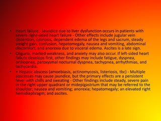
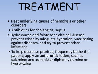
topic –
Differential
diagnosis of
jaundice
Subject –
Propodietics
DEFINITION
• Bile or liver problem causing yellowness
• A yellow discoloration of the skin, mucous
membranes, or sclera of the eyes, jaundice
indicates excessive levels of conjugated or
unconjugated bilirubin in the blood.
• In fair-skinned patients, it’s most noticeable
on the face, trunk, and sclera; in dark-skinned
patients, on the hard palate, sclera, and
conjunctiva.
• Jaundice is most apparent in natural sunlight.
In fact, it may be undetectable in artificial or
poor light. It’s commonly accompanied by
pruritus (because bile pigment damages
sensory nerves), dark urine, and clay-colored
stools.
• Jaundice develops from hyperbilirubinemia and may not be noticed
until the bilirubin exceeds 50 umol/L. – Scleral elastin has a high
affinity for bilirubin, and with a white background, it is a sensitive
indicator of jaundice. – Biliary obstruction gives a greenish skin tint
due to accumulation of biliverdin. – Hemolysis gives a lemon-yellow
tint when observed in natural light. – An orange-yellow color is
more consistent with hepatocellular disease. – Pseudojaundice may
be found in black patients with pigmented sclera, with carotinemia,
with uremia (a sallow yellowish pallor), and with quinacrine (a
yellow-green color).
• • Dark urine with green foam confirms a conjugated
hyperbilirubinemia and excludes hemolysis or a conjugating defect.
Unconjugated bilirubin is tightly bound to albumin, which prevents
glomerular filtration.
• Hyperbilirubinemia is due to - increased production of bilirubin -
impaired transport of bilirubin to the liver for excretion - and
decreased excretion of bilirubin. bilirubin
• Increased production.
• release of hemoglobin from the red cells and its subsequent
breakdown. – Common hereditary hemolytic anemias include the
hemoglobinopathies and abnormalities of RBC membranes and
enzymes. – Common acquired abnormalities include mechanical
trauma, antibody mediated damage, and other toxic or physical
insults. Thus, the hemolytic anemias are the principal cause of this
category of jaundice. These include: – hereditary spherocytosis –
Cooley anemia – Septicemia – autoimmune hemolytic anemia –
malaria.
• • Impaired transport CHF is the principal cause of this form of
jaundice, but it must be advanced enough to cause cardiac cirrhosis
• Hyperbilirubinemia is due to - increased production of bilirubin - impaired
transport of bilirubin to the liver for excretion - and decreased excretion of
bilirubin. bilirubin
• Decreased excretion This group of causes of jaundice is divided into conditions – in
which the liver is unable to transform unconjugated bilirubin to the conjugated
form
• Gilbert disease
• infectious hepatitis
• cirrhosis – conditions in which the liver cannot transfer the conjugated bilirubin into
the bile ducts
• Dubin–Johnson syndrome
• Rotor syndrome – and conditions that obstruct the bile ducts
• common duct stones
• Cholangitis
• chlorpromazine toxicity
• carcinomas of the pancreas and ampulla of Vater.
DIAGNOSTIC WORKUP
Physical examination
• Perform the physical examination in a room with natural light. Jaundice is best seen
in the periphery of ocular conjunctivae and oral mucous membranes
• Make sure that the orange-yellow hue is jaundice and not due to hypercarotenemia,
which is more prominent on the palms and soles and doesn’t affect the sclera.
• Inspect the patient’s skin for texture and dryness and for hyperpigmentation and
xanthomas.
• Look for spider angiomas or petechiae, clubbed fingers, testicular atrophy, palmar
erythema and gynecomastia.
• If the patient has heart failure, auscultate for arrhythmias, murmurs, and gallops as
well as crackles and abnormal bowel sounds.
• Palpate the lymph nodes for swelling and the abdomen for tenderness, pain, and
swelling.
• Palpate and percuss the liver and spleen for enlargement, and test for ascites with
the shifting dullness and fluid wave techniques.
• Obtain baseline data on the patient’s mental status: Slight changes in sensorium may
be an early sign of deteriorating hepatic function.
Lab and imaging
• Basic workup includes a CBC (hemolytic anemia,
infection), sedimentation rate, reticulocyte count, red
cell fragility test, urinalysis, chemistry panel, VDRL test,
ECG, a chest x-ray, and flat plate of the abdomen.
• If infectious hepatitis is suspected, a hepatitis profile,
febrile agglutinins, Monospot test, cytomegalic virus
antibody titer, and leptospirosis antibody titer should
be done.
• If lupoid hepatitis is suspected, a test for antinuclear
antibodies and a smooth muscle antibody should be
done.
Lab and imaging
• If hemochromatosis is suspected, a serum iron, iron-binding
capacity, and ferritin should be done.
• If hemolytic anemia is suspected, serum haptoglobins,
hemoglobin electrophoresis, and sickle cell preparations
may be done.
• If obstructive jaundice is suspected, then gallbladder
ultrasound should be done to rule out gallstones, and a CT
scan of the abdomen may be done to look for GI neoplasm.
An upper GI series may assist in finding a primary neoplasm
in the GI tract.
• Magnetic resonance cholangiopancreatography is useful for
common duct stone.
Lab and imaging
• ERCP or percutaneous transhepatic cholangiography will assist in determining
whether there is definitely obstructive jaundice and whether it is due to a
surgically resectable lesion.
• Peritoneoscopy can also be helpful.
• An exploratory laparotomy will probably be necessary regardless of whether one
performs the above tests.
• Cholangiopancreatography and endoscopic ultrasonography are two newer methods
that may be used to evaluate the biliary tree and pancreatic ducts, especially when
a neoplasm is suspected.
• Hepatocellular jaundice will often require a needle biopsy of the liver to pin down
the diagnosis.
• Antimitochondrial antibodies will need to be ordered to screen for biliary cirrhosis.
• Alpha 1-fetoprotein will help diagnose hepatocellular cancer. By the time you have
reached this point, you have gone to considerable expense in the diagnostic
workup. It would be much more prudent to ask for a gastroenterology consultation
before ordering all these expensive diagnostic tests.
• Viral hepatitis –Fatigue, anorexia, fever,
nausea, vomiting, dark urine, light-colored
(acholic) loose stools, RUQ pain,
hepatomegaly, and/or pruritis
• Alcoholic hepatitis –Associated with fever,
leukocytosis, and AST:ALT ratio >2
• Nonalcoholic steatohepatitis or nonalchoholic
fatty liver disease –Associated with obesity,
diabetes, hyperlipidemia and medications
Cholelithiasis
• commonly causes jaundice and biliary colic
• It’s characterized by severe, steady pain in the right
upper quadrant or epigastrium that radiates to the
right scapula or shoulder and intensifies over several
hours.
• Accompanying signs and symptoms include nausea and
vomiting, tachycardia, and restlessness. Occlusion of
the common bile duct causes a fever, chills, jaundice,
clay- colored stools, and abdominal tenderness.
• After consuming a fatty meal, the patient may
experience vague epigastric fullness and dyspepsia
Cholecystitis
• • produces nonobstructive jaundice in about 25% of
patients –RUQ pain, fever, leukocytosis – 4F Female,
fertile, fat, fourty – Murphy's sign: Pain upon palpation
of the gallbladder while taking a deep breath - Biliary
colic typically peaks abruptly, persisting for 2 to 4
hours.
• The pain then localizes to the right upper quadrant and
becomes constant. Local inflammation or passage of
stones to the common bile duct causes jaundice. -
Other findings include nausea, vomiting (usually
indicating the presence of a stone), a fever, profuse
diaphoresis, chills and, possibly, abdominal distention
and rigidity.
Drugs
• Estrogens produce canalicular cholestasis.
• Phenothiazines produce ductular cholestasis.
• Methyldopa causes autoimmune hemolytic
anemia.
• Hepatotoxins – Niacin – Acetaminophen –
Isoniazid – Phenytoin – Sulfonamides –
Ketoconazole – erythromycin estolate –
Chlorpromazine – Propylthiouracil – anabolic
steroids – Valproate – Amiodarone – vitamin A
and D (in high doses) – carbon tetrachloride –
amanita mushrooms.
• Heart failure - Jaundice due to liver dysfunction occurs in patients with
severe right-sided heart failure - Other effects include jugular vein
distention, cyanosis, dependent edema of the legs and sacrum, steady
weight gain, confusion, hepatomegaly, nausea and vomiting, abdominal
discomfort, and anorexia due to visceral edema. Ascites is a late sign.
• Oliguria, marked weakness, and anxiety may also occur. If left-sided heart
failure develops first, other findings may include fatigue, dyspnea,
orthopnea, paroxysmal nocturnal dyspnea, tachypnea, arrhythmias, and
tachycardia.
• • Hepatic abscess (amoebiasis, actinomycosis, listeriosis, tbc) - Multiple
abscesses may cause jaundice, but the primary effects are a persistent
fever with chills and sweating - Other findings include steady, severe pain
in the right upper quadrant or midepigastrium that may be referred to the
shoulder; nausea and vomiting; anorexia; hepatomegaly; an elevated right
hemidiaphragm; and ascites.
TREATMENT
• Treat underlying causes of hemolysis or other
disorders
• Antibiotics for cholangitis, sepsis
• Hydroxyurea and folate for sickle cell disease,
prevent crises by adequate hydration, vaccinating
against diseases, and try to prevent other
infections
• To help decrease pruritus, frequently bathe the
patient; apply an antipruritic lotion, such as
calamine; and administer diphenhydramine or
hydroxyzine
jaundice

jaundice

  • 1.
  • 2.
    DEFINITION • Bile orliver problem causing yellowness • A yellow discoloration of the skin, mucous membranes, or sclera of the eyes, jaundice indicates excessive levels of conjugated or unconjugated bilirubin in the blood. • In fair-skinned patients, it’s most noticeable on the face, trunk, and sclera; in dark-skinned patients, on the hard palate, sclera, and conjunctiva.
  • 3.
    • Jaundice ismost apparent in natural sunlight. In fact, it may be undetectable in artificial or poor light. It’s commonly accompanied by pruritus (because bile pigment damages sensory nerves), dark urine, and clay-colored stools.
  • 4.
    • Jaundice developsfrom hyperbilirubinemia and may not be noticed until the bilirubin exceeds 50 umol/L. – Scleral elastin has a high affinity for bilirubin, and with a white background, it is a sensitive indicator of jaundice. – Biliary obstruction gives a greenish skin tint due to accumulation of biliverdin. – Hemolysis gives a lemon-yellow tint when observed in natural light. – An orange-yellow color is more consistent with hepatocellular disease. – Pseudojaundice may be found in black patients with pigmented sclera, with carotinemia, with uremia (a sallow yellowish pallor), and with quinacrine (a yellow-green color). • • Dark urine with green foam confirms a conjugated hyperbilirubinemia and excludes hemolysis or a conjugating defect. Unconjugated bilirubin is tightly bound to albumin, which prevents glomerular filtration.
  • 5.
    • Hyperbilirubinemia isdue to - increased production of bilirubin - impaired transport of bilirubin to the liver for excretion - and decreased excretion of bilirubin. bilirubin • Increased production. • release of hemoglobin from the red cells and its subsequent breakdown. – Common hereditary hemolytic anemias include the hemoglobinopathies and abnormalities of RBC membranes and enzymes. – Common acquired abnormalities include mechanical trauma, antibody mediated damage, and other toxic or physical insults. Thus, the hemolytic anemias are the principal cause of this category of jaundice. These include: – hereditary spherocytosis – Cooley anemia – Septicemia – autoimmune hemolytic anemia – malaria. • • Impaired transport CHF is the principal cause of this form of jaundice, but it must be advanced enough to cause cardiac cirrhosis
  • 6.
    • Hyperbilirubinemia isdue to - increased production of bilirubin - impaired transport of bilirubin to the liver for excretion - and decreased excretion of bilirubin. bilirubin • Decreased excretion This group of causes of jaundice is divided into conditions – in which the liver is unable to transform unconjugated bilirubin to the conjugated form • Gilbert disease • infectious hepatitis • cirrhosis – conditions in which the liver cannot transfer the conjugated bilirubin into the bile ducts • Dubin–Johnson syndrome • Rotor syndrome – and conditions that obstruct the bile ducts • common duct stones • Cholangitis • chlorpromazine toxicity • carcinomas of the pancreas and ampulla of Vater.
  • 7.
    DIAGNOSTIC WORKUP Physical examination •Perform the physical examination in a room with natural light. Jaundice is best seen in the periphery of ocular conjunctivae and oral mucous membranes • Make sure that the orange-yellow hue is jaundice and not due to hypercarotenemia, which is more prominent on the palms and soles and doesn’t affect the sclera. • Inspect the patient’s skin for texture and dryness and for hyperpigmentation and xanthomas. • Look for spider angiomas or petechiae, clubbed fingers, testicular atrophy, palmar erythema and gynecomastia. • If the patient has heart failure, auscultate for arrhythmias, murmurs, and gallops as well as crackles and abnormal bowel sounds. • Palpate the lymph nodes for swelling and the abdomen for tenderness, pain, and swelling. • Palpate and percuss the liver and spleen for enlargement, and test for ascites with the shifting dullness and fluid wave techniques. • Obtain baseline data on the patient’s mental status: Slight changes in sensorium may be an early sign of deteriorating hepatic function.
  • 8.
    Lab and imaging •Basic workup includes a CBC (hemolytic anemia, infection), sedimentation rate, reticulocyte count, red cell fragility test, urinalysis, chemistry panel, VDRL test, ECG, a chest x-ray, and flat plate of the abdomen. • If infectious hepatitis is suspected, a hepatitis profile, febrile agglutinins, Monospot test, cytomegalic virus antibody titer, and leptospirosis antibody titer should be done. • If lupoid hepatitis is suspected, a test for antinuclear antibodies and a smooth muscle antibody should be done.
  • 9.
    Lab and imaging •If hemochromatosis is suspected, a serum iron, iron-binding capacity, and ferritin should be done. • If hemolytic anemia is suspected, serum haptoglobins, hemoglobin electrophoresis, and sickle cell preparations may be done. • If obstructive jaundice is suspected, then gallbladder ultrasound should be done to rule out gallstones, and a CT scan of the abdomen may be done to look for GI neoplasm. An upper GI series may assist in finding a primary neoplasm in the GI tract. • Magnetic resonance cholangiopancreatography is useful for common duct stone.
  • 10.
    Lab and imaging •ERCP or percutaneous transhepatic cholangiography will assist in determining whether there is definitely obstructive jaundice and whether it is due to a surgically resectable lesion. • Peritoneoscopy can also be helpful. • An exploratory laparotomy will probably be necessary regardless of whether one performs the above tests. • Cholangiopancreatography and endoscopic ultrasonography are two newer methods that may be used to evaluate the biliary tree and pancreatic ducts, especially when a neoplasm is suspected. • Hepatocellular jaundice will often require a needle biopsy of the liver to pin down the diagnosis. • Antimitochondrial antibodies will need to be ordered to screen for biliary cirrhosis. • Alpha 1-fetoprotein will help diagnose hepatocellular cancer. By the time you have reached this point, you have gone to considerable expense in the diagnostic workup. It would be much more prudent to ask for a gastroenterology consultation before ordering all these expensive diagnostic tests.
  • 11.
    • Viral hepatitis–Fatigue, anorexia, fever, nausea, vomiting, dark urine, light-colored (acholic) loose stools, RUQ pain, hepatomegaly, and/or pruritis • Alcoholic hepatitis –Associated with fever, leukocytosis, and AST:ALT ratio >2 • Nonalcoholic steatohepatitis or nonalchoholic fatty liver disease –Associated with obesity, diabetes, hyperlipidemia and medications
  • 12.
    Cholelithiasis • commonly causesjaundice and biliary colic • It’s characterized by severe, steady pain in the right upper quadrant or epigastrium that radiates to the right scapula or shoulder and intensifies over several hours. • Accompanying signs and symptoms include nausea and vomiting, tachycardia, and restlessness. Occlusion of the common bile duct causes a fever, chills, jaundice, clay- colored stools, and abdominal tenderness. • After consuming a fatty meal, the patient may experience vague epigastric fullness and dyspepsia
  • 13.
    Cholecystitis • • producesnonobstructive jaundice in about 25% of patients –RUQ pain, fever, leukocytosis – 4F Female, fertile, fat, fourty – Murphy's sign: Pain upon palpation of the gallbladder while taking a deep breath - Biliary colic typically peaks abruptly, persisting for 2 to 4 hours. • The pain then localizes to the right upper quadrant and becomes constant. Local inflammation or passage of stones to the common bile duct causes jaundice. - Other findings include nausea, vomiting (usually indicating the presence of a stone), a fever, profuse diaphoresis, chills and, possibly, abdominal distention and rigidity.
  • 14.
    Drugs • Estrogens producecanalicular cholestasis. • Phenothiazines produce ductular cholestasis. • Methyldopa causes autoimmune hemolytic anemia. • Hepatotoxins – Niacin – Acetaminophen – Isoniazid – Phenytoin – Sulfonamides – Ketoconazole – erythromycin estolate – Chlorpromazine – Propylthiouracil – anabolic steroids – Valproate – Amiodarone – vitamin A and D (in high doses) – carbon tetrachloride – amanita mushrooms.
  • 15.
    • Heart failure- Jaundice due to liver dysfunction occurs in patients with severe right-sided heart failure - Other effects include jugular vein distention, cyanosis, dependent edema of the legs and sacrum, steady weight gain, confusion, hepatomegaly, nausea and vomiting, abdominal discomfort, and anorexia due to visceral edema. Ascites is a late sign. • Oliguria, marked weakness, and anxiety may also occur. If left-sided heart failure develops first, other findings may include fatigue, dyspnea, orthopnea, paroxysmal nocturnal dyspnea, tachypnea, arrhythmias, and tachycardia. • • Hepatic abscess (amoebiasis, actinomycosis, listeriosis, tbc) - Multiple abscesses may cause jaundice, but the primary effects are a persistent fever with chills and sweating - Other findings include steady, severe pain in the right upper quadrant or midepigastrium that may be referred to the shoulder; nausea and vomiting; anorexia; hepatomegaly; an elevated right hemidiaphragm; and ascites.
  • 16.
    TREATMENT • Treat underlyingcauses of hemolysis or other disorders • Antibiotics for cholangitis, sepsis • Hydroxyurea and folate for sickle cell disease, prevent crises by adequate hydration, vaccinating against diseases, and try to prevent other infections • To help decrease pruritus, frequently bathe the patient; apply an antipruritic lotion, such as calamine; and administer diphenhydramine or hydroxyzine