This review article summarizes in vitro methods for assessing the bioaccessibility and bioavailability of nutrients from foods, including solubility assays, dialyzability methods, gastrointestinal models like TIM, and Caco-2 cell models. It focuses on how these methods have been applied to nutrients, carotenoids, and polyphenols. The review recommends which methods are best suited for answering bioaccessibility or bioavailability questions for specific nutrients. It also discusses the need for more validation studies comparing in vitro and in vivo results.
Lecture "The food metabolome" by C. Manach (INRA Clermont-Ferrand, France) at the 1st International workshop on "The Food metabolome and biomarkers for dietary exposure. Metabolomic approaches for biomarker discovery, validation and implementation" (Glasgow, 5th July, 2013)
This PPt Help Students For Improving Their Konwledge about Colon Drug Delivery. In this PPt I Covered All Essential Points About Colon Targeted Drug Delivery System.
colon drug delivery- advantage and disadvantage of colon delivery, anatomy of colon in healthy and diseased state , different approaches (conventional and new) for colon delivery, in vitro and in vivo evaluation
Introduction, Anatomy of colon, Advantage of colon drug delivery system, Disadvantage of colon drug delivery system, Factors influencing CDDS, Approaches of CDDS, Evaluation of CDDS
Targeted drug delivery into the colon is highly desirable for local treatment of a variety of bowel diseases such as ulcerative colitis, Crohn’s disease, amoebiasis, colonic cancer, local treatment of colonic pathologies, and systemic delivery of protein and peptide drugs, NSAIDs, steroids.
The colon is believed to be a suitable absorption site for peptides and protein drugs for the following reasons; (i) less diversity, and intensity of digestive enzymes, (ii) less proteolytic activity of colon mucosa than that of small intestine.
CRITERIA: Drugs used for local effects in colon against GIT diseases.
Drugs poorly absorbed from upper GIT.
Drugs for colon cancer.
Drugs that degrade in stomach and small intestine.
Drugs that undergo extensive first pass metabolism.
Drugs for targeting.
Health promoting properties encoded within fermented food microbiotaLaura Berry
Presented at the 2nd Probiotics Congress: USA. To find out more, visit:
www.global-engage.com
Paul Cotter from the APC Microbiome Institute and Teagasc presents sequencing based insights into health-promoting genes in fermented foods and discusses how we can harness this knowledge.
DRUG DELIVERY SYSTEM (gastro retentive drug delivery system)
Oral route is the most acceptable route for drug administration. Apart from conventional dosage forms several other forms were developed in order to enhance the drug delivery for prolonged time period and for delivering drug to a particular target site. Gastro-retentive drug delivery system (GRDDS) has gainned immense popularity in the field of oral drug delivery recently. it is a widely employed approach to retain the dosage form in the stomach for an extended period of time and release the drug slowly that can address many challenges associated with conventional oral delivery, including poor bioavailability. different innovative approaches are being applied to fabricate GRDDS. Gastroretentive drug delivery is an approach to prolong gastric residence time, there by targeting site-specific drugs release in the upper gastrointestinal tract (GIT) for local or systemic effects. It is obtained by retaining dosage form into stomach and by releasing the in controlled manner.
In vitro glucose absorption by everted gut sac methodKunj Fosi
Aim :- Aim is to find out the rate of glucose absorbed across an everted sac prepared from the rat intestine using the drugs Beta- caryophyllene and L-Arginine.
Result :- The combination of BCP & LA showed significant decrease in glucose absorption in small intestine.
Thus, we conclude that this combination approach (LA + BCP) possesses the new therapeutic regimen for the treatment of T2DM.
This presentation related to targeted drug delivery system particularly to colonic region. It includes various approaches for colonic delivery of drug.
Gastro retentive drug delivery is an approach to prolong gastric residence time.
Thereby targeting site-specific drug release in the upper gastrointestinal tract (GIT) for local or systemic effects
Gastro-retentive delivery is one of the site specific delivery of the drugs at stomach.
It is obtained by retaining dosage form into stomach and drug is being released at sustained manner to specific site either in stomach or intestine.
In the recent years, bio-based and biodegradable products have raised great interest since sustainable development policies tend to expand with the decreasing reserve of fossil fuel and the growing concern for the environment. Bio-Polymers are a form of polymers derived from plant sources such as sweet potatoes, soya bean oil, sugarcane, hemp oil, and corn starch. These polymers are naturally degraded by the action of microorganisms such as bacteria, fungi and algae. Bio-plastics can help alleviate the energy crisis as well as reduce the dependence on fossil fuels of our society. They have some remarkable properties which make it suitable for different applications. This paper tries to give an insight about Bio-plastics, their composition, preparation, properties, special cases, advantages disadvantages, commercial viability, its life cycle, marketing and pricing of these products.
As a result, the market of these environmentally friendly materials is in rapid expansion,
10 –20 % per year.
Bioavailability and Bioequivalence Studies (BABE) & Concept of BiowaiversJaspreet Guraya
The presentation gives an insight on BABE studies, mathematical and statistical procedures involved in designing these studies, the official guidelines regarding study design. In the later part it also discusses about biowaivers and their role.
1
5
Does Protein Affect the Metabolic Rate of ZebraFish
Statement of the Problem
Dietary ingredients, and nutrients as well as anti-nutritional remain important factors that affect its growth and development. However, there lacks proper nutritional control due to the absence of standardized reference diet (Boyle et al. 5354). Moreover, numerous epidemiological studies undertaken indicate that several prenatal are important in the growth and development of Zebrafish (Daniorerio)(Acheson et al. 456) As a way of providing a standardized dietary framework, a need emerges for the provision of specific dietary and nutritional standard aimed at improving the growth and development of Zebra fish. Thus, the necessity to investigate the growth and metabolic rate of Zebrafish (Daniorerio) when fed with different commercial diets.
BACKGROUND
The consumption of proteins in a diet could have significant effects on the body metabolism. The high calories contents of proteins commonly require more calories for breakdown that carbohydrates and this could have significant effects of various metabolic aspects. The amount nature of proteins makes them significantly different from other dietary constitutes that people consume routinely. In general, proteins cause an increase in the metabolic rate of organisms because of a variety of reasons Like for instance, Weight maintenance is actually a simple matter of the energy balance and also energy balance is attained when "energy in" is corresponding to "energy out
Thermic affects accounts for the numerous ways in which proteins increase the metabolic rate of the body. Due to the strong bond making the protein molecules, a lot of energy is required to break down the high protein that has been consumed. The thermic effect of proteins means that about 30% of the calories in proteins are burned in the process of digesting proteins(Acheson et al. 528). The heat generated in the process increases the general body heat output hence influencing metabolism.
The consumption of protein in diet has been established as a factor that increases the metabolic rate in human beings (Howard et al. 117).This is mainly because proteins require a lot of energy to be broken down and the body must provide such energy, hence it increase the metabolic rate. The process of protein synthesis in the body causes the amino acid structures of the proteins consumed to begin the process of building body muscles. The construction of these tissues in the body is a process that consumes high amounts of energy hence the body requires increased energy to undertaken the process. As a result on the increased energy requirements, high metabolic rate occurs in order to maintain the required amount of energy necessary to sustain the activities of breaking down amino acids to form muscle tissues.
Within marine environments, hypoxia episodes remain prevalent due to a variety of reasons. The presence of oxygen within the marine environmen ...
Role of Gut Microbiota in Lipid MetabolismSharafat Ali
It has become widely appreciated that our gut symbionts play integral roles in human health since perturbations of this bacterial community or the products they can produce have been associated with increased susceptibility to a variety of diseases.
Lactobacillus acidophilus CRL 1014 improved "gut health" in the SHIME(R) reactorEnrique Moreno Gonzalez
How to maintain “gut health” is a goal for scientists throughout the world. Therefore, microbiota management models for testing probiotics, prebiotics, and synbiotics have been developed.
Dietary Intervention with Yoghurt, Synbiotic Yogurt or Traditional Fermented ...Mostafa Gouda
Dietary Intervention with Yoghurt, Synbiotic
Yogurt or Traditional Fermented Sobya:
Bio-Potency among Male Adolescents Using
Five Bio-Markers of Relevance to Colonic
Metabolic Activities
his guideline should be read in conjunction with other ICH guidelines relevant to the
conduct of clinical trials (e.g., E2A (clinical safety data management), E3 (clinical study
reporting), E7 (geriatric populations), E8 (general considerations for clinical trials), E9
(statistical principles), and E11 (pediatric populations)).
This ICH GCP Guideline Integrated Addendum provides a unified standard for the European
Union, Japan, the United States, Canada, and Switzerland to facilitate the mutual acceptance
of data from clinical trials by the regulatory authorities in these jurisdictions. In the event of
any conflict between the E6(R1) text and the E6(R2) addendum text, the E6(R2) addendum
text should take priority.
Roti Bank Hyderabad: A Beacon of Hope and NourishmentRoti Bank
One of the top cities of India, Hyderabad is the capital of Telangana and home to some of the biggest companies. But the other aspect of the city is a huge chunk of population that is even deprived of the food and shelter. There are many people in Hyderabad that are not having access to
At Taste Of Middle East, we believe that food is not just about satisfying hunger, it's about experiencing different cultures and traditions. Our restaurant concept is based on selecting famous dishes from Iran, Turkey, Afghanistan, and other Arabic countries to give our customers an authentic taste of the Middle East
Piccola Cucina is regarded as the best restaurant in Brooklyn and as the best Italian restaurant in NYC. We offer authentic Italian cuisine with a Sicilian touch that elevates the entire fine dining experience. We’re the first result when someone searches for where to eat in Brooklyn or the best restaurant near me.
Ang Chong Yi Navigating Singaporean Flavors: A Journey from Cultural Heritage...Ang Chong Yi
In the heart of Singapore, where tradition meets modernity, He embarks on a culinary adventure that transcends borders. His mission? Ang Chong Yi Exploring the Cultural Heritage and Identity in Singaporean Cuisine. To explore the rich tapestry of flavours that define Singaporean cuisine while embracing innovative plant-based approaches. Join us as we follow his footsteps through bustling markets, hidden hawker stalls, and vibrant street corners.
Key Features of The Italian Restaurants.pdfmenafilo317
Filomena, a renowned Italian restaurant, is renowned for its authentic cuisine, warm environment, and exceptional service. Recognized for its homemade pasta, traditional dishes, and extensive wine selection, we provide a true taste of Italy. Its commitment to quality ingredients and classic recipes has made it a adored dining destination for Italian food enthusiasts.
1. REVIEW ARTICLE
published: 06 August 2012
doi: 10.3389/fphys.2012.00317
Application of in vitro bioaccessibility and bioavailability
methods for calcium, carotenoids, folate, iron, magnesium,
polyphenols, zinc, and vitamins B6, B12, D, and E
Paz Etcheverry1
*, Michael A. Grusak1
and Lisa E. Fleige2
1
Department of Pediatrics, USDA-ARS Children’s Nutrition Research Center, Baylor College of Medicine, Houston, TX, USA
2
Global Research and Development, PepsiCo, Barrington, IL, USA
Edited by:
Ravinder Abrol, California Institute of
Technology, USA
Reviewed by:
Sanqi Zhang, Xi’an Jiaotong
University, China
George Greeley, University of Texas
Medical Branch, USA
*Correspondence:
Paz Etcheverry, Department of
Pediatrics, USDA-ARS Children’s
Nutrition Research Center, Baylor
College of Medicine, 1100 Bates
Street, Houston, TX 77030, USA.
e-mail: paze@bcm.edu
A review of in vitro bioaccessibility and bioavailability methods for polyphenols and
selected nutrients is presented. The review focuses on in vitro solubility, dialyzability,
the dynamic gastrointestinal model (TIM)™, and Caco-2 cell models, the latter primarily
for uptake and transport, and a discussion of how these methods have been applied to
generate data for a range of nutrients, carotenoids, and polyphenols. Recommendations
are given regarding which methods are most justified for answering bioaccessibility or
bioavailability related questions for specific nutrients. The need for more validation studies
in which in vivo results are compared to in vitro results is also discussed.
Keywords: in vitro methods, minerals, vitamins, bioaccessibility, bioavailability
INTRODUCTION
Throughout the years, in vitro screening methods have been
developed and refined for the determination of nutrient bioac-
cessibility and bioavailability from foods. These are methods that
can provide useful information, especially when one considers
the vast number of factors that can affect nutrient absorp-
tion. Bioavailability, which is defined as the amount of an
ingested nutrient that is absorbed and available for physiologi-
cal functions, is dependent on digestion, release from the food
matrix, absorption by intestinal cells, and transport to body cells.
Bioaccessibility, which is the amount of an ingested nutrient
that is potentially available for absorption, is dependent only on
digestion and release from the food matrix.
It has to be kept in mind that bioavailability, which has a
physiological or metabolic endpoint, can never be measured in
its entirety by any of these in vitro methods. Furthermore, host
factors that can possibly influence nutrient absorption such as
nutrient status, age, genotype, physiological state (e.g., pregnancy,
lactation, and obesity), chronic and acute infectious disease states,
secretion of hydrochloric acid, gastric acid, and/or intrinsic fac-
tor, are impossible to factor in in vitro assays. Nonetheless, for
this review, we will use the term bioavailability in order to
retain the terminology used by many of the authors referenced
here. However, we urge readers to be cautious when interpreting
in vitro “bioavailability” data, and that they verify which aspect
of the bioavailability process is being assessed. In many cases,
researchers are only measuring uptake or absorption with their
in vitro method, yet refer to their analysis as bioavailability.
In vitro bioaccessibility/bioavailability methods are useful to
provide knowledge on possible interactions between nutrients
and/or food components, the effects of luminal factors (includ-
ing pH and enzymes), food preparation and processing practices,
nature of the food matrix etc., on either micronutrient absorba-
bility (a component of bioavailability) or on the potential for a
nutrient to be absorbed (i.e., bioaccessibility). In vitro methods
are less expensive, faster, and offer better controls of experimen-
tal variables than human or animal studies (Sandberg, 2005).
However, in vitro studies cannot be substituted for in vivo stud-
ies, and should be therefore regarded as a screening, ranking, or
categorizing tool.
In vitro METHODS
There are principally four in vitro methods for measuring bioac-
cessibility and/or bioavailability: solubility, dialyzability, or a
gastrointestinal model (e.g., TIM) for bioaccessibility, and the
Caco-2 models for bioavailability (Table 1).
In each of these methods, an in vitro digestion is conducted
to simulate the human digestive system via a two-step (some-
times a three-step) digestion that includes a gastric and intestinal
digestion. For the gastric digestion, pepsin (from porcine stom-
ach) is added prior to the acidification of the samples to pH 2
(to simulate the gastric pH of an adult) or to pH 4 (to simulate
the gastric pH of an infant). Acidification of the samples to pH
2 or 4 is important, because pepsin begins to denature itself and
thus will lose its activity at pH ≥ 5. Before the start of the intesti-
nal digestion, the samples are neutralized to pH 5.5–6 prior to
the addition of pancreatin (which consists of a cocktail of pancre-
atic enzymes such as pancreatic amylase, lipase, ribonuclease, and
proteases such as trypsin) and bile salts (which are emulsifiers),
and finally re-adjusted to pH 6.5–7. The third digestion step that
is sometimes introduced, and which precedes the gastric phase,
is the digestion by lingual alpha-amylase, which is an enzyme that
breaks apart the glycosidic bonds of starch molecules, i.e., amylose
and amylopectin. Once the food in question has been digested,
www.frontiersin.org August 2012 | Volume 3 | Article 317 | 1
2. Etcheverry et al. In vitro bioaccessibility/bioavailability methods
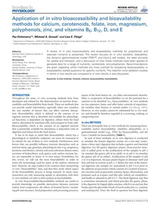
Table 1 | In vitro screening methods.
In vitro method End point Advantages Limitations
Solubility Measures bioaccessibility • Simple to do
• Relatively inexpensive
• Easy to conduct, every laboratory would
have the necessary equipment
• Sometimes not a reliable indicator of
bioavailability
• Cannot assess rate of uptake or
absorption or transport kinetics
• Cannot measure nutrient or food
component competition at the site of
absorption
Dialyzability Measures bioaccessibility • Simple to do
• Relatively inexpensive
• Easy to conduct, every laboratory would
have the necessary equipment
• Cannot assess rate of uptake or
absorption or transport kinetics
• Cannot measure nutrient or food
component competition at the site of
absorption
Gastrointestinal models Measures bioaccessibility.
However, when coupled to
intestinal cells, bioavailability
can also be measured
• Incorporates many digestion parameters
(peristalsis, churning, body temperature,
etc.,)
• Allows the collection of digest at any
step of the digestive system
• Expensive
• Few validation studies
Caco-2 cell model Measures bioavailability • Allows the study of nutrient or food
component competition at the site of
absorption
• Requires trained personnel with
knowledge of cell culture methods
bioaccessibility can either be measured via solubility, dialyzability
or gastrointestinal models.
For the solubility assay, the intestinal digests need to be cen-
trifuged, to yield a supernatant and precipitate. The nutrients or
compounds present in the supernatant represent the soluble com-
ponents and are measured by atomic absorption spectrophotom-
etry (AAS), mass spectrometry, spectrophotometry, inductively
coupled plasma atomic emission spectroscopy (ICP-AES), high
performance liquid chromatography (HPLC), or in the case of
radioactive compounds, by gamma or liquid scintillation count-
ing. Percent solubility is calculated as the amount of soluble
compound relative to the total amount of compound in the test
sample.
Dialyzability assays were introduced in 1981 by Miller et al. as
a means to estimate iron bioaccessibility from foods. The model,
which measures soluble minerals of low molecular weight, is
based on an equilibrium dialysis. It involves the addition of a
dialysis tubing of a certain molecular weight cut off (MWCO),
following the gastric digestion. The dialysis tubing or bag con-
tains a buffer, such as sodium bicarbonate, that slowly diffuses
out of the bag and neutralizes the peptic digest. After incubation,
pancreatin/bile is added and following another incubation total
dialyzable iron can thus be determined by measuring the amount
of mineral present in the dialysate. The whole premise of dialyz-
ability methods is that dialyzable compounds will be available for
absorption in the small intestine. This method has been applied
and slightly modified to study the bioaccessibility of a number of
micronutrients including calcium, zinc, and magnesium, among
others. An extension to this method involves the continuous-
flow dialysis system performed by means of a hollow-fibre system
(Wolters et al., 1993). As opposed to the in vitro methods based
on Miller et al. (1981), in which components that pass the dialysis
membrane are not removed, the continuous-flow dialysis system
takes the removal of dialysable components into account leading
probably to a better estimate of in vivo bioavailability.
A number of institutions and commercial groups have devel-
oped sophisticated gut models to simulate the human digestive
system (Afkhami et al., 2007; de Jong et al., 2007; Barmpalia-Davis
et al., 2008; van den Abbeele et al., 2010; Vardakou et al., 2011).
One commercial gastrointestinal model (TIM), which has been
developed by The Netherlands Organization (TNO) for Applied
Scientific Research, has been described in great detail by Minekus
et al. (1995, 1999). TNO’s intestinal model (TIM) is a very
sophisticated model since many parameters of the human diges-
tive system are simulated: e.g., body temperature, flow of saliva,
gastric- and pancreatic juice including digestive enzymes, and
bile, peristalsis and churning, gastrointestinal transit times, regu-
lation of gastric and intestinal pH, etc. The model consists of two
computer-controlled chambers, named TIM1 and TIM2. TIM1
comprises four compartments that represent the stomach, duo-
denum, jejunum, and ileum. Secretion of digestive juices and pH
adjustment in each section are simulated according to physiologi-
cal data. A dialysate component collects compounds and they rep-
resent the bioaccessible fraction. The material that exits the model
represents, on the other hand, the nonbioaccessible fraction and is
used to study colonic fermentation products in the TIM2 (Anson
et al., 2009). TIM2 represents the human large intestine, where the
colonic fermentation experiments are performed. The nonbioac-
cessible fraction generated from TIM1 can be inoculated with
active microbes obtained from humans. One of the main advan-
tages of the TIM system is the possibility of collecting samples at
any level of the gastrointestinal tract and at any time during diges-
tion (Etienne-Mesmin et al., 2011). Although this model mea-
sures bioaccessibility, bioavailability can also be measured if the
food digest at the end of the TIM1 digestion is added to human
intestinal cells and nutrient uptake is assessed (TNO, 2011).
Frontiers in Physiology | Gastrointestinal Sciences August 2012 | Volume 3 | Article 317 | 2
3. Etcheverry et al. In vitro bioaccessibility/bioavailability methods
Bioavailability (or more correctly, components of bioavailabil-
ity) can be assessed through the determination of nutrient uptake,
transport, or both by Caco-2 cells. Caco-2 cells belong to a human
epithelial cell line derived from a human colonic adenocarci-
noma. Even though they have a colonic origin, for reasons that
to this day are not understood, the cells behave very much like
intestinal cells upon culture. Uptake studies are performed with
cells grown on the surface of plastic dishes or wells, or alterna-
tively, if transport will also be measured, on Transwell inserts.
Transwell inserts allow the collection and measurement of nutri-
ents that have been absorbed through the apical membrane and
then released through the basolateral membrane. Following the
gastric digestion of the food, pancreatin/bile is added and the
digest is added to the cells. In vivo, cellular integrity is maintained
through the presence of an intestinal mucus layer. However,
in vitro, one of several methods must be used to prevent the
enzymatic degradation of the cells. One method is the introduc-
tion of a dialysis membrane secured with a silicone O-ring to a
plastic insert, which is placed on top of the cell monolayer. The
intestinal digest is placed on top of the dialysis membrane, thus
preventing the enzymes from reaching the cells (Gangloff et al.,
1996; Glahn et al., 1998). Another method involves heat treat-
ing the intestinal digests for 4 min at 100◦C in order to inhibit
the enzymes added during the experiment (Jovaní et al., 2001;
Frontela et al., 2009). This step, however, imposes a shortcoming
in the methodology, because heating the sample at 100◦C will also
likely denature food proteins, thus impacting (either positively
or negatively) bioavailability. Other methods involve the inacti-
vation of the enzymes by acidifying the intestinal digests to pH
2 (Frontela-Saseta et al., 2011) or by lowering the temperature of
the digests and subsequently filtering the samples (Au and Reddy,
2000). However, these steps are not physiologically representative
of in vivo conditions. The in vitro co-culture of Caco-2 and HT29-
MTX, a human mucus-producing cell line, might represent a
more physiological and realistic approach to in vivo conditions
(Mahler et al., 2009), as the generated mucus layer would protect
the Caco-2 cells from digestive enzymes. This approach has not
been used extensively; thus, more studies are needed to determine
its general applicability for various nutrients, and to evaluate the
consequences of incorporating an additional diffusional layer to
the apical membrane of the Caco-2 cells.
The Caco-2 uptake of some, but not all, dietary micronutri-
ents has been examined. In the case of carotenoids and other
fat soluble compounds, it is the Caco-2 uptake of either micel-
larized or soluble (but not necessarily micellarized) compounds
that is assessed. Iron uptake can be estimated via ferritin forma-
tion or 59Fe uptake (a radioisotope which had been allowed to
equilibrate with the food in question). Unlike ferritin formation,
which is an indicator of iron uptake, there are no biomarkers of
uptake for minerals like calcium and zinc. The use of metalloth-
ionein, a cytoplasmic protein that stores zinc, as an indicator of
zinc uptake has some potential. However, metallothionein can
also bind and store other metals like copper, selenium, cadmium,
mercury, silver, and arsenic (Bell and Vallee, 2009). Thus, the pro-
tein is not specific for zinc which questions the suitability of this
biomarker for measuring zinc bioavailability. Cellular calcium
and zinc uptake have been determined by measuring cell uptake
via atomic absorption spectroscopy. However, in this method
one cannot differentiate the calcium or zinc originally present
in the cells from the minerals that have been absorbed from
the digested food, since one is measuring total mineral content.
Alternatively, radioisotopic forms of the minerals can be used and
traced. However, this has certain complications that have to be
addressed such as radioactivity exposure, appropriate rinse solu-
tion to remove surface bound radioisotopes, increased costs, and
the possible lack of an equilibration between the isotope and the
endogenous mineral present in the food, among others.
Caco-2 transport studies require that the cells grow on
Transwell inserts containing semipermeable membranes, thus
allowing the formation of two chambers: an apical chamber
which receives the digested test meal and a basolateral cham-
ber where the transported compound can be collected and later
analyzed. Cell monolayer integrity on Transwell inserts has to
be monitored and most often is done by measuring transepithe-
lial electrical resistance (TEER) across the cell monolayer or by
measuring the amount of a nontransportable fluorescent com-
pound such as luciferase yellow. An optimal monolayer integrity
test result suggests that tight junctions between adjacent epithe-
lial cells exist, thus providing a good separation between the apical
and the basolateral chambers.
APPLICATIONS OF In vitro METHODS AND
RECOMMENDATIONS
CALCIUM
Calcium is a macromineral that plays an important role in
bone health, muscle contraction, blood clotting, nerve conduc-
tion, enzyme regulation, and possibly weight loss (Guéguen and
Pointillart, 2000; Tremblay and Gilbert, 2011). In humans, intesti-
nal calcium absorption is controlled by complex homeostatic
mechanisms involving calcitriol and the parathyroid hormone
(PTH). Calcitriol (1,25(OH2) vitamin D3) increases the syn-
thesis of a cytosolic calcium-binding protein (calbindin) result-
ing in increased calcium transport in intestinal cells (DeLuca,
1985). The PTH indirectly affects intestinal calcium absorption by
increasing the formation of calcitriol from its precursor, calcidiol
(25(OH) vitamin D3) (Raisz, 1981). This internal regulation of
intestinal absorption certainly makes it difficult to rely on in vitro
availability results as an estimation of calcium bioavailability.
However, regardless of the mechanism involved in calcium
homeostasis, calcium has to be soluble in the gastrointestinal tract
before it can be absorbed. Certain dietary factors can impact
calcium solubility, thereby affecting calcium bioavailability at
the absorptive surface of intestinal cells. Thus, in vitro meth-
ods might be useful to compare the bioaccessibility/bioavailability
of different calcium salts that are contained in dietary sup-
plements or when added as food fortificants (e.g., calcium
carbonate, calcium citrate, calcium phosphate, calcium glu-
conate, etc.). These methods can also be used to assess the
effects of the type of protein present in foods, the effect of
digestible carbohydrates such as lactose, and non-digestible car-
bohydrates such as fibers and carbohydrates gums, or plant
food components including phytate, and fructo-oligosaccharides
on calcium bioaccessibility/bioavailability (Cámara-Martos and
Amaro-López, 2002). Furthermore, calcium has the tendency to
www.frontiersin.org August 2012 | Volume 3 | Article 317 | 3
4. Etcheverry et al. In vitro bioaccessibility/bioavailability methods
bind to fatty acids in the lumen forming insoluble soaps. Thus,
studying which types of fatty acids (i.e., short vs. long chain, sat-
urated vs. unsaturated) lead to a more absorbable form of the
mineral will be very easy to conduct in an in vitro type of exper-
iment. Below are some of the dietary factors affecting calcium
bioavailability, which have been studied via in vitro methods.
Casein phosphopeptides
Casein phosphopeptides (CPPs) result from the enzymatic
hydrolysis of casein, the predominant protein found in cow’s
milk. CPPs contain clusters of phosphoserine residues, which can
effectively bind calcium, and inhibit formation of insoluble cal-
cium phosphates (Narva et al., 2003). Pure CPPs have been shown
to promote calcium absorption in in vitro assays using HT-29 cells
and Caco-2 cells (Ferraretto et al., 2003; Cosentino et al., 2010)
and in vivo. Erba et al. (2001) who studied the intestinal calcium
absorption in rats found that the absorption from CaCl2 solu-
tions decreased by 90% when in the presence of phosphate (Ca:Pi
molar ratio of 1:1), but decreased by only 40% from Ca-CPP at
the same Ca:Pi molar ratio.
On the other hand, Drago and Valencia (2004) who used an
in vitro dialyzability assay to measure bioaccessibility from infant
formulas found no increase in calcium dialyzability with increas-
ing casein concentration, perhaps due to an incomplete casein
proteolysis. Kennefick and Cashman (2000) similarly found no
effect of three different casein phosphopeptide preparations on
calcium dialyzability. A human study performed on nine Finnish
postmenopausal women who received milk and milk enriched
with CPPs, found no differences in serum calcium between the
two groups. According to the authors, a stimulatory effect of CPPs
on calcium absorption might have been observed had the sub-
jects been vitamin-D deficient (Narva et al., 2003). Likewise, a
calcium lactate drink supplemented with CPPs led to lower frac-
tional absorption of calcium in adults than the unsupplemented
kind (P = 0.015). Thus, there appears to be conflicting results on
the effects of CPPs both in in vivo and in in vitro experiments,
and more experiments are needed to clarify their role in mineral
bioavailability.
Phytate
Components in plant foods like phytate can form insoluble
complexes with calcium, thereby reducing its bioavailability.
Kennefick and Cashman (2000) reported that phytate had a more
pronounced negative effect on calcium solubility than oxalate,
wheat fibre-extract, barley fibre-extract, and casein. Liang et al.
(2010), who used an in vitro solubility assay to compare rice-
based foods from China, found that the high level of phytate in the
brown rice (ranging from 14.9 to 19.4 mg of phytic acid/gram of
rice) resulted in the lowest calcium solubility (12%) among all the
rice foods tested. Brown rice germination, a process that results
in phytate hydrolysis (Schlemmer et al., 2009), increased calcium
solubility from 12% to 18%. Not surprisingly, the calcium solubil-
ity of white rice, which was produced by milling and polishing of
the brown rice to remove the outer layer, increased with respect to
brown rice (16.2% vs. 12%). Rice noodles, which are soaked and
fermented prior to noodle making, had a percent calcium solubil-
ity ranging from 33.7% to 38.2% probably as a result of the low
levels of phytic acid present (ranging from 0.0 to 4.1 mg of phytic
acid/gram of rice noodles).
Phytate’s inhibitory role on calcium absorption is significant
only when the phytate to calcium molar ratio is above a certain
value; below that value, the inhibitory effect is trivial. According
to Frontela et al. (2009), the cut-off value is a molar ratio of 0.24.
The authors, who used an in vitro digestion/Caco-2 cell uptake
model to compare three different commercial cereals sold in
Spain, found that calcium uptake was higher from infant cereals
which had been dephytinized. However, results were significant
(P < 0.05) only for the infant cereal which contained the highest
phytate to calcium molar ratio. The other infant cereals tested had
a phytate to calcium molar ratio ≤ 0.18 (Frontela et al., 2009).
Using dialyzability assays, Kamchan et al. (2004) found that
vegetables containing the highest in vitro dialyzability for calcium
(20–39%) corresponded to the ones that contained the lowest lev-
els of phytate, fiber, and oxalate (e.g., kale, celery, collard, Chinese
cabbage, and soybean sprouts). On the other hand, low dialyz-
able calcium (2–7%) corresponded to samples with high levels
of oxalate and phytate (e.g., amaranth, white, and black sesame
seeds).
Carbohydrates
Soluble fibers may have negative or positive effects on calcium
absorption. In some European countries, carbohydrate gums such
as alginic acid, guar gum, and locust bean gum are used as thick-
eners in commercial anti-regurgitation milk formulas for infants
with evidence of gastroesophageal reflux (Bass and Chan, 2006).
Bosscher et al. (2000) found that the incorporation of locust
bean gum into an anti-regurgitation infant formula significantly
lowered calcium dialyzability (9.4% ± 0.7%; P < 0.01) in com-
parison with the corresponding nonthickened formula (13.3%
± 1.2%). According to the authors, locust bean gum appears to
affect calcium dialyzability by means of its physical properties to
act as a thickening agent, rather than to its chemical ability to
form complexes (Bosscher et al., 2003a). In another in vitro study,
calcium availability was similarly reduced after supplementation
with locust bean gum (11.9%) and high esterified pectin (11.7%),
but it increased by 30% after inulin supplementation (Bosscher
et al., 2003b). The ability of inulin to enhance calcium absorption
has also been shown both in human (Abrams et al., 2005, 2007;
Holloway et al., 2007) and animal (Coudray et al., 2005; Raschka
and Daniel, 2005) studies.
Maillard reaction products and other processing conditions
Maillard reaction products are compounds in foods or beverages
that are generated in the presence of heat, amino acids, and
reducing sugars. The Maillard reaction induces browning of
foods, has an effect on nutritive value, can have toxicological
implications (such as the formation of acrylamide), can produce
antioxidative components and it has also a large effect on flavor
(van Boekel, 2006). Furthermore, Maillard reaction products
may affect calcium bioavailability. Seiquer et al. (2010) used an
in vitro digestion/solubility assay to compare the effect on cal-
cium of thermally damaged milk, by comparing overheated milk
(three cycles of sterilization at 116◦C, 16 min) with ultra-high
temperature (UHT) milk (150◦C, 6 s). Calcium solubility was
Frontiers in Physiology | Gastrointestinal Sciences August 2012 | Volume 3 | Article 317 | 4
5. Etcheverry et al. In vitro bioaccessibility/bioavailability methods
lower from the overheated milk, which has higher concentrations
of Maillard reaction products, than from the UHT milk. The
results were validated against rat feeding trials. Feeding rats the
diet containing the overheated milk as the main protein source
led to significantly lower values of apparent calcium absorption
and retention than those found among animals fed the UHT
milk diet. On the other hand, Mesías et al. (2009) found no effect
of Maillard reaction products on Caco-2 calcium transport. The
authors used two diets: a “white diet (WD)” (low in Maillard
reaction products) and a “brown diet (BD)” (high in Maillard
reaction products). For the preparation of the WD, cooking
practices in which the Maillard reaction products develop (i.e.,
frying, toasting, and roasting) were avoided. The BD was rich
in processed foods (breakfast cereals, baked products, chocolate,
fried foods, toasted foods, and breaded foods, etc.,) with an
evident development of browning and, thus, rich in Maillard
reaction products. When 20 male adolescents were fed the two
diets using a randomized crossover trial, there were also no
differences in bioavailability (% calcium absorption; WD =
40.4%, BD = 38.2%) (Mesías et al., 2009).
Processing conditions were also tested. Viadel et al. (2006) used
the Caco-2 cell uptake model to assess the effect of cooking on
calcium availability. The bioavailability of calcium from cooked
white beans (Phaseolus vulgaris L.) was higher (calcium uptake
18.8%) than from the raw beans (3.6%). Repo-Carrasco-Valencia
et al. (2010) showed that boiled kañiwa (Chenopodium pallidi-
caule), a grain that grows in the Andes, had higher calcium
dialyzability values than the raw kañiwa. On the other hand, cal-
cium dialyzability was lower for the roasted and boiled quinoa
(Chenopodium quinoa) than in the raw quinoa. According to the
authors, cooking might increase the digestibility of the proteins
with which calcium is bound, thus increasing the release of the
mineral from any protein complexes. On the other hand, boiling
might lead to an increase in mineral loss into the water.
Calcium salts and organic acids
Using an in vitro digestion/Caco-2 cell model, Etcheverry et al.
(2005a) found no differences in calcium uptake results when
human milk fortifiers (i.e., supplements containing protein,
energy, minerals and an ample range of vitamins which are added
to expressed human milk) were supplemented with three types
of calcium salts: calcium glycerosphosphate gluconate, calcium
phosphate, and calcium chloride.
Rao et al. (2007) used an in vitro solubility assay to measure
calcium bioaccessibility from a commercial calcium-milk pro-
tein supplement. The results showed that the calcium present in
this supplement was readily released by enzymatic digestion: with
increasing pepsin concentration, more mineral was released from
the supplement. This was probably a result of the proteolytic role
that this enzyme has on the proteins present in this supplement,
such as β-lactoglobulin, α-lactalbumin, and lactoferrin. Both β-
lactoglobulin and α-lactalbumin have the ability to chelate/bind
calcium. Thus, the proteolytic digestion of these proteins might
liberate more calcium.
Organic acids might have an enhancing effect on calcium
absorption (Pak et al., 1987). Perales et al. (2005) used Caco-2
cells to compare calcium uptake from infant formulas and from
fruit juices containing milk and cereals (FMC). The calcium
uptake was higher from the FMC samples than from the infant
formulas, probably as a result of the presence of citric and malic
acids in the juices. Shiowatana et al. (2006) also found an enhanc-
ing effect of citric acid on calcium absorption using a continuous
flow dialysis system. The authors added organic acids to amaranth
leaves and found that the enhancement on calcium dialyzability
was most pronounced with the addition of citric acid followed by
tartaric, malic, and ascorbic acids. The authors pointed out that
the organic acids favorably affected calcium availability in spite of
the likely presence of oxalate and phytate in the amaranth leaves.
Bernardi et al. (2006) concluded that citric acid addition to a
cookie formulation made with seeds of algarrobo (Prosopis alba),
a leguminous tree, improved calcium dialyzability.
Recommended method
There are four methods for assessing calcium bioaccessibility
and/or bioavailability: solubility, dialyzability, Caco-2 cell uptake,
and transport. The Caco-2 cell model is a good model for predict-
ing calcium bioavailability in humans (Cashman, 2003). The cells
have features, including calbindin, vitamin D receptors, calcium
transport channels, etc., that are essential for the study of vita-
min D-mediated intestinal calcium absorption (Fleet et al., 2002).
Furthermore, the in vitro digestion/Caco-2 transport method has
been validated against human studies. When Mesías et al. (2009)
compared two diets with different content of Maillard reaction
products, the authors found no differences in calcium bioavail-
ability results when studied in humans or in Caco-2 cells. The
recommended method is therefore the in vitro digestion/Caco-2
uptake/transport method.
CAROTENOIDS
Carotenoids have received a lot of attention within the scientific
community not only because some of them possess pro-vitamin A
activity, meaning that they can be converted into retinoid forms,
but because they can also act as antioxidants. There are over
600 carotenoids in nature, and they are responsible for the red,
orange, and yellow colors of many fruits and vegetables. Beta-
carotene, α-carotene, and β-cryptoxanthin (carotenoids with
provitamin A activity), lycopene, lutein, and zeaxanthin (no
pro-vitamin A activity) (Gropper et al., 2009) are the six most
common dietary carotenoids. The consumption of carotenoids
is inversely related to the incidence of cardiovascular diseases,
cancer, cataracts, and age-related macular degeneration (Nagao,
2009), probably due to their antioxidant capabilities.
Food sources of carotenoids include plant foods such as car-
rots, sweet potatoes, tomatoes, kale, and spinach, to name a
few. Carotenoid availability from plant foods is dependent on
(1) factors that affect the food matrix in which the carotenoids
are present and (2) the presence of certain dietary components
(Yonekura and Nagao, 2007). In the food matrix, carotenoids
are usually associated with proteins: carotenes and lycopene are
found complexed to proteins in chromoplasts, whereas lutein is
located in chloroplasts (Garrett et al., 2000). Food processing
conditions (such as cooking, microwaving, and pasteurization)
as well as the enzymatic processes during digestion that soften
or break cell walls, disrupt the protein-carotenoid complexes,
www.frontiersin.org August 2012 | Volume 3 | Article 317 | 5
6. Etcheverry et al. In vitro bioaccessibility/bioavailability methods
favoring carotenoid release, and bioavailability (Parker, 1996).
Reduction in particle size (for instance through homogeniza-
tion, grinding, or milling) will similarly favor carotenoid absorp-
tion. Certain food components will also affect carotenoid
bioavailability. Once the carotenoid has been released from
the food, it is incorporated into lipid droplets before enter-
ing the micelles, thus the presence of dietary fat will favor
carotenoid absorption. On the other hand, the presence of
soluble fiber as well as plant sterols and stanols, will nega-
tively affect the absorption of carotenoids (Yonekura and Nagao,
2007).
Application of in vitro methods
A comprehensive literature search in PubMed revealed that there
are basically three main in vitro methods to determine the bioac-
cessibility and/or the bioavailability of carotenoids from foods.
An in vitro solubility method for measuring carotenoids has
been utilized for the bioaccessibility screening of multiple foods
(Hedrén et al., 2002a,b; Mulokozi et al., 2004). The method con-
sists of a digestion method that simulates the human digestive
system, followed by an assessment via HPLC of the types and
quantity of carotenoids released from the food. Following the
intestinal digestion, the samples are centrifuged, and the aqueous
portion is extracted with petroleum ether that is then evaporated.
The residue, containing the released carotenoids, is dissolved in
a mobile phase solvent (consisting of methanol, methyl-t-butyl
ether, and water) and filtered through a 0.45 μm pore size cellu-
lose membrane filter and subjected to reverse phase HPLC. This
method has been used after minor modifications to study the
effects of thermal processing (Lemmens et al., 2011) and particle
size (Lemmens et al., 2010) on β-carotene bioaccessibility from
carrots.
A modification of this method was introduced by Reboul et al.
(2006). What is essentially different in this method is that fol-
lowing the in vitro digestion the samples are ultracentrifuged at
very high speeds and the aqueous portion is collected and passed
through a 0.22 μm filter, thereby obtaining micelles. Thus, the
authors ultimately quantify the carotenoids present in micelles
(i.e., micellarized carotenoids) as a measure of bioaccessibility.
This method has been used to compare carotenoid bioaccessibil-
ity from durum wheat and egg pasta (Werner and Böhm, 2011)
and from different varieties and species of citrus fruits (Dhuique-
Mayer et al., 2007); and to assess the effect of thermal processing
on lycopene bioaccessibility from tomato pulp (Colle et al., 2010),
and others vegetables.
The study by Reboul et al. (2006) has been validated against
human studies. The in vivo bioaccessibility results were obtained
from a study published by Tyssandier et al. (2003). In this study,
Tyssandier et al. (2003) measured the percentage of carotenoids
recovered in the micellar phase (i.e., micellarized carotenoids)
from human duodenum during digestion of a carotenoid rich
meal. The meal contained sunflower oil, tomato puree (main
source of lycopene), chopped spinach (main source of lutein),
and carrot puree (main source of β-carotene). As reported by
Reboul et al. (2006), the bioaccessibility values from the in vivo
human results were in the same range as those measured after
the in vitro digestion model, with the exception of spinach lutein
bioaccessibility which was about fivefold higher in in vitro than
in in vivo studies.
Results from the solubility assay agree with what is expected to
occur in vivo. Cooking, which results in a more efficient release
of carotenoids from the food matrix by softening cell structures
so that digestive enzymes can work more efficiently, resulted in
higher β-carotene release from carrots compared to the uncooked
kind (Hedrén et al., 2002a). Homogenization, which represents
a mechanical disruption of the tissue, resulted in a sevenfold
and an almost fivefold improvement of β-carotene bioaccessibil-
ity from the raw and cooked carrot samples, respectively (Hedrén
et al., 2002a). Reboul et al. (2006) similarly found that percent
β-carotene bioaccessibility increased with the level of processing:
2.5–2.6% from canned or raw carrots, 4.4% from pureed carrots,
and 14.1% from carrot juice.
Addition of cooking oil to the carrots increased the percent
of β-carotene released from both the raw and cooked carrots,
but the results were more significant with homogenized sam-
ples (Hedrén et al., 2002a). Addition of oil similarly resulted in
higher bioaccessibility values from orange fleshed sweet potatoes
(Bengtsson et al., 2009a). Cooking green leafy vegetables (leaves of
amaranth (Amaranthus spp.), cowpea (Vigna unguiculata), sweet
potato (Ipomoea batatas), pumpkin (Cucurbita moschata), and
cassava (Manihot esculenta) in red palm oil instead of sunflower
oil, resulted in 1.7–2.5 times as much bioaccessible β-carotene
(Hedrén et al., 2002b).
Different cooking methods will affect in dissimilar manner
the release of carotenoid from foods. Microwaved orange fleshed
sweet potatoes resulted in lower β-carotene release either in the
absence or presence of oil (without oil: 23.7%; with oil: 27.5%)
than boiling or steaming (without oil: 38–40.7%, with oil: 45%)
(Bengtsson et al., 2009a). The authors concluded that the short
heating period for the microwaved samples was not sufficient to
obtain an adequate breakdown of the sweet potato cell matrix
and, subsequently, the release and transfer of β-carotene to the
supernatant/micellar fraction was impaired.
It has to be kept in mind that carotenoids are susceptible
to destruction by heat. Mulokozi et al. (2004) compared two
cooking methods on carotenoid bioaccessibility and retention
from diverse African vegetables: a traditional cooking method,
which consisted of boiling samples for 20–30 min in the absence
of oil, and a modified cooking method, which consisted of
reduced boiling times, and thus a potential for reduced carotenoid
destruction. Bioaccessibility of β-carotene from the traditional
cooking method ranged from 5% to 26% and from 18% to 77%
from the modified method. Losses of β-carotene were 14–51%
from vegetables prepared via traditional methods and 6–34%
when prepared with the modified method. Thus, while cooking
will increase carotene release and bioaccessibility from the food
matrix, it will also lead to a reduction in carotene concentration,
due to destruction of the molecule.
Lycopene and β-carotene appear to be sensitive to digestive
conditions. Déat et al. (2009) found there was a 25% loss of
lycopene in a simulated gastrointestinal TIM model that mea-
sured bioaccessibility from a meal containing red tomatoes and
sunflower oil. While lycopene appeared to be stable in the gas-
tric and duodenal compartments, it was in part degraded in the
Frontiers in Physiology | Gastrointestinal Sciences August 2012 | Volume 3 | Article 317 | 6
7. Etcheverry et al. In vitro bioaccessibility/bioavailability methods
terminal parts of the small intestine. Blanquet-Diot et al. (2009)
also showed that lycopene, along with β-carotene, were sensitive
to destruction. Recovery percentages of β-carotene were lower
for a red tomato-containing meal than from a yellow tomato-
containing meal (P < 0.05). On the other hand, zeaxanthin and
lutein were stable during in vitro digestion.
Garrett et al. (1999) were basically the pioneers in the devel-
opment of the Caco-2 method for carotenoid bioavailability. The
method relies on an in vitro digestion followed by the addition
of the aqueous, filtered portion of the digestate (which would be
representative of micellarized carotenoids) to Caco-2 cells. The
cells are then harvested in phosphate buffered saline, contain-
ing ethanol, and BHT (butylated hydroxytoluene, an antioxidant)
and stored at −20◦C. On the day of the carotenoid analysis,
the carotenoids are extracted from cells with a series of ace-
tone and/or hexane additions. The pooled hexane extract is then
evaporated to dryness, reconstituted and analyzed by reverse-
phase HPLC.
The method by Garrett et al. (1999) has been used to study the
bioavailability of carotenoids from vegetables (Huo et al., 2007),
spinach puree (Ferruzzi et al., 2001), and orange fleshed melons
(Fleshman et al., 2011), among others. A very similar method
was introduced by Liu et al. (2004). In this method the authors
measured both bioaccessibility and bioavailability, but they did
not ultracentrifuge nor did they filter the samples, thus they did
not necessarily add micellarized carotenoids to the Caco-2. The
authors found that cooking corn samples enhanced the amount
of lutein (0.9 fold) and zeaxanthin (1.2-fold) taken up by the cells
compared to the raw grain (Liu et al., 2004).
A concern with this bioavailability method has been the sta-
bility of the micellar carotenoids during the incubation time with
Caco-2 cells. Some of these bioavailability studies have incuba-
tion times as long as 6 (Garrett et al., 2000) or 8 h (Liu et al.,
2004). Oxidative reactions might modify and affect the quantity
of carotenoids during their exposure to the Caco-2 cells, thus it
is important to keep incubation time to a minimum while not
affecting the sensitivity of this assay. Garrett et al. (2000) observed
that the addition of 500 μmol/L α-tocopherol to the medium
might confer protection against oxidation and thus improve the
stability of carotenoids.
Interestingly, Biehler et al. (2011) found that the addition of
calcium, iron, and zinc significantly reduced both micellarization
and Caco-2 uptake of total carotenoids from a spinach meal by up
to 55% (Ca) and 90% (Fe, Zn), respectively. The minerals, which
had been added at concentrations ranging from 3.8 to 25 mM,
can presumably interact with free fatty acids, forming insoluble
soaps, and with bile acids, thus compromising carotenoid emul-
sification. Also, minerals might reduce the size of the micelles,
resulting in a marked and significant decrease of carotenoids in
the micelles. Bengtsson et al. (2009b) also found that iron inhibits
β-carotene uptake by Caco-2 cells, and that an inverse relation-
ship between the beta-carotene uptake and iron concentration
in the test solution exists (r2 = 0.93, P < 0.05). With the addi-
tion of ferrous chloride (30 μM), the beta-carotene uptake was
significantly reduced (P < 0.05), on average by 22%.
An extension to the above method involves transport stud-
ies in Caco-2 cells in which the cells are grown on Transwell
inserts. Only a couple of transport studies have been conducted
(O’Sullivan et al., 2008, 2010).
Recommended method and other comments
In all of the above methods, carotenoid bioaccessibility can be
assessed; however, the Caco-2 method allows the measurement
of both bioaccessibility and bioavailability. There are basically two
in vitro solubility methods: one that measures soluble carotenoids
and one that measures soluble micellarized carotenoids. In the
first method there is always the possibility of overestimating the
true bioaccessibility of carotenoids, because in the supernatant
one is measuring carotenoids which are not micellarized as well
as micellarized carotenoids. Micellarized carotenoids are obtained
by measuring the fraction of the food carotenoid incorporated
into the micelles (obtained from ultracentrifugation and filtra-
tion of the aqueous component through a 0.22 μM pore size
membrane).
It is important to choose an in vitro method for carotenoid
bioaccessibility that includes the extraction and measurement of
carotenoids in micelles, the form in which the carotenoids will
ultimately be absorbed by the intestinal cells. This is important
for various reasons. First, there are compounds in foods that
impair the transfer of carotenoids from the food matrix into
the micelles, such as sucrose polyester, the structure in Olestra
(Weststrate and van het Hof, 1995), fibers such as alginates, cel-
lulose, and pectins (Yonekura and Nagao, 2009) plant sterols
and stanols (Yonekura and Nagao, 2007) and divalent cations
(Biehler et al., 2011). By the first solubility method, one could
never assess this impairment in the carotenoid transfer from the
food matrix to the micelle. Second, isomers of the same com-
pound may incorporate into the micelle differently. For example,
cis lycopene is more likely to be incorporated into micelles than
trans lycopene, resulting in a higher bioavailability from the cis
form than from the trans form. This might be as a result of a
greater tendency for the trans isomer to form aggregates or due
to its slightly lower solubility (Boileau et al., 1999; Failla et al.,
2008). A higher micellarization was similarly reported for cis β-
carotene than for trans β-carotene (Ferruzzi et al., 2006). This is of
importance if different foods contain different amounts or ratios
of cis and trans carotenoids. Third, different carotenoids might
compete with each other at the level of entry into the micelle
(van Het Hof et al., 2000) and different carotenoids might be
incorporated into micelles differently. For instance, according to
Garrett et al. (1999), the differential transfer of the carotenoids
into micelles is dependent on their hydrophilicity. Carotenoids
that have been released from the food matrix but are embedded
in the very core of the fat droplet will not transfer to the micelle
with the same ease as those carotenoids that are associated with
the surface of the oil droplet. Thus, carotenoids like lutein are
likely to be micellarized to a greater extent than α-carotene and
β-carotene (O’Sullivan et al., 2010). Consequently, it is important
to follow an in vitro digestion model that uses micelles to measure
bioaccessibility.
An important question to ask is whether carotenoid bioac-
cessibility is a reliable predictor of bioavailability. According to
O’Sullivan et al. (2010) and Garrett et al. (2000), this might
indeed be the case: the amount of carotenoids present in the plant
www.frontiersin.org August 2012 | Volume 3 | Article 317 | 7
8. Etcheverry et al. In vitro bioaccessibility/bioavailability methods
food and in their respective micelles will reflect the amount accu-
mulated (a measure of uptake) and also secreted (a measure of
transport) by Caco-2 cells. Thus, a measure of bioaccessibility
might be sufficient as an estimation of how bioavailable the
carotenoid is from the food in question.
When studying cellular carotenoid transport it is impor-
tant to note that the presence of the cytosolic enzyme (β-
C 15,15 -oxygenase) responsible for the cleavage of β-carotene
into retinoids could affect the amount of carotenoids being
released and consequently measured at the basolateral end. This
is of no concern, however, when working with the parent line
(HTB 37) of Caco-2 cells as this cell line does not produce the
enzyme. However, in two clones of Caco-2 cells, PF11 and TC7,
β-C 15,15 -oxygenase has been detected (During et al., 1998).
It is very difficult to compare results from different carotenoid
in vitro bioavailability studies. As noted previously, one of the
most important factors limiting the availability of carotenoids
from foods is their release from the food matrix (Parker, 1996).
Thus, not only will the species, cultivar, growth conditions, har-
vest method, storage conditions affect carotenoid levels in the
food, the processing conditions will most certainly affect the
bioaccessibility data. Added to this is the wide inter- and, even
intra-, variations of different research laboratories in prepar-
ing the samples for in vitro digestion experiments, making the
carotenoid bioavailability results very difficult, and almost impos-
sible, to compare and make sense of.
Another problem one finds when reviewing the literature is
the lack of homogeneity among different labs in presenting the
data. For the most part, the results of carotenoid bioavailability
are expressed as a percentage of the amount taken up by the cells,
relative to the total amount of carotenoids in the micelles that are
given to the cells. However, some authors express results in terms
of the amount of absorbed carotenoids per cell protein. It would
be advisable to present the data both as a percentage and as an
absolute amount absorbed. A higher percent carotenoid uptake
from one test meal versus another does not translate into a higher
carotenoid amount taken up by the cells if the test meals have dif-
ferent carotenoid concentrations to begin with, or if the amount
of carotenoids in the micelles is different.
FOLATE
Folate is a very important vitamin for pregnant women and those
of childbearing age due to its role in the prevention of neural tube
defects, which can lead to congenital malformations like spina
bifida and/or anencephaly where the brain has not developed.
Worldwide, spina bifida and anencephaly are estimated to affect
225,000 children a year (Oakley, 2002). Folate also plays a role in
the prevention of certain cancers (Rampersaud et al., 2002; Oaks
et al., 2010; Williams et al., 2012), and of neurodegenerative and
neuropsychiatric diseases, including Alzheimer’s, dementia and
depression (Kronenberg et al., 2009).
Food folate is present in orange juice, dark green leafy vegeta-
bles, dried beans and peas, asparagus, strawberries, and peanuts
and exists as a pteroylglutamatyl form, which can have up to 9
glutamate residues (Gropper et al., 2009). The main pteroylglu-
tamates in food are 5-methyl tetrahydrofolate (THF; 5-CH3-H4-
folate) and 10-formyl THF (Gropper et al., 2009). The synthetic
form of the vitamin, folic acid, is found in supplements as well
as in fortified foods (Rampersaud et al., 2003) and exists as a
monoglutamate. In the US, fortification of foods (such as bread,
cereal, flour, pasta, and grain products) with folic acid was man-
dated by the Food and Drug Administration in 1998 in an attempt
to prevent neural tube defects and other diseases.
Folate bioavailability is dependent on several factors includ-
ing the intestinal deconjugation of polyglutamate folate, the
stability of the vitamin before ingestion (i.e., during process-
ing) and during digestion, the presence of compounds which
might impact its stability, and the food matrix (McNulty and
Pentieva, 2004). For folate to be absorbed, it has to be converted
into its monoglutamate form by the brush border enzyme glu-
tamate carboxypeptidase II (GCPII), also known as pteroylglu-
tamate hydrolase, poly(glutamic acid) hydrolyse II, etc. Organic
acids such as citric, malic, and phytic acid have been shown to
inhibit this enzyme, thus reducing the bioavailability of polyglu-
tamyl folates (Wei and Gregory, 1998). Furthermore, compounds
in beans, banana, and spinach cause a moderate inhibition of
the enzyme (35%), whereas tomato (46%) and orange juice
(80%) cause a more drastic inhibition (Bhandari and Gregory,
1990).
Folate is a vitamin that can be unstable. Irradiation (Galán
et al., 2010) and glycation, which is the binding of a protein
or lipid molecule to a sugar molecule (Munyaka et al., 2010),
have been shown to increase folate losses. Oxidation of folate,
which results in inactive pterin and p-aminobenzoylglutamate
compounds, is influenced by factors such as amount of oxy-
gen present, temperature, pressure, pH, light, metal ions, and
the duration of exposure to oxidants. Some compounds with
antioxidant capabilities, such as ascorbic acid (AA), have an effect
of stabilizing the vitamin, thereby increasing its bioaccessibility
(Öhrvik et al., 2010).
The food matrix also plays a role. In a study by Castenmiller
et al. (2000), the authors found that consumption of minced
spinach, as opposed to whole leaf spinach, led to higher plasma
folate levels in individuals. Similarly, microwaved chopped
spinach led to higher plasma folate levels than microwaved whole
spinach (van het Hof et al., 1999). Dietary fibers such as cellu-
lose, lignin, pectin, sodium alginate, and wheat bran, appear not
to affect folic acid bioavailability (Ristow et al., 1982).
The gastrointestinal model for measuring bioaccessibility of folate
Without a doubt, the method that has been used the most, in
the past decade, to measure folate bioaccessibility is the dynamic
gastrointestinal model (TIM) (Arkbåge et al., 2003; Verwei et al.,
2003; Ohrvik and Witthöft, 2008; Öhrvik et al., 2010). It has been
used to study both folate and folic acid bioaccessibility from foods
like orange juice, breads, milk, and yogurt. Using this model,
Verwei et al. (2003) found that folate binding proteins (FBPs)
added to milk samples have different binding characteristics for
folic acid and for 5-CH3-H4-folate. During gastric passage, a large
fraction of folic acid remains bound to FBPs, whereas a large
fraction of 5-CH3-H4-folate dissociates from the FBP, increas-
ing the bioaccessibility of the vitamin. Fortification of milk with
5-CH3-H4-folate leads to higher folate bioaccessibility (∼70%)
than that fortified with folic acid (∼60%). The authors attributed
Frontiers in Physiology | Gastrointestinal Sciences August 2012 | Volume 3 | Article 317 | 8
9. Etcheverry et al. In vitro bioaccessibility/bioavailability methods
this difference to a lower binding affinity of FBP for 5-CH3-H4-
folate compared with folic acid at the pH range of 5–7.4. A lower
binding affinity could result in a higher release or dissociation of
the folate compound from the folate-FBP complex during gastric
passage and/or through the duodenum.
Arkbåge et al. (2003) also found a more pronounced inhibitory
role of FBPs on folic acid than on folate (P < 0.05). In the absence
of FPBs, folate bioaccessibility was 82% from yogurt fortified
with folic acid and 5-CH3-H4-folate (Arkbåge et al., 2003). When
FBPs were added, folic acid bioaccessibility decreased to 34% and
5-CH3-H4-folate bioaccessibility decreased to 54%. Interestingly,
this study also found that FBPs were somewhat resistant to the
digestive enzymes in the stomach and small intestine, and this
resistance was dependent on the folate form present in yogurt.
The FBP stability in yogurt fortified with folic acid (34%) was
twice as high as the FBP stability in yogurt fortified with 5-CH3-
H4-folate (17%). Thus, a relationship between the inhibitory
effect of FBP on the bioaccessibility of folic acid and 5-CH3-H4-
folate, and the FBP stability in folic acid and 5-CH3-H4-folate
fortified yogurt appears to exist (Arkbåge et al., 2003).
While the TIM method allows the removal of digested mate-
rial (along with the subsequent determination of folate) at any
step of the digestion model, it only measures bioaccessibility,
and not absorption. Absorption ultimately depends on the abil-
ity of the brush border enzyme glutamate carboxypeptidase II to
deconjugate the polyglutamate forms of folate.
A method which incorporates the brush border enzyme
In 1998, Seyoum and Selhub incorporated a method in which the
susceptibility of food folates to glutamate carboxypeptidase II was
studied. In this method, the food was subjected to a peptic diges-
tion at low pH and then incubated with a porcine jejunal brush
border membrane extract which contained the hydrolase enzyme.
The folate bioavailability index was assessed by comparing the
concentration of the monoglutamyl folate in the experimental
group to the total folate concentration in the control group as
follows:
Folate bioavailability index = (M/T) × 100
where M is the monoglutamyl folate concentration after treat-
ment and T is the total folate concentration (5-CH3-H4-folate)
in the control group.
The authors compared the folate bioavailability indices with
the indices of bioavailability for the same foods (egg yolk, cow’s
liver, lettuce, lima beans, orange juice, cabbage, and baker’s yeast)
reported in human studies (Tamura and Stokstad, 1973; Babu and
Srikantia, 1976). The results showed that the two sets of indices
have a significant correlation (P = 0.068). Thus, this method
measures the potential for food folates to be absorbed.
Recommended method
The main in vitro method which has been used to assess folate
bioaccessibility is the dynamic TIM. This model mimics the
human digestive system in a way that cannot be replicated by
other in vitro systems. Effects like churning, peristaltic move-
ments, flow of saliva, etc., are all replicated and controlled in
the TIM. However, this model only measures bioaccessibility, and
not absorption. Absorption of dietary folate ultimately depends
on the ability of an intestinal enzyme located on the cell surface
(called glutamate carboxypeptidase II) to deconjugate the polyg-
lutamate form to the monoglutamate form. Thus, it is important
not to rely solely on bioaccessibility results since absorption
would ultimately depend on the deconjugation of folate and the
effect that certain food components might have on the activity of
glutamate carboxypeptidase II. Further studies which incorporate
the susceptibility of food folates to the intestinal enzyme (Seyoum
and Selhub, 1998) should be conducted.
IRON
Iron deficiency is one of the leading risk factors for death world-
wide, affecting an estimated two billion people (Zimmermann
and Hurrell, 2007). The high prevalence of iron deficiency in
the developing world has substantial health and economic costs,
including poor pregnancy outcome, impaired school perfor-
mance, and decreased productivity.
In humans, iron bioavailability is affected by dietary, luminal,
and systemic factors. Dietary factors affect the solubility, the
oxidation state of the mineral, or both, and include the iron
absorption enhancers (AA, meat, poultry, and fish) and iron
absorption inhibitors (phytate, egg yolk protein and egg yolk
phosvitin, casein, conglycinin, albumin, and certain polyphe-
nols). Calcium has been shown to impair both nonheme and
heme iron absorption, which makes it different from the pre-
vious inhibitors that only affect nonheme iron absorption. The
influence of vitamin A, carotenoids, nondigestible carbohydrates
such as inulin (Hurrell and Egli, 2010), and oxalates on iron
absorption remains unresolved. Luminal factors of the host
include impaired hydrochloric acid and gastric secretions which
could potentially reduce the digestive ability of the stomach and
the solubility of the mineral as well as malabsorption syndromes
that can affect iron absorption such as steatorrhea and tropical
sprue (Beard et al., 1996). Systemic factors include iron status
of the host, physiological state (e.g., pregnancy and obesity),
genetics (e.g., hemochromatosis, thalassemias and related
hemoglobinopathies), hormonal secretion (e.g., hepcidin), and
chronic and acute infectious disease states.
Amongst all the nutrients in this review, the amount of infor-
mation available for estimating iron bioavailability is certainly the
most voluminous. Also, it is the micronutrient that has been vali-
dated the most against human studies (e.g., Au and Reddy, 2000;
Yun et al., 2004). Below is a list of factors that have been studied
in in vitro experiments.
Phytate
Several in vitro studies have been performed on the effect of phy-
tate and dephytinization on iron bioavailability from plant-based
foods. Afify et al. (2011) used an in vitro digestion/solubility assay
to measure iron bioaccessibility from three white sorghum vari-
eties (Sorghum bicolor L.). Iron solubility was 8.02–13.60% for
the raw sorghum grains, 14.62–20.75% for the soaked grains, and
16.67–20.63% for the germinated grains. Soaking and germina-
tion are processes that activate the endogenous phytase present in
the plant material. Soaking may also lead to a phytate reduction
www.frontiersin.org August 2012 | Volume 3 | Article 317 | 9
10. Etcheverry et al. In vitro bioaccessibility/bioavailability methods
through water solubilization and subsequent leaching (from the
food) of some phytic acid salts. Interestingly, after soaking and
germination the iron content in the seed significantly decreased,
which could be attributed to leaching of iron ions into the soaking
medium (Afify et al., 2011).
Caco-2 iron uptake from infant cereals was improved after
treatment with exogenous phytases (Frontela et al., 2009).
Likewise, iron solubility of whole faba bean flours was signifi-
cantly improved by phytate degradation (Luo et al., 2010). Total
dephytinization of dehulled faba bean flour led to an increase in
iron solubility, but dephytinization of hull flour had no effect on
iron solubility. This is because the hull is rich in fiber and tan-
nins, but has a low content of phytate compared to the dehulled
faba bean. Phytate is more localized in the cotyledon of the
bean. Treatment with endogenous phytases (achieved by incu-
bating the samples at 55◦C in the presence of acetate buffer)
significantly decreased (P < 0.05) the total iron content of faba
bean flour from 3.52 to 3.15 mg/100 g because of iron leaching
into the medium. By contrast, when exogenous phytases were
added, the total iron content was apparently less affected, proba-
bly because it was complexed with the added proteins (Luo et al.,
2010).
Pynaert et al. (2006) compared processed vs. unprocessed
complementary foods (CF) in Tanzania. The processed CF
consisted of germinated, autoclaved and dried finger millet,
kidney beans, roasted peanuts, and mango puree. The same
ingredients in identical proportions were used for the unpro-
cessed CF. Iron solubility was higher in the processed samples
(19%) than in the unprocessed samples (5%) (P < 0.001). The
in vitro solubility results, however, did not agree with a field trial
in which no improvement in iron status could be demonstrated
in children who were fed the processed food (Mamiro et al.,
2004). The reduction in phytates by 34% and improvement in
iron solubility to 19% due to processing might not have been
enough to compensate for the rather low iron content of the
complementary food.
Engle-Stone et al. (2005) studied iron bioavailability from an
iron-phytic acid (PA) solution (1 Fe:20 PA molar ratios) with dif-
ferent amounts of AA added to achieve Fe: AA molar ratios of 1:0,
1, 5, 10, 20, 40, and 100. Caco-2 iron uptake from the 1:20 molar
ratio of iron to phytic acid decreased Caco-2 cell ferritin forma-
tion by 91% in comparison to the control (i.e., Fe without PA).
When AA was added (1:20:1 molar ratio of FeCl3:PA:AA) iron
uptake increased by 180% relative to the control (i.e., Fe without
PA or AA). Additional AA increased cell ferritin formation, but
the effect was maximal at a 1:20:10 molar ratio of FeCl3:PA:AA.
Clearly, the AA was able to partially reverse the effects of phytate
inhibition under these conditions.
On the other hand, Beiseigel et al. (2007) found no differences
in Caco-2 ferritin formation between two maize varieties, one of
which contained more phytate (7% more) than the other. Adding
AA to the two maize samples, significantly enhanced iron uptake
from 2% to 7%. When Caco-2 values were compared to absorp-
tion values obtained from female participants who were fed the
maize samples in the presence and absence of AA, the authors
found that the Caco-2 model accurately predicted relative iron
absorption from the maize meals (Beiseigel et al., 2007).
Iron salts
The bioaccessibility/bioavailability of different iron salts was also
studied. Kapsokefalou et al. (2005) reported that iron dialyzability
was higher in pasteurized milk samples fortified with iron
pyrophosphate, ferrous lactate and ferrous bis-glycinate (P <
0.05) than with ferrous sulfate and ferrous gluconate. However,
in commercial pasteurized and UHT milk products, there were
no differences in dialyzable iron in products fortified with ferrous
lactate or ferrous sulfate. Zhu et al. (2009) also found increased
Caco-2 iron uptake from pure ferric pyrophosphate than from
any pure iron compounds or chelates. Exposure of iron to pH
2 followed by adjustment to pH 7 markedly decreased FeSO4
bioavailability but had a smaller effect on bioavailabilities from
ferric pyrosphosphate and sodium iron(III) ethylenediaminete-
traacetate (NaFeEDTA), suggesting that these chelating agents
minimize the effects of pH on iron bioavailability.
Kloots et al. (2004) found that the iron dialyzability was higher
in chapatis (a typical Indian bread) prepared from whole-grain
wheat flour fortified with NaFeEDTA or SunActive® Fe (ferric
pyrophosphate) than those fortified with ferrous sulfate. Iron
dialyzability from whole-grain wheat flour baked into chapatis
was similar for all added iron sources (ferrous sulfate, ferrous
lactate, ferrous fumarate, ferric pyrophosphate, carbonyl iron,
electrolytic iron, Ferrochel® amino acid chelate, ferric amino acid
chelate taste free [TF], and Lipofer™ which is a complex of ferric
pyrophosphate, starch, and lecithin).
The effectiveness of disodium EDTA (Na2EDTA) on enhanc-
ing iron bioaccessibility was studied by Walter et al. (2003). Flour
tortillas were fortified with different iron salts in the presence
and absence of Na2EDTA. Iron dialyzability from flour tortillas
fortified with reduced iron alone, reduced iron- Na2EDTA, fer-
rous fumarate Na2EDTA and native iron plus Na2EDTA were 8.8,
15.3, 10.2, and 18.2%, respectively. Native iron from corn-masa
flour had a dialyzability of 1.4%, but upon addition of Na2EDTA,
it increased to 18.2%. Like AA, Na2EDTA may combine with
the iron fortificant, thereby enhancing dialyzability, with the
advantage that it is stable during storage and processing. The
authors conducted human iron absorption studies using the same
flour tortillas used in the in vitro solubility studies. The human
bioavailability results closely paralleled the ranks obtained in the
dialyzability studies. The in vitro dialyzability and in vivo human
absorption results were highly correlated (r = 0.89, P < 0.001).
Protein
The variable effects of different proteins on iron bioaccessibil-
ity and/or bioavailability was assessed. Bosscher et al. (2001a)
found that iron dialyzability was reduced by soluble dietary fiber.
However, the inhibitory effect of soluble dietary fiber was more
pronounced in casein than in whey-based formulas. Iron dialyz-
ability from casein- and whey-based formulas supplemented with
0.42 g of locust-bean gum/100 mL were 0.32% and 1.45% (P <
0.05), respectively. Drago and Valencia (2004) similarly found a
more pronounced inhibitory effect of casein than whey on iron
dialyzability.
Hypoallergenic formulas (which were based on protein
hydrolysates) resulted in the highest iron dialyzability values, fol-
lowed by a preterm formula, the followup and soy, an adapted
Frontiers in Physiology | Gastrointestinal Sciences August 2012 | Volume 3 | Article 317 | 10
11. Etcheverry et al. In vitro bioaccessibility/bioavailability methods
formula, and finally one without lactose (García et al., 1998). No
differences, however, were observed in formulas having whey or
casein as the main protein fraction.
The addition of milk to fortified fruit beverages containing
either iron or iron and zinc had a positive effect on iron uptake by
Caco-2 cells. There was a significant (P < 0.05) threefold increase
in ferritin formation in samples with milk vs. no-milk added sam-
ples. Intact bovine milk proteins may maintain iron in a soluble
form in the digestive tract, but inhibit its absorption unless the
proteins are hydrolyzed. The increase in iron uptake could have
been due to the effect of CPPs formed during gastrointestinal
digestion (Cilla et al., 2008).
Polyphenols
In the presence of tannic acid (TA), Caco-2 iron uptake was sig-
nificantly inhibited (98%) in comparison to the control (i.e., Fe
without TA). An increase in cellular iron uptake was observed
when AA was added at a molar ratio of 1:1:1000 Fe:TA:AA.
However, the ferritin formation (i.e., iron uptake) at the 1:1:1000
Fe:TA:AA ratios was only half the ferritin observed for the control
(i.e., Fe without TA or AA) (Engle-Stone et al., 2005).
Using the Caco-2 iron uptake assay, Miret et al. (2010) stud-
ied different food matrices (water, dough, powdered drink, and
chocolate) containing one of the following iron forms: iron sul-
fate, hemoglobin, or sodium iron chlorophyllin, a water-soluble
semisynthetic chlorophyll derivative where the magnesium in
the porphyrin ring has been substituted by iron. Iron uptake
from hemoglobin was not reduced by the dough but was signifi-
cantly reduced by the powdered drink and chocolate (a source of
polyphenols). This was interesting, since polyphenols are known
to inhibit nonheme iron, not heme iron, absorption. According
to the authors, polyphenols from wine and tea have been shown
to increase pepsin activity, and this could influence the digestion
of hemoglobin and the solubility of the released heme. Peptides
derived from hemoglobin digestion are known to maintain heme
solubility and to allow heme uptake. Extensive digestion of the
peptides could decrease heme solubility and consequently, heme-
iron bioavailability. Iron uptake from sodium iron chlorophyllin
was significantly reduced by the dough and powdered drink
but not by chocolate. However, the iron uptake of hemoglobin
and sodium iron chlorophyllin was significantly higher than that
of FeSO4.
A handful of studies have been conducted using the in vitro
digestion/Caco-2 uptake model to compare white and red com-
mon bean (Phaseolus vulgaris L.) (Hu et al., 2006; Laparra et al.,
2008; Tako et al., 2009; Tako and Glahn, 2010). All of them
showed that Caco-2 iron uptake was lower from the red beans
than from the white beans, probably due to the higher presence
of polyphenolic compounds in the colored beans (Tako et al.,
2009) that included flavonoids such as kaempferol and astragalin
(Laparra et al., 2008). Animal trials were conducted with 1-week-
old chicks (Gallus gallus) fed white beans and red beans with
and without iron for 8 weeks. Following the 8 weeks, divalent
metal transporter 1 (DMT1; iron-uptake-transporter), duodenal-
cytochrome-B (Dcytb; iron reductase), and ferroportin (iron-
exporter) expressions were higher (P < 0.05) in the intestines of
the group fed red beans vs. other groups (i.e., groups fed red beans
+ Fe, white beans, or white beans + Fe). Higher expression of
DMT1, Dcytb and ferroportin (as was seen in the red bean group)
is indicative of a more iron deficient state (Tako and Glahn, 2010).
Iron absorption from white beans was also higher in anemic
piglets compared to red beans (14–16% vs. 9–10.5%, P < 0.05)
(Tako et al., 2009).
The low cellular iron uptake results are supported by the lower
dialyzability of Fe from colored beans (1.5-2.7%) than white
beans (12.1-18.8%) (Laparra et al., 2008). Interestingly, there was
no significant difference in iron uptake from red and black beans,
in spite of differences in iron concentration. The MIB465 sam-
ple contained 49.7% more Fe (up to 30 μg g−1 of bean, dw)
than DOR500, but both of the black bean (DOR500 and MIB465)
genotypes exhibited no significant (P < 0.05) difference in Fe
uptake (Laparra et al., 2008).
Beiseigel et al. (2007) found that following an in vitro diges-
tion, Caco-2 cell uptake was higher from cooked great northern
beans, which are white in color, than from cooked pinto beans,
which are a mottled red color. Caco-2 ferritin values increased
when the beans were mixed with orange juice, a source of AA.
Human subjects were also fed the cooked beans with and with-
out orange juice. When the in vitro data were compared to the
in vivo data, the authors found that the Caco-2 cells inaccurately
predicted lower iron bioavailability from pinto beans than from
great northern beans, and a lesser enhancing effect of AA with
pinto beans than with great northern beans.
Recommended method
Solubility, dialyzability, Caco-2 uptake and/or transport assays
have all been used as iron bioaccessibility/bioavailability screening
methods. It is important, however, to be cautious about solubility
assays. A review by Miller and Berner (1989) concluded that dis-
crepancies do exist between in vitro iron solubility and in vivo iron
absorption results, especially when the effects of protein on iron
bioavailability are being assessed. On the other hand, the authors
stated that iron solubility appears to be a reliable indicator of AA
effects on bioavailability. Dialyzability (Walter et al., 2003) and
Caco-2 uptake studies (Au and Reddy, 2000; Yun et al., 2004)
have been validated against human absorption results. However,
a significant drawback to the dialyzability method is that when
iron diffuses into the dialysis bag, during the intestinal diges-
tion phase, a significant amount of the iron immediately becomes
insoluble at the higher pH (van Campen and Glahn, 1999), which
might significantly affect results. The in vitro digestion/Caco-2
uptake model is the recommended bioavailability method for
iron, because it is an assay that can provide more information
than bioaccessibility studies alone, such as the impact of food
components on absorption rate and efficiency, and the possible
competition amongst nutrients or between nutrients and food
components for the same absorptive site.
MAGNESIUM
To the best of our knowledge, not much research has been
conducted on magnesium bioaccessibility/bioavailability in spite
of the fact that magnesium deficiency is a concern in the US.
According to the 2005–2006 National Health and Nutrition
Examination Survey (NHANES), 60% of all adult Americans
www.frontiersin.org August 2012 | Volume 3 | Article 317 | 11
12. Etcheverry et al. In vitro bioaccessibility/bioavailability methods
do not meet the estimated average requirements (EAR) for
this mineral (Moshfegh et al., 2009). A low magnesium status
is associated with numerous pathological conditions, including
atherosclerosis, hypertension, osteoporosis, diabetes mellitus, and
some cancers (colon, breast), which has led the scientific com-
munity to conclude that magnesium deficiency is a greater nutri-
tional problem than currently recognized (Nielsen, 2010). Fiber,
protein, and phosphorus appear to affect magnesium bioavail-
ability from foods (Institute of Medicine, 1997).
Using an in vitro digestion/solubility assay, Wróbel et al.
(1999) found that magnesium solubility from Mexican maize tor-
tillas was low (32.4%). This was probably a result of the high
fiber/phytate content present in the maize tortillas. When Walter
et al. (1998) supplemented diets containing maize, soybean meal,
and corn starch with 0, 1, 2, 3, and 4% citric acid, they found
an enhancing effect of citric acid on magnesium dialyzability.
Magnesium dialyzability significantly increased with the addition
of 1% and 2% citric acid. However, there were no differences in
the percentage of dialyzed magnesium between the 2, 3 or 4%
citric acid. Authors explained that the enhancing effect of citric
acid on magnesium might be due to the high solubility of cer-
tain citrates formed in the digest after the addition of citric acid.
Furthermore, there might be a ligand competition between the
citrate and the phytate present in the meal.
Comments
None of the methods currently used to assess magnesium
bioaccessibility/bioavailability have been validated against human
absorption studies, and no method has been used extensively.
Thus, there are insufficient data to make a recommendation on
the most appropriate bioavailability/bioaccessibility method for
this particular nutrient.
POLYPHENOLS
Of all the food components in this review, polyphenols
comprise without a doubt the largest group of compounds.
Polyphenols consist of several thousand compounds found
in fruits, vegetables, and beverages. The polyphenols can be
classified as flavonoids and non-flavonoids. Flavonoids consist
of the flavonols, flavones, isoflavones, flavanones, anthocyani-
dins, and flavanols. The non-flavonoids comprise the phenolic
acids (hydrobenzoic and hydroxycinnamic acids), lignans, and
stillbenes (Table 2).
Polyphenols, unlike the other food components in this review,
are not considered nutrients since they are not essential in our
diet, in spite of the many health benefits they possess. Polyphenols
have been associated with the prevention of cardiovascular heart
disease, cancers, neurodegenerative diseases, and gastrointestinal
disorders (González-Gallego et al., 2010).
Polyphenolic concentration in fruits and vegetables is depen-
dent on many factors. In cherries, for example, the anthocyanidin
and phenolic concentration is dependent on the cultivar, matu-
rity, geographic location, and environmental factors such as light,
temperature, and various stresses (Fazzari et al., 2008). Other
factors that can affect polyphenolic concentration include soil
type, rainfall, fruit yield per tree, whether cultured in greenhouses
or in fields, etc. (Manach et al., 2004). Storage will also affect
polyphenolic concentration (resulting in acceptable organoleptic
changes like in black tea and in undesirable characteristics like the
browning of fruits) as well as culinary methods (peeling, cooking)
and industrial food processes (Manach et al., 2004).
The bioavailability of polyphenols is dependent on the food
matrix and whether they can be released following digestion
(Anson et al., 2009). Food polyphenols are usually bound to a
carbohydrate moiety, forming glycones; without the sugar moiety,
the simple polyphenol structure is an aglycone. During gastroin-
testinal digestion, the polyphenol is detached from the sugar
resulting in a more absorbable compound. The bioavailability
of some polyphenols, like quercetin and hesperidin, are strongly
affected by the type of attached sugar (Scholz and Williamson,
2007). The presence of protein in a food matrix has been shown to
form a complex with procyanidins, reducing the bioaccessibility
Table 2 | Polyphenols in foods.
Polyphenol Compounds Examples Food sources
Flavonoids Flavonols Kaempferol, quercetin, myricetin Onions, kale, broccoli, apples, cherries, fennel,
sorrel, berries, tea
Flavones Apigenin, luteolin, diosmetin Parsley, thyme, celery, sweet red pepper
Isoflavones Daidzein, genistein Soya bean, legumes
Flavanones Naringenin, eriodictyol, hesperidin Citrus fruits, prunes
Anthocyanidins Pelargonidin, cyanidin, delphinidin, petunidin,
malvidin
Cherries, grapes
Flavanols Catechins, gallocatechin Tea, apple, cocoa
Phenolic acids Hydroxybenzoic acid Protocatechuic acid, gallic acid, p-hydroxybenzoic
acid
Blackberry, raspberry, black currant, strawberry
Hydroxycinnamic acids Coumaric acid, caffeic acid, ferulic acid, synaptic
acid, chlorogenic acid
Blueberry, kiwi, cherry, aubergine, apple, pear,
chicory, artichoke, potato, corn flour, cider, coffee
Lignans Secoisolariciresinol Linseed, lentils, garlic, asparagus, carrots, pears,
prunes
Stillbenes Resveratrol Grapes, pomegranate, groundnut
Adapted from Ross and Kasum (2002); Manach et al. (2004); Singh et al. (2008).
Frontiers in Physiology | Gastrointestinal Sciences August 2012 | Volume 3 | Article 317 | 12
13. Etcheverry et al. In vitro bioaccessibility/bioavailability methods
of the compound (Keogh et al., 2007). Ferulic acid, one of the
most abundant polyphenols in wheat grain, has a low bioavail-
ability due to the fact that most of the ferulic acid cannot be
released from the food matrix (Anson et al., 2009).
When absorbed, polyphenols are subjected to processes like
methylation, sulfation, and glucuronidation inside intestinal cells.
Those that are not absorbed will reach the colon where the
microflora will hydrolyze the glycosides into aglycones and con-
vert them into aromatic acids such as hydroxyphenylacetic acids
from flavonols, hydroxyphenylpropionic acids from flavones and
flavanones, and phenylvalerolactones and hydroxyphenylpropi-
onic acids from flavanols, to name a few (Manach et al., 2004;
D’Archivio et al., 2007). Some absorption of the polyphenols and
enzymatic products might occur in the large intestine.
Bioaccessibility studies
The main in vitro bioavailability method that has been used
repeatedly to measure the bioaccessibility of polyphenols is
in vitro solubility. This method has been used to test the
bioaccessibility of various polyphenols in extra virgin olive oil
(Dinnella et al., 2007), orange (Gil-Izquierdo et al., 2001, 2003)
and pomegranate juices (Pérez-Vicente et al., 2002), broccoli
(Vallejo et al., 2004), cocoa liquor (Ortega et al., 2009), and
raspberries (McDougall et al., 2005), among other foods. Fazzari
et al. (2008) studied the polyphenol bioaccessibility of cherries
(Prunus avium L.) of different degree of maturity. The authors
found that the percent polyphenol bioaccessibility was higher in
immature cherries (i.e., picked 1 week early) than the mature
or overmature (i.e., picked 1 week late) cherries. Because imma-
ture cherries had a lower concentration of polyphenols, the actual
bioavailable amounts of these compounds were lower than for
mature and overmature fruit (Fazzari et al., 2008).
McDougall et al. (2005) studied the bioaccessibility of
polyphenols from raspberries (Rubus idaeus L., variety Glen
Ample) in the presence of different food matrices. Results showed
that co-digestion of raspberries with commonly combined food-
stuffs such as bread, breakfast cereal, ice cream, and cooked
minced beef gave different patterns. Phenol bioaccessibility was
slightly decreased by co-digestion with ice cream and cereal,
whereas bread had no effect and minced beef caused an increase.
Anthocyanin bioaccessibility was either unaffected or increased
by co-digestion with the foodstuffs. Thus, anthocyanins may bind
to food matrices during digestion, protecting them from degrada-
tion and increasing their bioaccessibility (McDougall et al., 2005).
Bioavailability studies
Only one in vitro polyphenol bioavailability study using Caco-2
cells has been conducted. This study assessed the absorption of
resveratrol from boiled and roasted peanuts (Chukwumah et al.,
2011). Digests of roasted peanuts showed higher resveratrol trans-
port as opposed to boiled peanuts, even though bioaccessibility
results were higher for boiled than for roasted peanut, which sup-
ports the idea that a higher amount does not necessarily imply
higher bioavailability.
It is important to note that Caco-2 cells are able to metabolize
some polyphenols. Kern et al. (2003) found that after a 24 h expo-
sure of hydroxycinnamates to differentiated Caco-2 cells, several
metabolites were generated including ferullic acid-sulfate, synap-
tic acids-sulfate, p-coumaric acid-sulfate, and methyl ferulate-
sulfate. Similarly, incubation in the presence of diferulates
resulted in free acid metabolites. Furthermore, after a 2 h incuba-
tion, only 10% of the original methyl ferulate (a hydroxycinnamic
acid) was present in the media, disappearing completely by 4 h of
incubation. Yi et al. (2006) who added anthocyanins from blue-
berries to Caco-2 cells grown on Transwell membranes, suggested
that anthocyanins can be degraded and demethylated during
absorption and transport by the cells.
Caco-2 cells therefore have the capacity to carry out processes
like glucuronidation, sulfation and methylation which are normal
metabolic processes that polyphenols undergo both in the small
intestine and in the liver. Thus, it appears that in order to assess
uptake or transport in this human cell line, following the incu-
bation it would be appropriate not only to measure the original
polyphenol present but also any possible metabolite/degradation
products that might have resulted from it. This is something that
would be very challenging to do if one is not aware of all the
possible metabolic and degradation products that might arise.
Comments
For polyphenols, there is not a substantial amount of evidence
as to which method is the most appropriate for measuring
bioaccessibility/bioavailability. In general, in vitro methods are
somewhat limited for the assessment of polyphenol bioaccessibil-
ity/bioavailability due to the active participation of the colon in
the digestion and absorption of these compounds. An exception
would be certain gastrointestinal models, like TNO’s TIM model,
which allow the incorporation of colonic fermentation experi-
ments. Certainly, the in vitro solubility method has been utilized
more frequently over the past 10 years than the other methods,
and is more economical. No method has been validated against
human absorption studies. Thus, there are insufficient data to
make a recommendation on the most appropriate bioavailabil-
ity/bioaccessibility method for this particular phytochemical.
VITAMIN B6
Vitamin B6 comprises a group of six related compounds: pyri-
doxal (PL), pyridoxine (PN), pyridoxamine (PM), and their
respective 5 -phosphates (PLP, PNP, and PMP) (Institute of
Medicine, 1998). The major forms in animal tissues are PLP
and PMP; plant-derived foods contain primarily PN and PNP,
sometimes in the form of a glucoside (Institute of Medicine,
1998). Vitamin B6 plays a role in modulating the actions of
steroid and other hormones, glycogen degradation and amino
acid metabolism. Because of its central importance in amino acid
metabolism, requirements and reference intakes for vitamin B6
are usually expressed per gram of protein intake (Bender, 1994).
While overt vitamin B6 deficiency is not a frequent finding nowa-
days in medical practice, evidence suggests that insufficiency of
this vitamin is rather widespread in a quite large portion of the
American population, especially in the elderly and in individuals
with an alcohol addiction (Friso et al., 2012).
The known factors that impair the bioavailability of vitamin
B6 from various foods include reactions that occur during food
processing, reaction products that are formed in the presence of
www.frontiersin.org August 2012 | Volume 3 | Article 317 | 13
14. Etcheverry et al. In vitro bioaccessibility/bioavailability methods
amino acids, fiber type and content, and the presence of a vita-
min B6 glucoside (Reynolds, 1988). Pyridoxine-5 -β-D-glucoside
(PN-glucoside) is a major naturally occurring form of vitamin
B6 in fruits, vegetables and cereal-grains. The bioavailability of
PN-glucoside as a source of vitamin B6 depends primarily on the
extent of in vivo enzymatic hydrolysis (Nakano et al., 1997).
Ekanayake and Nelson (1986) measured the vitamin B6 bioac-
cessibility of a synthetic meal composed of casein (vitamin-free),
Alphacell (non-nutritive bulk), maize oil, dextrose, and waxy
maize starch. They were fortified to 20, 60 and 100% of the US
recommended daily allowance levels for vitamin B6. The same
diet was fed to rats. Vitamin B6 bioaccessibility results determined
by this method showed a good correlation with the rat bioassay.
Comments
Based on an extensive literature search, there are currently two
methods for assessing vitamin B6 bioavailability and/or bioac-
cessibility: solubility and uptake by Caco-2 cells. No method has
been validated against human absorption studies. Thus, there are
insufficient data to make a recommendation on the most appro-
priate bioavailability/bioaccessibility method for this particular
nutrient.
VITAMIN B12
Vitamin B12, also known as cobalamin, is a water soluble vitamin
that belongs to a group of compounds called “corrinoids” because
of their corrin nucleus. Food sources of vitamin B12 are animal
products including meat, meat products, poultry, fish, shellfish,
and eggs. Milk and milk products contain less of the vitamin
(Gropper et al., 2009). Its functions include homocysteine regu-
lations, which may help decrease heart disease risk, and red blood
cell production.
In foods, vitamin B12 is bound to proteins. The release of the
vitamin from the food proteins is achieved by the gastric action of
pepsin and hydrochloric acid. Vitamin B12 released from foods is
first bound to haptocorrin (a protein found in saliva and gastric
juice) also referred to as cobalophilin (Watanabe, 2007) or R pro-
tein (Quadros, 2010). In the duodenum, the complex is disrupted
thanks to the action of pancreatic proteases. IF (intrinsic factor), a
glycoprotein released by the stomach cells is associated with vita-
min B12. The IF-vitamin B12 complex is then absorbed intact by
intestinal cells in the distal ileum (Watanabe, 2007) through a
receptor called cubilin (Quadros, 2010).
Vitamin B12 deficiency is common in people of all ages who
consume a low intake of animal-source foods, and no vitamin B12
supplements or fortified foods. Malabsorption caused by atrophic
gastritis or Helicobacter pylori infection, pancreatic or intestinal
pathology, and gastric acid-reducing medications are likely to
contribute to a deficiency (Park and Johnson, 2006). While vita-
min B12 deficiency is more prevalent in developing nations, it
is also prevalent in wealthier countries, among vegans and the
elderly (Allen, 2010). In the US and UK, approximately 6% of
those aged 60 or over are vitamin B12 deficient (Allen, 2009). The
prevalence of vitamin B12 deficiency increases with advanced age,
mainly because atrophic gastritis decreases the production of the
acid and digestive enzymes needed to cleave the protein-bound
vitamin B12 (Park and Johnson, 2006).
In vitro method
Because bioavailability of dietary vitamin B12 is dependent on
the complex production and release of proteins from the mouth
and stomach (i.e., haptocorrins and intrinsic factor), it is no
surprise that there are no in vitro methods for studying vita-
min B12 bioavailability from foods. There is one in vitro method
which measures bioaccessibility (Miyamoto et al., 2009). In this
method, the authors studied the bioaccessibility of vitamin B12
from Korean purple lavers, an edible alga, which appears to con-
tain more vitamin B12 than other edible algae. Bioaccessibility
was based on a gastric and intestinal digestion of the sample.
Following the intestinal digestion, the samples were centrifuged
and the soluble fraction was applied to a Sephadex G-50 fine
gel filtration column. The macromolecular and free B12 fractions
were estimated with blue dextran and pure B12 by measuring
absorbance at 600 and 551 nm, respectively. The results indicated
that the dried purple laver could be well digested only under the
pH 2.0 conditions, but not under the pH 4.0 and 7.0 conditions.
Under the pH 2.0 and 4.0 conditions, about half of the B12 found
in the dried purple laver was soluble. Release of B12 from the pur-
ple laver was significantly decreased under the pH 7.0 conditions,
a pH that serves as a model for severe atrophic gastritis, which
prevails in elderly people (Miyamoto et al., 2009).
Comments
Unlike other nutrients, bioaccessibility of vitamin B12 does not
equal bioavailability due to the complex physiological process
involved in B12 absorption. An extensive literature review, did not
reveal any in vitro method for measuring vitamin B12 bioavailabil-
ity from foods except for the one previously discussed. The rec-
ommended method is to conduct absorption studies (using either
fecal excretion or body retention methods) in human subjects.
VITAMIN D
Vitamin D, which was first identified as a vitamin early in the
twentieth century, is now recognized as a pro-hormone. A unique
aspect of vitamin D as a nutrient is that it can be synthesized by
the human body through the action of sunlight, a characteristic
that has made it challenging to develop dietary reference intake
values (Institute of Medicine. Food, and Nutrition Board., 2011).
Vitamin D, also known as calciferol, comprises a group of
fat-soluble seco-sterols. The two major forms are vitamin D2
and vitamin D3. Vitamin D2 (ergocalciferol) is made by plants,
whereas vitamin D3 (cholecalciferol) is synthesized in the skin of
humans from 7-dehydrocholesterol and is also consumed in the
diet via the intake of animal-based foods. Both vitamin D3 and
vitamin D2 are synthesized commercially and found in dietary
supplements or fortified foods (Institute of Medicine. Food,
and Nutrition Board., 2011). Circulating vitamin D3 is metab-
olized in the liver, by the enzyme vitamin D-25-hydroxylase, to
25(OH)D3, which is not biologically active. Activation requires its
conversion to 1,25(OH)2D3 in the kidney by the enzyme
25(OH)D-1α-hydroxylase. Production of 1,25(OH)2D3 is tightly
regulated by a number of factors, the most important of which are
serum phosphorus and PTH levels (Tsiaras and Weinstock, 2011).
It is unclear whether vitamins D2 and D3 are metabolized in the
same manner.
Frontiers in Physiology | Gastrointestinal Sciences August 2012 | Volume 3 | Article 317 | 14
15. Etcheverry et al. In vitro bioaccessibility/bioavailability methods
Like other fat soluble vitamins, vitamin D is most absorbable
when lipids are present. Other compounds, like dietary sterols,
might affect vitamin D3 absorption. Using a Caco-2 cell model,
Goncalves et al. (2011) found that vitamin D3 uptake was neg-
atively affected by the presence of cholesterol and phytosterols
in the mixed micelles. Sterols decreased the efficiency of vita-
min D3 uptake by Caco-2 cells in a dose-dependent manner.
These data were strengthened by the fact that phytosterols also
significantly decreased vitamin D3 uptake in mouse intestinal
fragments. According to the authors, the presence of sterols in
the mixed micelles might have led to a different micellar struc-
ture that was less efficiently absorbed. Another possibility is a
competition for uptake via a common membrane transporter.
NPC1L1 (a transporter) has been described as the main choles-
terol and phytosterol transporter in the small intestine but has
also been involved in cholecalciferol uptake (Goncalves et al.,
2011).
Comments
The study by Goncalves et al. (2011) is not a “standard” method
for measuring vitamin D3 bioavailability from foods. For starters,
the study did not include an in vitro digestion of the sam-
ples (mostly because pure vitamin D3 was used, as opposed
to vitamin D3 from foods), but that could easily be incorpo-
rated into the methodology. Bioavailability of vitamin D3, unlike
the bioavailability of other nutrients, is not exclusively depen-
dent on the release of the nutrient from the food matrix (a
measure of bioaccessibility) or on the absorption of the vita-
min by intestinal cells (a measure of bioavailability). Vitamin
D3 bioavailability is also dependent on the metabolism of the
vitamin which includes the conversion of vitamin D3 into
1,25(OH)2D3. Thus, there is insufficient data to make a recom-
mendation.
VITAMIN E
Vitamin E includes eight naturally occurring fat-soluble nutri-
ents: α-tocopherol, β-tocopherol, γ-tocopherol, δ-tocopherol,
and the tocotrienols (α-, β-, γ- and δ-). Alpha-tocopherol has
the highest biological activity and the highest molar concen-
tration of lipid soluble antioxidant in man. The commercially
available synthetic forms of vitamin E are comprised of approx-
imately an equal mixture of eight stereoisomeric forms of α-
tocopherol, either unesterified or usually as the ester of acetate,
succinate, or nicotinate. Supplements can contain either the nat-
ural RRR- or the synthetic (all rac) α-tocopherol (Brigelius-Flohé
and Traber, 1999). In addition to its role as a potent antioxi-
dant, vitamin E is involved in physiological processes, ranging
from immune function and control of inflammation to regulation
of gene expression and cognitive performance (Dror and Allen,
2011).
A handful of in vitro studies have been conducted to assess
vitamin E release from foods. A comparative study of commer-
cial pastas made with and without eggs showed that vitamin
E bioaccessibility from pure durum wheat pasta (on average,
70.0 ± 4.2%) was significantly higher than that from egg pasta
(on average, 49.4 ± 5.1%). The bioaccessibility of α-tocopherol
tended to be higher than that of γ-tocopherol and β-tocopherol.
The bioaccessibility of β-tocotrienol was higher than that of α-
tocotrienol (Werner and Böhm, 2011). Granado-Lorencio et al.
(2009) studied vitamin C-fortified juices with and without milk
and iron. Vitamin E bioaccessibility was higher in the presence
of milk and iron. The amount of α-tocopherol transferred into
the micellar phase was slightly higher (although not statistically
significant) in the presence of milk and in the presence of milk
and iron than when the fruit juice was tested alone. However,
the in vitro and in vivo results were inconsistent because the juice
with the apparently higher in vitro bioaccessibility (fruit juice +
milk + iron) showed the lowest serum response in thirty young
women. Reboul et al. (2006) studied the bioaccessibility of a meal
composed of boiled potatoes, minced beef, and olive oil, along
with different foods rich in vitamin E (wheat germ oil, sunflower
oil, hazelnut, almonds, wheatgerm, lettuce, Camembert cheese,
apples, carrot, white wheat bread, fresh bananas, and cow’s milk).
They found that vitamin E bioaccessibility was extremely vari-
able, ranging from 0.47% (from apple) to almost 100% (banana,
white bread, and lettuce). With the exception of apple as source,
α-tocopherol showed similar bioaccessibility (when sourced from
almonds, wheat germ, cheese, and hazelnut) or higher bioaccessi-
bility (when sourced from bananas, bread, lettuce, and milk) than
γ-tocopherol.
Déat et al. (2009), who used the TIM coupled to Caco-2 cells,
found that the percentages of α-tocopherol absorbed were signif-
icantly lower from a vitamin E-containing meal compared to the
pure compound. The meal (as opposed to the pure compound)
provides other components that may change the uptake behav-
ior of vitamin E (other tocopherols, lecithins, etc.). Furthermore,
other compounds present in the test meal might have com-
peted with d-α-tocopherol for absorption through the SR-BI
transporter (a transporter located on the intestinal cell surface).
Comments
Two methods have been used to measure vitamin E bioaccessi-
bility: solubility and the TIM developed by TNO. The valida-
tion studies performed using solubility assays showed inconsis-
tent results (Granado-Lorencio et al., 2009). The gastrointestinal
model has not been used extensively, and has yet to be validated
against human absorption data.
ZINC
Zinc is a trace mineral with roles in cell growth and replica-
tion, bone formation, skin integrity, immune system function,
and sexual maturation (Gropper et al., 2009). Its deficiency is very
prevalent in the world, along with deficiencies in iron, vitamin
A, iodine, and selenium (Etcheverry et al., 2005b). Populations
with zinc deficiency are more likely to have infants born with
neural tube defects (Dey et al., 2010), have higher incidences of
infant and child mortality attributed to respiratory tract pneu-
monia (Barnett et al., 2010) and diarrhea (Luabeya et al., 2007),
and exhibit a high incidence of child stunting (Umeta et al.,
2003).
The bioavailability of zinc from foods is dependent on the
presence of dietary components in the intestinal lumen. For
example, iron inhibits zinc only when consumed in the form of
supplements, in the absence of food, and when the iron to zinc
www.frontiersin.org August 2012 | Volume 3 | Article 317 | 15