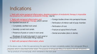
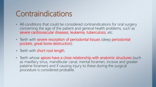
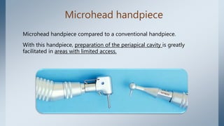
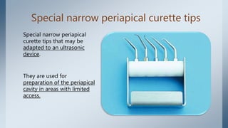
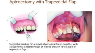
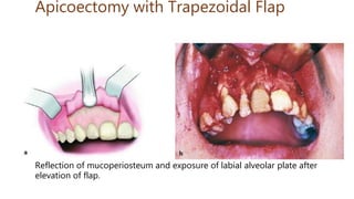
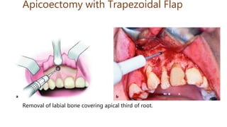
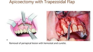
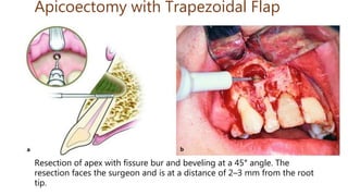
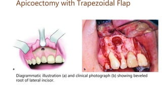
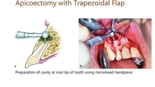
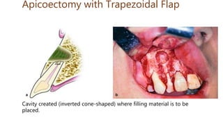
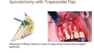
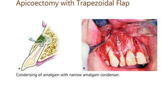
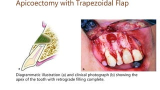
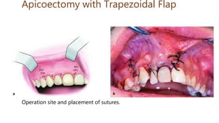
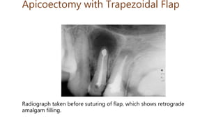
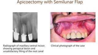
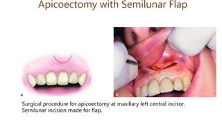
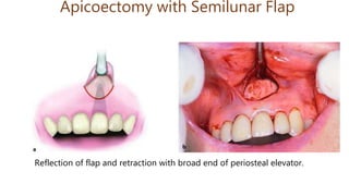
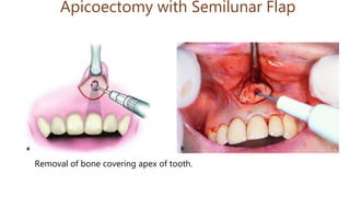
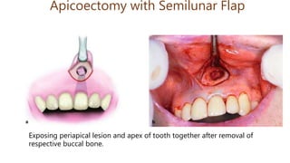
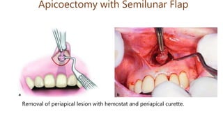
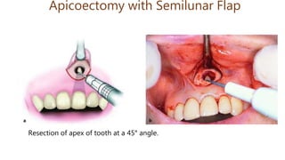
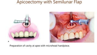
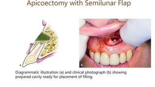
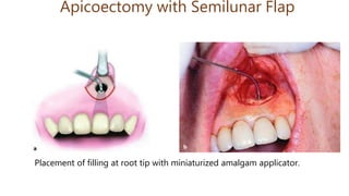
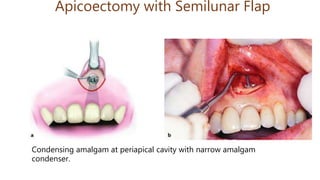
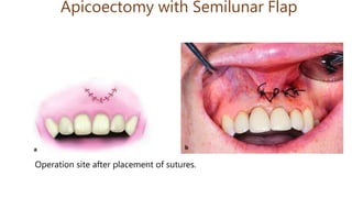
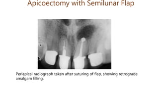
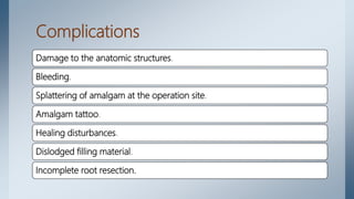
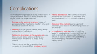
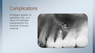
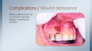
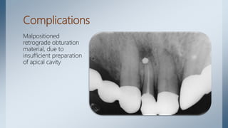
Apicoectomy
By
Muhammed M. Nasser
Outline
Introduction
Indications
Contraindications
Armamentarium
Surgical Technique
Complications
Introduction
• Apicoectomy is the surgical resection of the root tip of a tooth and its
removal together with the pathological periapical tissues.
• Accessory root canals and additional apical foramina are also
removed in this way, which may occur in the periapical area and
which may be considered responsible for failure of an endodontic
therapy.
Indications
1. Teeth with active periapical inflammation, despite the
presence of a satisfactory endodontic therapy.
2. Teeth with periapical inflammation and unsatisfactory
endodontic therapy.
3. Teeth with periapical inflammation, where completion of
endodontic therapy is impossible.
Indications
1. Teeth with active periapical inflammation, despite
the presence of a satisfactory endodontic therapy.
2. Teeth with periapical inflammation and
unsatisfactory endodontic therapy, which cannot
be repeated because of:
– Completely calcified root canal.
– Severely curved root canals.
– Presence of posts or cores in root canal.
– Breakage of small instrument in root canal or
the presence of irretrievable filling material.
3. Teeth with periapical inflammation, where
completion of endodontic therapy is impossible
due to:
– Foreign bodies driven into periapical tissues.
– Perforation of inferior wall of pulp chamber.
– Perforation of root.
– Fracture at apical third of tooth.
– Dental anomalies (dens in dente).
In the above cases, if after the apicoectomy the apex has not been completely sealed, then retrograde filling is
required, which is described further down. The purpose of retrograde filling is to obstruct the exit of bacteria
the by-products of nonvital pulp, which remained in the root canal
Contraindications
• All conditions that could be considered contraindications for oral surgery
concerning the age of the patient and general health problems, such as
severe cardiovascular diseases, leukemia, tuberculosis, etc.
• Teeth with severe resorption of periodontal tissues (deep periodontal
pockets, great bone destruction).
• Teeth with short root length.
• Teeth whose apices have a close relationship with anatomic structures (such
as maxillary sinus, mandibular canal, mental foramen, incisive and greater
palatine foramen) and if causing injury to these during the surgical
procedure is considered probable.
Armamentarium
• Microhead handpiece (straight
and contra-angle)and microbur.
• Special narrow periapical curette
tips for preparation of the
periapical cavity.
• Apical retrograde micro-mirror
and micro-explorers.
• Local anesthetic syringe and
cartridges.
• Scalpel handle.
• Scalpel blade (no. 15).
• Mirror.
• Periosteal elevator.
• Cotton pliers.
• Small hemostat.
• Suction tips (small, large).
• Irrigation receptacle.
Armamentarium
• Needle holder.
• Retractors.
• Periodontal curette.
• Periapical curette.
• Appropriate burs (round, fissure,
inverted cone).
• Miniaturized amalgam applicator
for retrograde fillings.
• Narrow amalgam condensers.
• Scissors, needles and no. 3–0 and
4–0 sutures.
• Metal endodontic ruler.
• Gauze and cotton rolls/pellets.
• Syringe for irrigating surgical
field.
• Saline solution.
Microhead handpiece
Microhead handpiece compared to a conventional handpiece.
With this handpiece, preparation of the periapical cavity is greatly
facilitated in areas with limited access.
Special narrow periapical curette tips
Special narrow periapical
curette tips that may be
adapted to an ultrasonic
device.
They are used for
preparation of the periapical
cavity in areas with limited
access.
Apical retrograde micromirror and
micro-explorers
Apical retrograde
micromirror and
micro-explorers for
determining the
dimensions of the
created periapical
cavity
Miniaturized amalgam applicator
Miniaturized
amalgam applicator
for retrograde
fillings, with a knob
that controls
amalgam increment
size
Miniaturized amalgam applicator
Miniaturized
amalgam
applicator
compared to a
standard amalgam
carrier
Instruments and materials for
retrograde filling
Instruments and materials
for retrograde filling.
* Amalgam capsule (top
left).
* Miniaturized amalgam
applicator (top right).
* Narrow amalgam
condensers, with tips
appropriately shaped so
that they may enter
narrow areas easily
(bottom)
Surgical Technique
The procedure for apicoectomy includes the following steps:
1. Designing of flap.
2. Localization of apex, exposure of the periapical area and removal of
pathological tissue.
3. Resection of apex of tooth.
4. Retrograde filling, if deemed necessary.
5. Wound cleansing and suturing.
Designing of Flap
Flap design depends on various factors, which mainly include
• position of the tooth
• presence of a periodontal pocket
• presence of a prosthetic restoration
• the extent of the periapical lesion.
There are three types of flaps principally used for apicoectomy:
• Semilunar.
• Triangular.
• Trapezoidal.
Designing of Flap
• The semilunar flap is indicated for surgical procedures of limited extent and is usually created
at the anterior region of the maxilla, which is where most apicoectomies are performed. In
order to ensure optimal wound healing, the incision must be made at a distance from the
presumed borders of the bony defect, so that the flap is repositioned over healthy bone.
• If there is an extensive bony defect, especially towards the alveolar crest, then the triangular or
trapezoidal flap is preferred. It must be noted that the pathological lesion, which has
perforated the bone and has become attached to the periosteum, must be separated from the
flap with a scalpel. In case of a fistula, the fistulous tract must also be excised near the bone,
because, if it is excised at the mucosa, then there is risk of even greater perforation, resulting in
disturbances of the healing process.
• When the apicoectomy is performed at the anterior region (e.g., maxillary lateral incisor) and
there is an extensive bony defect near the alveolar crest, the surgical procedure is performed
using a trapezoidal flap. The incision for creating the flap begins at the mesial aspect of the
central incisor and, after continuing around the cervical lines of the teeth, ends at the distal
aspect of the canine. With a periosteal elevator, the mucoperiosteum is then carefully reflected
upwards.
Localization and Exposure of Apex
• When the periapical lesion has perforated the buccal bone, localization and exposure of the
root tip is easy, after removing the pathological tissues with a curette.
• If the buccal bone covering the lesion has not been completely destroyed, but is very thin, then
its surface is detected with an explorer or dental curette, whereupon, due to decreased bone
density, the underlying bone is easily removed and the apex localized.
• When the buccal bone remains completely intact, then the root tip may be located with a
radiograph. More specifically, after taking a radiograph, the length of the root is determined
with a sterilized endodontic file or metal endodontic ruler. The length measured is then
transferred to the surgical field, determining the exact position of the root tip. Afterwards, with
a round bur and a steady stream of saline solution, the bone covering the root tip is removed
peripherally, creating an osseous window until the apex of the tooth is exposed.
Localization and Exposure of Apex
• If the overlying bone is thin and the pathological lesion is large, the osseous window is
enlarged with a blunt bur or a rongeur. Enough bone is removed until easy access to the entire
lesion is permitted.
• A curette is then used to remove pathological tissue and every foreign body or filling material,
while resection of the root tip follows.
Resection of Apex of Tooth
• The apex is resected (2–3 mm of the total root length) with a narrow fissure bur and beveled at
a 45° angle to the long axis of the tooth. For the best possible visualization of the root tip, the
beveled surface must be facing the dental surgeon.
• After this procedure, the cavity is inspected and all pathological tissue is meticulously removed
by curettage, especially in the area behind the apex of the tooth.
• If the entire root canal is not completely filled with filling material or if the seal is inadequate,
then retrograde filling is deemed necessary.
Retrograde Filling
• After beveling of the apex and curettage of periapical tissues, gauze impregnated with
adrenaline to minimize bleeding is placed in the bony defect.
• A microhead handpiece with a narrow round microbur is then used to prepare a cavity
approximately 2 mm long, with a diameter slightly larger than that of the root canal. The cavity
may be enlarged at its base using an inverted cone-shaped bur to undercut the preparation for
better retention of the filling material. During preparation of the cavity, the dentist must pay
careful attention to the width of the cavity, which must be as narrow as possible, because there
is a risk of weakening the root tip and causing a fracture (which may not even be perceived)
during condensing.
• After drying the bone cavity with gauze or a cotton pellet, sterile gauze is packed inside the
bone deficit and around the apex of the tooth, in such a way that only the prepared cavity of
the root end is exposed. Splattering of amalgam is thus avoided at the periapical region.
• The amalgam is placed inside the cavity with the miniaturized amalgam applicator and is
condensed with the narrow amalgam condenser. The excess amalgam is carefully removed and
the filling is smoothed with the usual instruments.
Retrograde filling material
• Amalgam is the most commonly used retrograde filling material.
• Recently, though, IRM and Super-EBA cement have been considered
choice materials, with preparation of the cavity being performed in
exactly the same way as that for amalgam.
Wound Cleansing and Suturing of Flap
• After placement of the amalgam, the gauze is carefully removed from the bony defect and,
after copious irrigation with saline solution, a radiographic examination is performed to
determine if there is amalgam splattering in the surrounding tissues. The flap is repositioned
and interrupted sutures are placed.
• Healing of the periapical area is checked every 6–12 months radiographically, until ossification
of the cavity is ascertained. In order to evaluate the result, a preoperative radiograph is
necessary, which will be compared to the postoperative radiographs later.
• When apicoectomy is performed in the anterior region (e.g., maxillary central incisor) and the
size of the lesion is small, and when there are esthetic crowns on the anterior teeth, the
semilunar flap is preferred. The procedure in such a case is similar to that of the previously
mentioned surgical procedure employing the trapezoidal flap.
Apicoectomy with Trapezoidal Flap
Extensive periapical lesion at maxillary
right lateral incisor. Indication for
apicoectomy.
Apicoectomy with Trapezoidal Flap
Clinical photograph. Arrow points to
possible location of lesion.
Surgical procedure for removal of periapical lesion, together with
apicoectomy at lateral incisor of maxilla. Incision for creation of
trapezoidal flap.
Apicoectomy with Trapezoidal Flap
Reflection of mucoperiosteum and exposure of labial alveolar plate after
elevation of flap.
Apicoectomy with Trapezoidal Flap
Removal of labial bone covering apical third of root.
Apicoectomy with Trapezoidal Flap
Removal of periapical lesion with hemostat and curette.
Apicoectomy with Trapezoidal Flap
Resection of apex with fissure bur and beveling at a 45° angle. The
resection faces the surgeon and is at a distance of 2–3 mm from the root
tip.
Apicoectomy with Trapezoidal Flap
Diagrammatic illustration (a) and clinical photograph (b) showing beveled
root of lateral incisor.
Apicoectomy with Trapezoidal Flap
Preparation of cavity at root tip of tooth using microhead handpiece.
Apicoectomy with Trapezoidal Flap
Cavity created (inverted cone-shaped) where filling material is to be
placed.
Apicoectomy with Trapezoidal Flap
Placement of filling material in cavity of apex using miniaturized amalgam
applicator.
Apicoectomy with Trapezoidal Flap
Condensing of amalgam with narrow amalgam condenser.
Apicoectomy with Trapezoidal Flap
Diagrammatic illustration (a) and clinical photograph (b) showing the
apex of the tooth with retrograde filling complete.
Apicoectomy with Trapezoidal Flap
Operation site and placement of sutures.
Apicoectomy with Trapezoidal Flap
Radiograph taken before suturing of flap, which shows retrograde
amalgam filling.
Apicoectomy with Semilunar Flap
Radiograph of maxillary central incisor,
showing periapical lesion and
unsatisfactory filling of the root canal
Clinical photograph of the case
Apicoectomy with Semilunar Flap
Surgical procedure for apicoectomy at maxillary left central incisor.
Semilunar incision made for flap.
Apicoectomy with Semilunar Flap
Reflection of flap and retraction with broad end of periosteal elevator.
Apicoectomy with Semilunar Flap
Removal of bone covering apex of tooth.
Apicoectomy with Semilunar Flap
Exposing periapical lesion and apex of tooth together after removal of
respective buccal bone.
Apicoectomy with Semilunar Flap
Removal of periapical lesion with hemostat and periapical curette.
Apicoectomy with Semilunar Flap
Resection of apex of tooth at a 45° angle.
Apicoectomy with Semilunar Flap
Preparation of cavity at apex with microhead handpiece.
Apicoectomy with Semilunar Flap
Diagrammatic illustration (a) and clinical photograph (b) showing
prepared cavity ready for placement of filling.
Apicoectomy with Semilunar Flap
Placement of filling at root tip with miniaturized amalgam applicator.
Apicoectomy with Semilunar Flap
Condensing amalgam at periapical cavity with narrow amalgam
condenser.
Apicoectomy with Semilunar Flap
Operation site after placement of sutures.
Apicoectomy with Semilunar Flap
Periapical radiograph taken after suturing of flap, showing retrograde
amalgam filling.
Complications
Damage to the anatomic structures.
Bleeding.
Splattering of amalgam at the operation site.
Amalgam tattoo.
Healing disturbances.
Dislodged filling material.
Incomplete root resection.
Complications
The most common perioperative and postoperative
complications that may occur during and after the
surgical procedure, respectively, are:
• Damage to the anatomic structures in case of
penetration of the nasal cavity, maxillary sinus and
mandibular canal with the bur.
• Bleeding from the greater palatine artery during
apicoectomy of palatal root.
• Splattering of amalgam at the operation site, due
to inadequate apical isolation and improper
manipulations for removal of excess filling
material.
• Staining of mucosa due to amalgam that
remained at the surgical field (amalgam tattoo).
• Healing disturbances, if the semilunar incision is
made over the bony deficit or if the flap, after
reapproximation, is not positioned on healthy
bone.
• Dislodged filling material due to superficial
placement, as a result of insufficient preparation of
apical cavity.
• Incomplete root resection, due to insufficient
access or visualization and misjudged length of
root. As a result, the apical portion of the root
remains in position and the retrograde filling is
placed improperly, with all the resulting
consequences.
Complications
Amalgam splatter at
operation site, as a
result of improper
manipulations for
removal of excess
material
Complications / amalgam tattoo
Complications / Wound dehiscence
Wound dehiscence, as
a result of improper
design of semilunar
incision
Complications
Malpositioned
retrograde obturation
material, due to
insufficient preparation
of apical cavity
Reference
• Oral Surgery/ Fragiskos D. Fragiskos
HANK YOU

Apicoectomy

  • 1.
  • 2.
  • 4.
    Introduction • Apicoectomy isthe surgical resection of the root tip of a tooth and its removal together with the pathological periapical tissues. • Accessory root canals and additional apical foramina are also removed in this way, which may occur in the periapical area and which may be considered responsible for failure of an endodontic therapy.
  • 6.
    Indications 1. Teeth withactive periapical inflammation, despite the presence of a satisfactory endodontic therapy. 2. Teeth with periapical inflammation and unsatisfactory endodontic therapy. 3. Teeth with periapical inflammation, where completion of endodontic therapy is impossible.
  • 7.
    Indications 1. Teeth withactive periapical inflammation, despite the presence of a satisfactory endodontic therapy. 2. Teeth with periapical inflammation and unsatisfactory endodontic therapy, which cannot be repeated because of: – Completely calcified root canal. – Severely curved root canals. – Presence of posts or cores in root canal. – Breakage of small instrument in root canal or the presence of irretrievable filling material. 3. Teeth with periapical inflammation, where completion of endodontic therapy is impossible due to: – Foreign bodies driven into periapical tissues. – Perforation of inferior wall of pulp chamber. – Perforation of root. – Fracture at apical third of tooth. – Dental anomalies (dens in dente). In the above cases, if after the apicoectomy the apex has not been completely sealed, then retrograde filling is required, which is described further down. The purpose of retrograde filling is to obstruct the exit of bacteria the by-products of nonvital pulp, which remained in the root canal
  • 9.
    Contraindications • All conditionsthat could be considered contraindications for oral surgery concerning the age of the patient and general health problems, such as severe cardiovascular diseases, leukemia, tuberculosis, etc. • Teeth with severe resorption of periodontal tissues (deep periodontal pockets, great bone destruction). • Teeth with short root length. • Teeth whose apices have a close relationship with anatomic structures (such as maxillary sinus, mandibular canal, mental foramen, incisive and greater palatine foramen) and if causing injury to these during the surgical procedure is considered probable.
  • 11.
    Armamentarium • Microhead handpiece(straight and contra-angle)and microbur. • Special narrow periapical curette tips for preparation of the periapical cavity. • Apical retrograde micro-mirror and micro-explorers. • Local anesthetic syringe and cartridges. • Scalpel handle. • Scalpel blade (no. 15). • Mirror. • Periosteal elevator. • Cotton pliers. • Small hemostat. • Suction tips (small, large). • Irrigation receptacle.
  • 12.
    Armamentarium • Needle holder. •Retractors. • Periodontal curette. • Periapical curette. • Appropriate burs (round, fissure, inverted cone). • Miniaturized amalgam applicator for retrograde fillings. • Narrow amalgam condensers. • Scissors, needles and no. 3–0 and 4–0 sutures. • Metal endodontic ruler. • Gauze and cotton rolls/pellets. • Syringe for irrigating surgical field. • Saline solution.
  • 13.
    Microhead handpiece Microhead handpiececompared to a conventional handpiece. With this handpiece, preparation of the periapical cavity is greatly facilitated in areas with limited access.
  • 14.
    Special narrow periapicalcurette tips Special narrow periapical curette tips that may be adapted to an ultrasonic device. They are used for preparation of the periapical cavity in areas with limited access.
  • 15.
    Apical retrograde micromirrorand micro-explorers Apical retrograde micromirror and micro-explorers for determining the dimensions of the created periapical cavity
  • 16.
    Miniaturized amalgam applicator Miniaturized amalgamapplicator for retrograde fillings, with a knob that controls amalgam increment size
  • 17.
  • 18.
    Instruments and materialsfor retrograde filling Instruments and materials for retrograde filling. * Amalgam capsule (top left). * Miniaturized amalgam applicator (top right). * Narrow amalgam condensers, with tips appropriately shaped so that they may enter narrow areas easily (bottom)
  • 20.
    Surgical Technique The procedurefor apicoectomy includes the following steps: 1. Designing of flap. 2. Localization of apex, exposure of the periapical area and removal of pathological tissue. 3. Resection of apex of tooth. 4. Retrograde filling, if deemed necessary. 5. Wound cleansing and suturing.
  • 21.
    Designing of Flap Flapdesign depends on various factors, which mainly include • position of the tooth • presence of a periodontal pocket • presence of a prosthetic restoration • the extent of the periapical lesion. There are three types of flaps principally used for apicoectomy: • Semilunar. • Triangular. • Trapezoidal.
  • 22.
    Designing of Flap •The semilunar flap is indicated for surgical procedures of limited extent and is usually created at the anterior region of the maxilla, which is where most apicoectomies are performed. In order to ensure optimal wound healing, the incision must be made at a distance from the presumed borders of the bony defect, so that the flap is repositioned over healthy bone. • If there is an extensive bony defect, especially towards the alveolar crest, then the triangular or trapezoidal flap is preferred. It must be noted that the pathological lesion, which has perforated the bone and has become attached to the periosteum, must be separated from the flap with a scalpel. In case of a fistula, the fistulous tract must also be excised near the bone, because, if it is excised at the mucosa, then there is risk of even greater perforation, resulting in disturbances of the healing process. • When the apicoectomy is performed at the anterior region (e.g., maxillary lateral incisor) and there is an extensive bony defect near the alveolar crest, the surgical procedure is performed using a trapezoidal flap. The incision for creating the flap begins at the mesial aspect of the central incisor and, after continuing around the cervical lines of the teeth, ends at the distal aspect of the canine. With a periosteal elevator, the mucoperiosteum is then carefully reflected upwards.
  • 23.
    Localization and Exposureof Apex • When the periapical lesion has perforated the buccal bone, localization and exposure of the root tip is easy, after removing the pathological tissues with a curette. • If the buccal bone covering the lesion has not been completely destroyed, but is very thin, then its surface is detected with an explorer or dental curette, whereupon, due to decreased bone density, the underlying bone is easily removed and the apex localized. • When the buccal bone remains completely intact, then the root tip may be located with a radiograph. More specifically, after taking a radiograph, the length of the root is determined with a sterilized endodontic file or metal endodontic ruler. The length measured is then transferred to the surgical field, determining the exact position of the root tip. Afterwards, with a round bur and a steady stream of saline solution, the bone covering the root tip is removed peripherally, creating an osseous window until the apex of the tooth is exposed.
  • 24.
    Localization and Exposureof Apex • If the overlying bone is thin and the pathological lesion is large, the osseous window is enlarged with a blunt bur or a rongeur. Enough bone is removed until easy access to the entire lesion is permitted. • A curette is then used to remove pathological tissue and every foreign body or filling material, while resection of the root tip follows.
  • 25.
    Resection of Apexof Tooth • The apex is resected (2–3 mm of the total root length) with a narrow fissure bur and beveled at a 45° angle to the long axis of the tooth. For the best possible visualization of the root tip, the beveled surface must be facing the dental surgeon. • After this procedure, the cavity is inspected and all pathological tissue is meticulously removed by curettage, especially in the area behind the apex of the tooth. • If the entire root canal is not completely filled with filling material or if the seal is inadequate, then retrograde filling is deemed necessary.
  • 26.
    Retrograde Filling • Afterbeveling of the apex and curettage of periapical tissues, gauze impregnated with adrenaline to minimize bleeding is placed in the bony defect. • A microhead handpiece with a narrow round microbur is then used to prepare a cavity approximately 2 mm long, with a diameter slightly larger than that of the root canal. The cavity may be enlarged at its base using an inverted cone-shaped bur to undercut the preparation for better retention of the filling material. During preparation of the cavity, the dentist must pay careful attention to the width of the cavity, which must be as narrow as possible, because there is a risk of weakening the root tip and causing a fracture (which may not even be perceived) during condensing. • After drying the bone cavity with gauze or a cotton pellet, sterile gauze is packed inside the bone deficit and around the apex of the tooth, in such a way that only the prepared cavity of the root end is exposed. Splattering of amalgam is thus avoided at the periapical region. • The amalgam is placed inside the cavity with the miniaturized amalgam applicator and is condensed with the narrow amalgam condenser. The excess amalgam is carefully removed and the filling is smoothed with the usual instruments.
  • 27.
    Retrograde filling material •Amalgam is the most commonly used retrograde filling material. • Recently, though, IRM and Super-EBA cement have been considered choice materials, with preparation of the cavity being performed in exactly the same way as that for amalgam.
  • 28.
    Wound Cleansing andSuturing of Flap • After placement of the amalgam, the gauze is carefully removed from the bony defect and, after copious irrigation with saline solution, a radiographic examination is performed to determine if there is amalgam splattering in the surrounding tissues. The flap is repositioned and interrupted sutures are placed. • Healing of the periapical area is checked every 6–12 months radiographically, until ossification of the cavity is ascertained. In order to evaluate the result, a preoperative radiograph is necessary, which will be compared to the postoperative radiographs later. • When apicoectomy is performed in the anterior region (e.g., maxillary central incisor) and the size of the lesion is small, and when there are esthetic crowns on the anterior teeth, the semilunar flap is preferred. The procedure in such a case is similar to that of the previously mentioned surgical procedure employing the trapezoidal flap.
  • 29.
  • 30.
    Extensive periapical lesionat maxillary right lateral incisor. Indication for apicoectomy. Apicoectomy with Trapezoidal Flap Clinical photograph. Arrow points to possible location of lesion.
  • 31.
    Surgical procedure forremoval of periapical lesion, together with apicoectomy at lateral incisor of maxilla. Incision for creation of trapezoidal flap. Apicoectomy with Trapezoidal Flap
  • 32.
    Apicoectomy with TrapezoidalFlap Reflection of mucoperiosteum and exposure of labial alveolar plate after elevation of flap.
  • 33.
    Apicoectomy with TrapezoidalFlap Removal of labial bone covering apical third of root.
  • 34.
    Apicoectomy with TrapezoidalFlap Removal of periapical lesion with hemostat and curette.
  • 35.
    Apicoectomy with TrapezoidalFlap Resection of apex with fissure bur and beveling at a 45° angle. The resection faces the surgeon and is at a distance of 2–3 mm from the root tip.
  • 36.
    Apicoectomy with TrapezoidalFlap Diagrammatic illustration (a) and clinical photograph (b) showing beveled root of lateral incisor.
  • 37.
    Apicoectomy with TrapezoidalFlap Preparation of cavity at root tip of tooth using microhead handpiece.
  • 38.
    Apicoectomy with TrapezoidalFlap Cavity created (inverted cone-shaped) where filling material is to be placed.
  • 39.
    Apicoectomy with TrapezoidalFlap Placement of filling material in cavity of apex using miniaturized amalgam applicator.
  • 40.
    Apicoectomy with TrapezoidalFlap Condensing of amalgam with narrow amalgam condenser.
  • 41.
    Apicoectomy with TrapezoidalFlap Diagrammatic illustration (a) and clinical photograph (b) showing the apex of the tooth with retrograde filling complete.
  • 42.
    Apicoectomy with TrapezoidalFlap Operation site and placement of sutures.
  • 43.
    Apicoectomy with TrapezoidalFlap Radiograph taken before suturing of flap, which shows retrograde amalgam filling.
  • 44.
  • 45.
    Apicoectomy with SemilunarFlap Radiograph of maxillary central incisor, showing periapical lesion and unsatisfactory filling of the root canal Clinical photograph of the case
  • 46.
    Apicoectomy with SemilunarFlap Surgical procedure for apicoectomy at maxillary left central incisor. Semilunar incision made for flap.
  • 47.
    Apicoectomy with SemilunarFlap Reflection of flap and retraction with broad end of periosteal elevator.
  • 48.
    Apicoectomy with SemilunarFlap Removal of bone covering apex of tooth.
  • 49.
    Apicoectomy with SemilunarFlap Exposing periapical lesion and apex of tooth together after removal of respective buccal bone.
  • 50.
    Apicoectomy with SemilunarFlap Removal of periapical lesion with hemostat and periapical curette.
  • 51.
    Apicoectomy with SemilunarFlap Resection of apex of tooth at a 45° angle.
  • 52.
    Apicoectomy with SemilunarFlap Preparation of cavity at apex with microhead handpiece.
  • 53.
    Apicoectomy with SemilunarFlap Diagrammatic illustration (a) and clinical photograph (b) showing prepared cavity ready for placement of filling.
  • 54.
    Apicoectomy with SemilunarFlap Placement of filling at root tip with miniaturized amalgam applicator.
  • 55.
    Apicoectomy with SemilunarFlap Condensing amalgam at periapical cavity with narrow amalgam condenser.
  • 56.
    Apicoectomy with SemilunarFlap Operation site after placement of sutures.
  • 57.
    Apicoectomy with SemilunarFlap Periapical radiograph taken after suturing of flap, showing retrograde amalgam filling.
  • 59.
    Complications Damage to theanatomic structures. Bleeding. Splattering of amalgam at the operation site. Amalgam tattoo. Healing disturbances. Dislodged filling material. Incomplete root resection.
  • 60.
    Complications The most commonperioperative and postoperative complications that may occur during and after the surgical procedure, respectively, are: • Damage to the anatomic structures in case of penetration of the nasal cavity, maxillary sinus and mandibular canal with the bur. • Bleeding from the greater palatine artery during apicoectomy of palatal root. • Splattering of amalgam at the operation site, due to inadequate apical isolation and improper manipulations for removal of excess filling material. • Staining of mucosa due to amalgam that remained at the surgical field (amalgam tattoo). • Healing disturbances, if the semilunar incision is made over the bony deficit or if the flap, after reapproximation, is not positioned on healthy bone. • Dislodged filling material due to superficial placement, as a result of insufficient preparation of apical cavity. • Incomplete root resection, due to insufficient access or visualization and misjudged length of root. As a result, the apical portion of the root remains in position and the retrograde filling is placed improperly, with all the resulting consequences.
  • 61.
    Complications Amalgam splatter at operationsite, as a result of improper manipulations for removal of excess material
  • 62.
  • 63.
    Complications / Wounddehiscence Wound dehiscence, as a result of improper design of semilunar incision
  • 64.
    Complications Malpositioned retrograde obturation material, dueto insufficient preparation of apical cavity
  • 65.
    Reference • Oral Surgery/Fragiskos D. Fragiskos
  • 66.