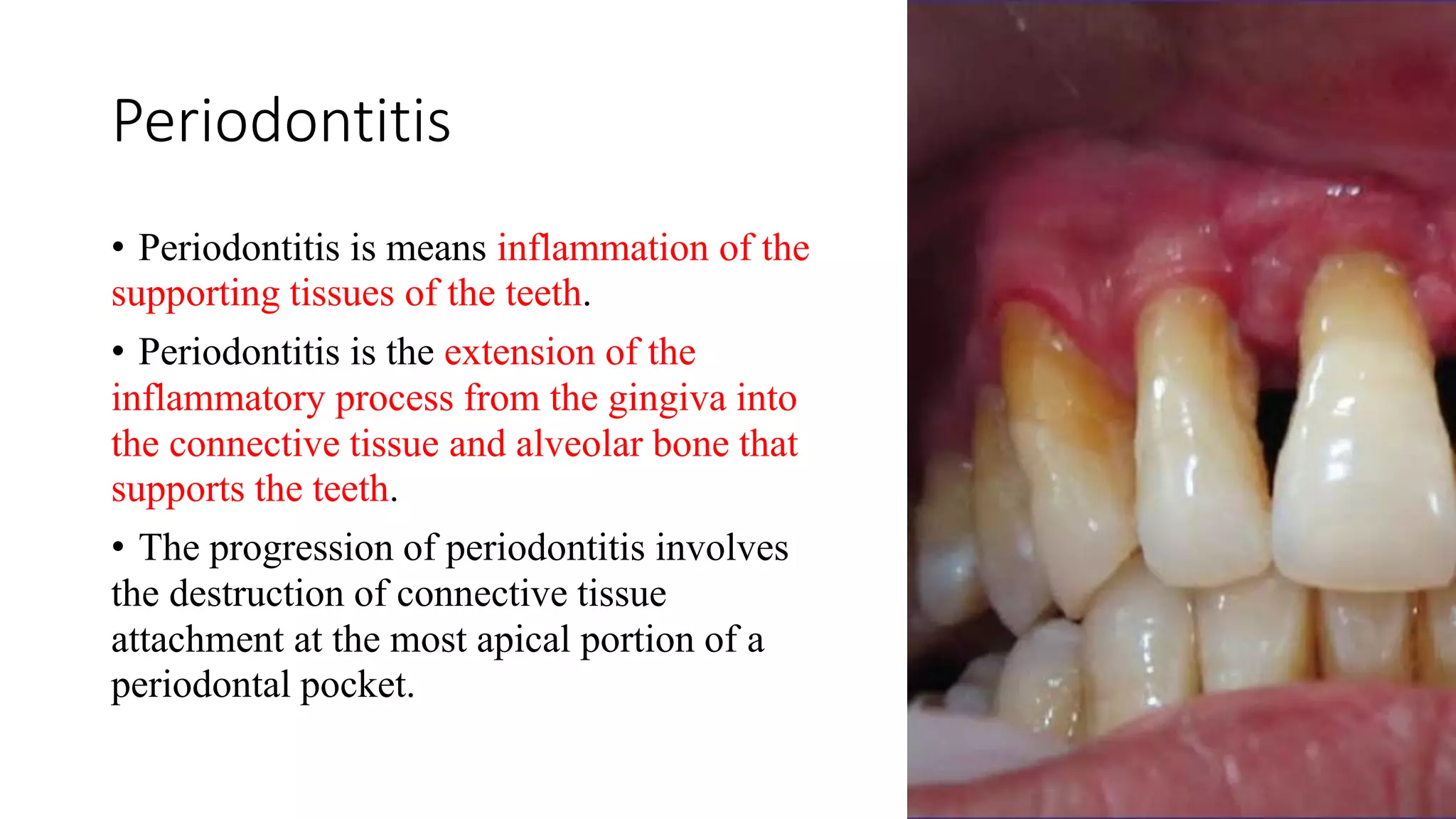
Welcome to Indian Dental Academy
The Indian Dental Academy is the Leader in continuing dental education , training dentists in all aspects of dentistry and offering a wide range of dental certified courses in different formats.
Indian dental academy has a unique training program & curriculum that provides students with exceptional clinical skills and enabling them to return to their office with high level confidence and start treating patients
State of the art comprehensive training-Faculty of world wide repute &Very affordable.
Necrotising periodontal diseases, Necrotising periodontal diseases as a manifestation of systemic diseases.
By Dr. Ritam Kundu, MDS PGT, Dr. R. Ahmed Dental College & Hospital, Kolkata, India.
The defense mechanism of gingiva includes GCF, Saliva, epithelial barrier and connective tissue cells. All these protect the periodontium from bacterial invasion.
explained here is bone loos and patterns of bone loos in alveolar bone to various insults . Dr Harshavardhan pawal also gives emphasis on rate on bone loss and radius of action .
This presentation is helpful for the dental student interested in dealing with the infectious disease AIDS.
The material also includes evidence based article on the relation of the HIV stage on periodontal status.
the most common viral infections that affects the maxillofacial area
sources( burket's oral medicine 11th ed,oral and maxillofacial pathology neville 2e )
In this talk we will discuss the most common findings associated with scleroderma. We will discuss some of the methods your dental team can utilize to help manage your condition, and also some ways that you can help yourself and your dental team manage your condition. We will discuss some unique methods for maintaining your oral health care and will conclude with an open Q&A session.
DIAGNOSTIC SETUP FOR REMOVABLE PARTIAL DENTURE /prosthodontic coursesIndian dental academy
The Indian Dental Academy is the Leader in continuing dental education , training dentists in all aspects of dentistry and
offering a wide range of dental certified courses in different formats.for more details please visit
www.indiandentalacademy.com
Talk about:
- Types of occlusal forces and Periodontal Response to External Forces
- Clinical & Radiographic
Signs of Trauma from Occlusion
- Therapy & Treatment Outcomes for this condition.
Talk about
- Structure of the Scalp {Skin, Connective tissue, Aponeurosis (epicranial), Loose areolar tissue, Pericranium}.
- Muscles of the Scalp {occipitofrontalis muscle}.
- Sensory Nerve Supply of the Scalp
- Arterial Supply of the Scalp
- Venous Drainage of the Scalp
- Lymph Drainage of the Scalp
This Handbook content summarized information about General Surgery; Started with Classification of Surgical Diseases & Classification of surgical specialties, Then talk about Case History, General Examination, Investigations...
Also discussed ATLS (Advance Trauma Life Support), Fracture & Dislocation of lower jaw, Differential diagnosis of Neck lump, Ulcers, Tumor, Surgical wound & Infection, Wound healing, Oroantral Fistula, Hemorrhage, General Anaesthesia (GA), Fluid and Electrolyte Management of the Surgical Patient.
Macroeconomics- Movie Location
This will be used as part of your Personal Professional Portfolio once graded.
Objective:
Prepare a presentation or a paper using research, basic comparative analysis, data organization and application of economic information. You will make an informed assessment of an economic climate outside of the United States to accomplish an entertainment industry objective.
The Roman Empire A Historical Colossus.pdfkaushalkr1407
The Roman Empire, a vast and enduring power, stands as one of history's most remarkable civilizations, leaving an indelible imprint on the world. It emerged from the Roman Republic, transitioning into an imperial powerhouse under the leadership of Augustus Caesar in 27 BCE. This transformation marked the beginning of an era defined by unprecedented territorial expansion, architectural marvels, and profound cultural influence.
The empire's roots lie in the city of Rome, founded, according to legend, by Romulus in 753 BCE. Over centuries, Rome evolved from a small settlement to a formidable republic, characterized by a complex political system with elected officials and checks on power. However, internal strife, class conflicts, and military ambitions paved the way for the end of the Republic. Julius Caesar’s dictatorship and subsequent assassination in 44 BCE created a power vacuum, leading to a civil war. Octavian, later Augustus, emerged victorious, heralding the Roman Empire’s birth.
Under Augustus, the empire experienced the Pax Romana, a 200-year period of relative peace and stability. Augustus reformed the military, established efficient administrative systems, and initiated grand construction projects. The empire's borders expanded, encompassing territories from Britain to Egypt and from Spain to the Euphrates. Roman legions, renowned for their discipline and engineering prowess, secured and maintained these vast territories, building roads, fortifications, and cities that facilitated control and integration.
The Roman Empire’s society was hierarchical, with a rigid class system. At the top were the patricians, wealthy elites who held significant political power. Below them were the plebeians, free citizens with limited political influence, and the vast numbers of slaves who formed the backbone of the economy. The family unit was central, governed by the paterfamilias, the male head who held absolute authority.
Culturally, the Romans were eclectic, absorbing and adapting elements from the civilizations they encountered, particularly the Greeks. Roman art, literature, and philosophy reflected this synthesis, creating a rich cultural tapestry. Latin, the Roman language, became the lingua franca of the Western world, influencing numerous modern languages.
Roman architecture and engineering achievements were monumental. They perfected the arch, vault, and dome, constructing enduring structures like the Colosseum, Pantheon, and aqueducts. These engineering marvels not only showcased Roman ingenuity but also served practical purposes, from public entertainment to water supply.
Unit 8 - Information and Communication Technology (Paper I).pdfThiyagu K
This slides describes the basic concepts of ICT, basics of Email, Emerging Technology and Digital Initiatives in Education. This presentations aligns with the UGC Paper I syllabus.
Acetabularia Information For Class 9 .docxvaibhavrinwa19
Acetabularia acetabulum is a single-celled green alga that in its vegetative state is morphologically differentiated into a basal rhizoid and an axially elongated stalk, which bears whorls of branching hairs. The single diploid nucleus resides in the rhizoid.
A Strategic Approach: GenAI in EducationPeter Windle
Artificial Intelligence (AI) technologies such as Generative AI, Image Generators and Large Language Models have had a dramatic impact on teaching, learning and assessment over the past 18 months. The most immediate threat AI posed was to Academic Integrity with Higher Education Institutes (HEIs) focusing their efforts on combating the use of GenAI in assessment. Guidelines were developed for staff and students, policies put in place too. Innovative educators have forged paths in the use of Generative AI for teaching, learning and assessments leading to pockets of transformation springing up across HEIs, often with little or no top-down guidance, support or direction.
This Gasta posits a strategic approach to integrating AI into HEIs to prepare staff, students and the curriculum for an evolving world and workplace. We will highlight the advantages of working with these technologies beyond the realm of teaching, learning and assessment by considering prompt engineering skills, industry impact, curriculum changes, and the need for staff upskilling. In contrast, not engaging strategically with Generative AI poses risks, including falling behind peers, missed opportunities and failing to ensure our graduates remain employable. The rapid evolution of AI technologies necessitates a proactive and strategic approach if we are to remain relevant.
2024.06.01 Introducing a competency framework for languag learning materials ...Sandy Millin
http://sandymillin.wordpress.com/iateflwebinar2024
Published classroom materials form the basis of syllabuses, drive teacher professional development, and have a potentially huge influence on learners, teachers and education systems. All teachers also create their own materials, whether a few sentences on a blackboard, a highly-structured fully-realised online course, or anything in between. Despite this, the knowledge and skills needed to create effective language learning materials are rarely part of teacher training, and are mostly learnt by trial and error.
Knowledge and skills frameworks, generally called competency frameworks, for ELT teachers, trainers and managers have existed for a few years now. However, until I created one for my MA dissertation, there wasn’t one drawing together what we need to know and do to be able to effectively produce language learning materials.
This webinar will introduce you to my framework, highlighting the key competencies I identified from my research. It will also show how anybody involved in language teaching (any language, not just English!), teacher training, managing schools or developing language learning materials can benefit from using the framework.
Read| The latest issue of The Challenger is here! We are thrilled to announce that our school paper has qualified for the NATIONAL SCHOOLS PRESS CONFERENCE (NSPC) 2024. Thank you for your unwavering support and trust. Dive into the stories that made us stand out!
Welcome to TechSoup New Member Orientation and Q&A (May 2024).pdfTechSoup
In this webinar you will learn how your organization can access TechSoup's wide variety of product discount and donation programs. From hardware to software, we'll give you a tour of the tools available to help your nonprofit with productivity, collaboration, financial management, donor tracking, security, and more.
Instructions for Submissions thorugh G- Classroom.pptxJheel Barad
This presentation provides a briefing on how to upload submissions and documents in Google Classroom. It was prepared as part of an orientation for new Sainik School in-service teacher trainees. As a training officer, my goal is to ensure that you are comfortable and proficient with this essential tool for managing assignments and fostering student engagement.
3. Gingival Diseases
Gingivitis is inflammation of the
gingival tissue.
Gingivitis is characterized by areas of
redness and swelling, and there is a
tendency for the gingiva to bleed easily.
Gingivitis is limited to the epithelium
and gingival connective tissues.
It is important to note that there is no
tissue recession or loss of connective
tissue or bone.
4. Gingival Diseases
Other types of gingivitis are associated with:
- Puberty.
- Pregnancy.
- Use of birth control medications.
Gingivitis is painless and often unrecognized until a
dental professional emphasizes its importance.
Improved daily oral hygiene practices will reverse
gingivitis.
6. Periodontitis
• Periodontitis is means inflammation of the
supporting tissues of the teeth.
• Periodontitis is the extension of the
inflammatory process from the gingiva into
the connective tissue and alveolar bone that
supports the teeth.
• The progression of periodontitis involves
the destruction of connective tissue
attachment at the most apical portion of a
periodontal pocket.
7. Causes of Periodontal Diseases
Dental plaque is the major factor in causing periodontal disease.
Dental calculus provides a surface for plaque to attach.
- Subgingival calculus.
- Supragingival calculus.
Supragingival calculus:
located coronal to the gingival margin and therefore visible in the oral cavity.
Subgingival calculus:
located below the crest of the marginal gingiva and therefore not visible on
routine clinical examination
8. • Calculus (CA) penetrates the
tooth surface and is
embedded within the
cementum (C).
Note the plaque (P) attached
to the calculus.
11. Signs and Symptoms of Periodontal Disease
• Red, swollen, or tender gingiva.
• Bleeding gingiva while brushing or
flossing.
• Loose or separating teeth.
• Pain or pressure when chewing.
• Pus around the teeth or gingival.
12. • Recognizing a departure from normal
in the periodontium and
distinguishing one disease from
another.
• Based on information obtained from:
- The medical and dental histories.
- Clinical and radiographic
examination of the patient.
- Laboratory findings.
Periodontal Diagnosis
13. Why we do diagnosis?
• To determine whether the disease is present or not.
• Identify its types.
• Extent.
• Severity.
• Distribution.
• Underlying pathological process and their causes.
14. Clinical diagnosis /First visit
• Overall apprasial of the patient.
• Medical history.
• Dental history.
• Social history
• Radiographic survey.
• Cast.
• Clinical photograph.
15. Overall apprasial of the patient
• Mental and emotional status.
• Attitude.
• Physiologic age.
16. Medical history
Helps with diagnosis of oral
manifestations of systemic
disease.
And may detect systemic
conditions that may be affecting
the periodontal tissue response.
Oral infection may affect severity
of some systemic disease.
• Most of the medical history is obtained
the first visit.
• It can be supplemented by pertinent
questioning at subsequent visits.
• The health history can be obtained
verbally by questioning the patient and
recording his or her responses on a
blank piece of paper or by means of a
printed questionnaire that the patient
completes.
17. Medical history
It includes:
1. If the patient is under the care of a physician.
2. hospitalizations and operations.
3. all medications.
4. All medical problems.
5. Abnormal bleeding tendencies.
6. The patient’s allergy history.
7. onset of puberty and for females, menopause, menstrual disorders, pregnancies, and
miscarriages.
8. A family medical history.
18. * What is the relevance of a patient’s medical
history to his or her periodontal care?
• Medical problems can increase susceptibility to periodontal diseases (e.g., diabetes and
HIV).
• Medical problems can have periodontal and other oral manifestations (e.g., leukaemia and
mucocutaneous disorders).
• Prescribed and nonprescribed medications can have oral and periodontal side effects (e.g.,
calcium channel blocker or phenytoin).
• precautions to be taken to avoid complications during or after treatment (e.g., antibiotic
prophylaxis and patients on certain medications such as anticoagulants).
• Treating certain conditions may present additional risk to the dental team (e.g.,
communicable diseases) and may alter the way treatment is provided (e.g., not using
ultrasonic instrumentation to avoid producing aerosol)
19. Dental history
Current illness, dental history includes:
- Dental visit.
- Oral hygiene.
- Orthodontic treatment.
- Pain in the teeth or in the gingiva.
- Bleeding gum.
- Bad taste.
- Do the tooth feel loose or insecure.
- Dental habits.
- Previous periodontal problems.
- Removable prosthesis
- Implants
20. Social History
1) Details of habits such as tobacco use, including smokeless tobacco, and alcohol
consumption.
NOTE A smoking history should include not only whether patients are current smokers but
also if they have smoked in the past, when they quit smoking, how many they smoked or
are smoking now, and whether are they interested in quitting.
2) A social history may also give information about the patient’s occupation that is both
useful for interacting with the patient and may indicate specific dental issues:
* e.g., wind instrument player and importance of aesthetics in a job.
* It can also provide information about the patient’s ability to attend appointments
regularly.
* Identify issues such as stress in a patient’s life.
21. Cast
used to study
- Position of gingival margin.
- Inclination of teeth
- Contact relationship
- Food impaction area
- provide a view of the lingual–cuspal relationships.
- Casts are important records of the dentition before it is altered by treatment.
- Visual aids during discussions with the patient
- Useful for pretreatment and posttreatment.
- Reference at recall visits.
- Determine the position of implant placement.
22. Radiographic survey
- 14 intra-oral periapical films + 4 posterior bitewing films
- Panoramic radiograph
They are helpful for the detection of developmental anomalies,
pathologic lesions of the teeth and jaws, and fractures as well as for the
dental screening examinations of large groups.
They provide an informative overall radiographic picture of the
distribution and severity of bone destruction with periodontal disease.
25. Radiographic survey
Radiographs do not:
- Show periodontal pockets
- Distinguish between
successfully treated and
untreated cases
- Show structures on buccal,
lingual, and labial aspect of
tooth
- Record tooth mobility
Full mouth series Look At:
- lamina dura
- crown: root ratio
- interproximal bone
- hypercementosis
- furcation involvement
- periapical pathology
- overhangs
- calculus
- resorption
28. Oral cavity examination
- Oral hygiene.
- Oral malodor
- The lips
- The floor of the mouth
- The tongue
- The palate
- The vestibule
- The oropharyngeal region
- The oral mucosa in the lateral and apical areas of the tooth may be palpated for
tenderness to detect periapical and periodontal abscesses.
29. Oral Hygiene
The cleanliness of the oral cavity is appraised in terms of the extent of
accumulated food debris, plaque, and tooth surface stains.
* Disclosing solution may be used to detect plaque that would otherwise
be unnoticed.
* The amount of plaque detected, however, is not necessarily related to
the severity of the disease present. For example, aggressive periodontitis
is a destructive type of periodontitis in which plaque is minimal.
Qualitative assessments of plaque are more meaningful.
30. Mal odor
• Oral malodor which is
also termed fetor oris, or
halitosis, is a foul or
offensive odor that
emanates from the oral
cavity. Mouth odors may
be of diagnostic
significance, and their
origin may be either oral
or extraoral (remote).
31. Mal odor
may be due to:
• ANUG
• Food impaction
• Caries
• Dehydration
• Smoking
• Dentures
• Diabetes
• Infections
• Alcohol
32. Lymph node
* Because periodontal, periapical, and other oral diseases may result in
lymph node changes, the diagnostician should routinely examine and
evaluate the lymph nodes of the head and neck.
* Lymph nodes can become enlarged as a result of an infectious
episode, malignant metastases, or residual fibrotic changes.
* Inflammatory nodes become enlarged, palpable, tender, and fairly
immobile. The overlying skin may be red and warm. Patients are often
aware of the presence of “swollen glands.”
Primary herpetic gingivostomatitis, necrotizing ulcerative gingivitis,
and acute periodontal abscesses may produce lymph node enlargement.
* After successful therapy, lymph nodes return to normal in a matter of
days to weeks.
33. Examination of the Teeth and Implants
The teeth are examined for:
Caries, poor restorations, developmental defects, anomalies of tooth form
Wasting disease
Dental stain
Hypersensitivity
Proximal contact relationship
Tooth mobility
Trauma from occlusion
Pathologic migration of teeth
Sensitivity to percussion
Dentition with jaw closed.
The
stability,
position,
and number
of implants
and their
relationship
to the
adjacent
natural
dentition
are also
examined.
34. Wasting Disease of the Teeth
• Wasting is defined as any gradual loss of tooth substance, which is characterized by the formation of smooth,
polished surfaces without regard to the possible mechanism of this loss.
• wedge- shaped defect (enamel, dentine then cementum)
Erosion
• occlusal wear result from functional contact with opposing
teeth
Attrition
• saucer-shaped defect starts on cementum then dentine
Abrasion
• occlusal loading surfaces causing flexure and mechanical
microfractures and tooth substances loss in the cervical area
Abfraction
35.
36. Erosion
* Erosion is a sharply defined wedge-shaped depression in the cervical area of the
facial tooth surface.
* The etiology of erosion is not known. Decalcification by acidic beverages or citrus
fruits in combination with the effect of acid salivary secretion are suggested causes.
37. Abrasion
Abrasion refers to the loss of tooth substance that is induced by
mechanical wear other than that of mastication. Abrasion results in
saucer-shaped or wedge-shaped indentations with a smooth, shiny
surface. Abrasion starts on the exposed cementum surfaces rather than
on the enamel, and it extends to involve the dentin of the root. A sharp
“ditching” around the cementoenamel junction appears to be the result
of the softer cemental surface as compared with the much harder enamel
surface. The involvement of the roots is followed by the undermining of
the enamel.
* Toothbrushing and the action of clasps are frequently mentioned, but
aggressive toothbrushing is the most common cause.
* Occasionally, abrasion of the incisal edges occurs as a result of habits
such as holding objects (e.g., bobby pins, tacks) between the teeth.
38. Attrition
• is occlusal wear that results from functional contacts
with opposing teeth.
• Such physical wear patterns may occur on incisal,
occlusal, and approximal tooth surfaces.
• Occlusal or incisal surfaces worn by attrition are
called facets. When active tooth grinding occurs, the
enamel rods are fractured and become highly
reflective to light. Thus, shiny, smooth, and
curviplanar facets are usually the best indicator of
ongoing frictional activity. If dentin is exposed, a
yellowish brown discoloration is frequently present.
39. Dental Stains
• Dental stains are
pigmented deposits
on the teeth.
• They should be
carefully examined
to determine their
origin.
40. Hypersensitivity
• Root surfaces exposed
by gingival recession may
be hypersensitive to
thermal changes or tactile
stimulation.
• Patients often direct the
clinician to the sensitive
areas. These may be
located by gentle
exploration with a probe
or cold air.
42. Tooth Mobility
All teeth have a slight degree of physiologic mobility, which varies for different teeth and at different
times of the day.
* It is greatest when arising in the morning, and it progressively decreases.
The increased mobility in the morning is attributed to slight extrusion of the tooth as a result of
limited occlusal contact during sleep. During the waking hours, mobility is reduced by chewing and
swallowing forces, which intrude the teeth in the sockets. These 24-hour variations are less marked in
persons with a healthy periodontium than in those with occlusal habits such as bruxism and
clenching.
* Single-rooted teeth have more mobility than multi-rooted teeth, with incisors having the most
mobility.
43. Tooth Mobility
1. Loss of tooth support (bone loss) can result in mobility: The amount of mobility depends on the
severity and distribution of bone loss at individual root surfaces, the length and shape of the roots,
and the root size as compared with that of the crown. A tooth with short, tapered roots is more likely
to loosen than one with normal-size or bulbous roots with the same amount of bone loss.
* One should carefully evaluate postorthodontic cases for possible apical shortening of the root,
which may lead to excessive mobility.
2. Trauma from occlusion (i.e., injury produced by excessive occlusal forces or incurred as a result
of abnormal occlusal habits such as bruxism and clenching) is a common cause of tooth mobility.
Mobility produced by trauma from occlusion occurs initially as a result of resorption of the cortical
layer of bone, which leads to reduced fiber support, and later as an adaptation phenomenon that
results in a widened periodontal space.
44. Tooth Mobility
3. Extension of inflammation from the gingiva or from the periapex into the periodontal
ligament results in changes that increase mobility. The spread of inflammation from an acute
periapical abscess may increase tooth mobility in the absence of periodontal disease.
4. Periodontal surgery temporarily increases tooth mobility immediately after the intervention
and for a short period.
5. Tooth mobility is increased during pregnancy, and it is sometimes associated with the menstrual
cycle or the use of hormonal contraceptives. This is unrelated to periodontal disease, and it occurs
presumably because of physicochemical changes in the periodontal tissues.
6. Pathologic processes of the jaws that destroy the alveolar bone or the roots of the teeth can
also result in mobility. Osteomyelitis and tumors of the jaws belong in this category.
45. Tooth Mobility
Normal mobility
Less than 1 mm
More than 1 mm
Severe mobility faciolingually and
mesiodistally combined with vertical
displacement
46. Trauma from occlusion
• tissue injury produced by occlusal forces.
• The criterion that determines that an occlusion is traumatic is whether it causes damage in
the periodontal tissues; therefore, the diagnosis of trauma from occlusion is made from the
condition of the periodontal tissues.
• Periodontal findings that suggest the presence of trauma from occlusion include:
- excessive tooth mobility
- widened periodontal space
- vertical or angular bone destruction
- infrabony pockets
- pathologic migration
47. Pathological migration
• Alterations in tooth position should be carefully noted, particularly
with a view toward identifying abnormal forces, a tongue-thrusting
habit, or other habits that may be contributing factors.
• Premature tooth contacts in the posterior region that deflect the
mandible anteriorly contribute to the destruction of the periodontium
of the maxillary anterior teeth and to pathologic migration.
• The loss of posterior teeth can lead to the facial “flaring” of the
maxillary anterior dentition. This is due to the increased trauma that
the mandibular anterior dentition places against the palatal surface of
the maxillary anterior dentition.
• Pathologic migration of the anterior teeth in young persons may be
a sign of localized aggressive (juvenile) periodontitis.
48. Sensitivity to Percussion
• Sensitivity to percussion is a
feature of acute
inflammation of the
periodontal ligament.
• Gentle percussion of a
tooth at different angles to
the long axis often helps with
the localization of the site of
inflammatory involvement.
49. Examination of periodontium
• The periodontal examination should be systematic, starting in the molar region in
either the maxilla or the mandible and proceeding around the arch.
• It is important to detect the earliest signs of gingival and periodontal disease.
50. Plaque and Calculus
• The presence of supragingival plaque and calculus can be directly observed, and the amount can be
measured with a calibrated probe.
• For the detection of subgingival calculus, each tooth surface is carefully checked to the level of the
gingival attachment with a no. 17 explorer. Warm air may be used to deflect the gingiva and to aid in
the visualization of the calculus.
• Although radiographs may sometimes reveal heavy calculus deposits interproximally and even on
the facial and lingual surfaces, they cannot be relied on for the thorough detection of calculus.
51. Gingiva
* The gingiva must be dried before accurate observations can be made.
* In addition to visual examination and exploration with instruments, firm but
gentle palpation should be used to detect pathologic alterations in normal resilience
as well as to locate areas of exudate.
* Features of the gingiva to consider include color, size, contour, consistency,
position, ease of bleeding, and pain.
* Clinically, gingival inflammation can produce two basic types of tissue response:
edematous and fibrotic.
Edematous tissue response is characterized by a smooth, glossy, soft, red
gingiva.
With the fibrotic tissue response, some of the characteristics of normalcy
persist; the gingiva is more firm, stippled, and opaque; it is usually thicker, and
the margin appears rounded.
52. Periodontal Pockets
Examination for periodontal
pockets must include:
• their presence and
distribution on each tooth
surface
• the pocket depth
• the level of attachment on
the root
• the type of pocket (i.e.,
suprabony or infrabony).
54. Signs and Symptoms
• Although probing is the only reliable method of detecting pockets.
• Clinical signs such as:
1) color changes (i.e., a bluish-red marginal gingiva or a bluish-red vertical zone
that extends from the gingival margin to the attached gingiva).
2) A “rolled” edge separating the gingival margin from the tooth surface.
3) An enlarged, edematous gingiva may suggest their presence.
4) The presence of bleeding, suppuration, may also denote the presence of a
pocket.
55. Signs and Symptoms
• Periodontal pockets are generally painless, but they may give rise to
symptoms such as:
1) localized or sometimes radiating pain.
2) the sensation of pressure after eating that gradually diminishes.
3) foul taste in localized areas.
4) sensitivity to hot and cold.
5) toothache in the absence of caries is also sometimes present.
56. Detection of Pockets
• The only accurate method of detecting and measuring periodontal pockets is careful exploration
with a periodontal probe.
• Pockets are not detected by radiographic examination. The periodontal pocket is a soft-tissue
change. Radiographs indicate areas of bone loss in which pockets may be suspected, but they do not
show pocket presence or depth, and consequently they show no difference before and after pocket
elimination unless bone has been modified.
• Gutta-percha points or calibrated silver points can be used with the radiograph to assist with
determining the level of attachment of the periodontal pockets. They may be used effectively for
individual pockets or in clinical research, but their routine use throughout the mouth would be
difficult to manage. Clinical examination and probing are more direct and efficient.
57. Pocket Probing
There are two different pocket depths: (1) the biologic or
histologic depth; and (2) the clinical or probing depth.
The biologic depth is the distance between the gingival
margin and the base of the pocket (i.e., the coronal end
of the junctional epithelium). This can be measured only
in carefully prepared and adequately oriented histologic
sections.
The probing depth is the distance to which a probe
penetrates into the pocket.
58. Pocket Probing
Probe penetration can vary, depending on:
1) the force of introduction.
2) the shape and size of the probe tip.
3) the direction of penetration.
4) the resistance of the tissues.
5) the convexity of the crown.
6) the degree of tissue inflammation.
Several studies have been made to determine the depth of penetration of a probe in a sulcus or
pocket. The probing forces have been explored by several investigators forces of 0.75 N have been
found to be well tolerated and accurate.
59. Probing Technique
• The probe should be inserted parallel to the vertical axis of the tooth and “walked”
circumferentially around each surface of each tooth to detect the areas of deepest
penetration in addition, special attention should be directed to detecting the
presence of interdental craters and furcation involvements.
• To detect an interdental crater, the probe should be placed obliquely from both the
facial and lingual surfaces to explore the deepest point of the pocket located
beneath the contact point.
• In multi-rooted teeth, the possibility of furcation involvement should be carefully
explored. The use of specially designed probes (e.g., Nabers probes) allows for an
easier and more accurate exploration of the horizontal component of furcation
lesions
60. Level of Attachment Versus Pocket Depth
• Pocket depth is the distance between the base of
the pocket and the gingival margin. It may change
from time to time, even in patients with untreated
periodontal disease, as a result of changes in the
position of the gingival margin. Therefore, it may
be unrelated to the existing attachment of the tooth.
• The level of attachment is the distance between
the base of the pocket and a fixed point on the
crown, such as the cementoenamel junction (CEJ).
Changes in the level of attachment can be the result
of a gain or loss of attachment, and they can afford
a better indication of the degree of periodontal
destruction or gain.
61. Determining the Level of Attachment
When the gingival margin is located on the anatomic crown, the level of
attachment is determined by subtracting from the depth of the pocket the distance
from the gingival margin to the CEJ. If both are the same, the loss of attachment
is zero.
When the gingival margin coincides with the CEJ, the loss of attachment equals
the pocket depth.
When the gingival margin is located apical to the CEJ, the loss of attachment is
greater than the pocket depth. Therefore, the distance between the CEJ and the
gingival margin should be added to the pocket depth. Drawing the gingival
margin on the chart where pocket depths are entered helps to clarify this
important point.
62.
63. Bleeding on Probing
• The insertion of a probe to the bottom of the pocket elicits bleeding
if the gingiva is inflamed and if the pocket epithelium is atrophic or
ulcerated. Noninflamed sites rarely bleed. In most cases, bleeding on
probing is an earlier sign of inflammation than gingival color changes.
• Depending on the severity of inflammation, bleeding can vary from
a tenuous red line along the gingival sulcus to profuse bleeding. If
periodontal treatment is successful, bleeding on probing will cease. To
test for bleeding after probing, the probe is carefully introduced to the
bottom of the pocket and gently moved laterally along the pocket wall.
Sometimes bleeding appears immediately after the removal of the
probe; other times, it may be delayed for a few seconds. Therefore, the
clinician should recheck for bleeding 30 to 60 seconds after probing.
64. Amount of Attached Gingiva
• It is important to establish the relationship between the bottom of the pocket and
the mucogingival line. The width of the attached gingiva is the distance between the
mucogingival junction and the projection on the external surface of the bottom of
the gingival sulcus or the periodontal pocket.
• The width of the attached gingiva is determined by subtracting the sulcus or
pocket depth from the total width of the gingiva (i.e., the gingival margin to the
mucogingival line). This is done by stretching the lip or cheek to demarcate the
mucogingival line while the pocket is being probed. The amount of attached gingiva
is generally considered to be insufficient when the stretching of the lip or cheek
induces the movement of the free gingival margin.
65. Degree of Gingival Recession
• During periodontal examination, it is necessary to record the data
regarding the amount of gingival recession.
• This measurement is taken with a periodontal probe from the CEJ to
the gingival crest, and it is drawn on the patient’s chart.
66. Alveolar Bone Loss
• Alveolar bone levels are evaluated via both clinical and radiographic examination.
• Probing is helpful for determining the following: (1) the height and contour of the
facial and lingual bones, which are obscured on the radiograph by the roots; and (2)
the architecture of the interdental bone.
• Transgingival probing, which is performed after the area is anesthetized, is a more
accurate method of evaluation, and it provides additional information about bone
architecture.
• A panoramic radiograph demonstrating generalized horizontal bone loss
supplemented with a periapical of the UL1 showing vertical bone loss almost to the
apex.
67. Alveolar Bone Loss
Is there a difference between horizontal and vertical bone loss?
• If the level of the bone is essentially equal interdentally, it is called horizontal bone
loss and measured as the percentage of bone lost (e.g., 20% of the original bone
height is lost).
• Vertical/angular bone loss occurs when one tooth has lost more bone than the tooth
next to it and is suggested when the bone crest is more apical to the CEJ adjacent to
one tooth than to the other
• Bone loss around an individual tooth can also be a combination of a horizontal
component and a vertical component and is still expressed as the total amount of
bone lost from the CEJ.
68. Suppuration
• The presence of an abundant number of neutrophils in the gingival fluid
transforms it into a purulent exudate.
• Several studies have evaluated the association between suppuration and the
progression of periodontitis, and they have reported that this sign is present in a very
low percentage of diseased sites (i.e., 3% to 5%). Therefore, it is not by itself a good
indicator. Clinically, the presence of exudate in a periodontal pocket is determined by
placing the ball of the index finger along the lateral aspect of the marginal gingiva
and applying pressure in a rolling motion toward the crown (Fig.5).
• Visual examination without digital pressure is not enough. The purulent exudate is
formed in the inner pocket wall, and therefore the external appearance may give no
indication of its presence. Exudate formation does not occur in all periodontal
pockets, but digital pressure often reveals it in pockets where its presence is not
suspected.
69. Periodontal Abscess
• A periodontal abscess is a localized
accumulation of exudate within the
gingival wall of a periodontal
pocket.
• Periodontal abscesses may be acute
or chronic.
70. The acute periodontal abscess
The acute periodontal abscess appears as an ovoid elevation of the gingiva along the lateral aspect of
the root. The gingiva is edematous and red, with a smooth, shiny surface. The shape and consistency
of the elevated area vary; the area may be domelike and relatively firm, or it may be pointed and soft.
In most cases, exudate may be expressed from the gingival margin with gentle digital pressure.
The acute periodontal abscess is accompanied by symptoms such as:
* Throbbing, radiating pain, and tenderness of the gingiva to palpation.
* Other symptoms may include sensitivity of the tooth to palpation; tooth mobility and
lymphadenitis.
* Less frequently, systemic effects such as fever, leukocytosis, and malaise.
* Occasionally the patient may have symptoms of an acute periodontal abscess without any notable
clinical lesion or radiographic changes.
71. The chronic periodontal abscess
The chronic periodontal abscess usually presents a sinus that opens onto the gingival mucosa along
the length of the root. There may be a history of intermittent exudation. The orifice of the sinus may
appear as a difficult-to-detect pinpoint opening, which, when probed, reveals a sinus tract that leads
deep into the periodontium.
The chronic periodontal abscess is usually asymptomatic. However, the patient may report episodes
of dull, gnawing pain; a slight elevation of the tooth; and a desire to bite down and grind the tooth.
The chronic periodontal abscess often undergoes acute exacerbations, with all of the associated
symptoms.
72. Periodontal Abscess and Gingival Abscess
• The principal differences between the periodontal
abscess and the gingival abscess are location and
history.
• The gingival abscess is confined to the marginal
gingiva, and it often occurs in previously disease-
free areas. It is usually an acute inflammatory
response to the forcing of foreign material into the
gingiva.
• The periodontal abscess involves the supporting
periodontal structures, and it generally occurs during
the course of chronic destructive periodontitis.
73. Periodontal Abscess and Periapical Abscess
If the tooth is nonvital, the lesion is most likely periapical. However, a previously nonvital tooth can have a deep
periodontal pocket that can abscess.
Moreover, a deep periodontal pocket can extend to the apex and cause pulpal involvement and necrosis.
An apical abscess may spread along the lateral aspect of the root to the gingival margin.
Radiographic findings are helpful for differentiating between a periodontal lesion and a periapical lesion.
Early acute periodontal and periapical abscesses present no radiographic changes. Ordinarily, a radiolucent area
along the lateral surface of the root suggests the presence of a periodontal abscess, whereas apical rarefaction
suggests a periapical abscess.
Clinical findings such as the presence of extensive caries, pocket formation, lack of tooth vitality, and the
existence of continuity between the gingival margin and the abscess area often prove to be of greater diagnostic
value than the radiographic appearance.
75. Definitions
• Risk assessment is defined by numerous components.
• Disease risk is the probability that an individual will develop a specific disease in a given period. The
risk of developing the disease will vary from individual to individual.
• The term “risk factor” is used to describe something that is causally related to increased risk of disease.
• The term “risk marker” is a factor that, although associated with increased risk of disease, is not
necessarily causally related. For example, age is a well-recognized marker of disease in that there is an
increased prevalence of periodontitis in older people. However, this is not necessarily because getting
older increases one’s susceptibility to disease but, rather, because periodontal attachment loss is
cumulative, so more people in older age groups are likely to have the disease.
• The term risk determinant /background characteristic, which is sometimes substituted for the term
risk factor, should be reserved for those risk factors that cannot be modified.
• Risk indicators are risk factors that have been identified in cross-sectional studies but not confirmed
through longitudinal studies.
76. Categories of Risk Elements for Periodontal Disease
Risk Factors
• Tobacco
smoking
• Diabetes
• Pathogenic
bacteria and
microbial
tooth
deposits
Risk Determinants /
Background
Characteristics
• Genetic factors
• Age
• Gender
• Socioeconomic
status
• Stress
Risk Indicators
• Human
immunodeficiency
virus (HIV)/acquired
immunodeficiency
syndrome (AIDS)
• Osteoporosis
• Infrequent dental
visits
Risk Markers /
Predictors
• Previous
history of
periodontal
disease
• Bleeding on
probing
77. Risk Factors
Smoking
• Smoking is one of the most important risk factors for periodontitis.
• In patients who regularly smoke more than 10 cigarettes daily, it has been estimated that the relative risk of
periodontitis may be as high as fivefold.
• The effect of smoking is both time and dose dependent.
• It appears that the effects of smoking are largely due to:
1) a systemic effect resulting in an impaired inflammatory respond reduced neutrophil numbers and function at the
inflammatory site.
2) some studies have suggested an increased number of pathogenic bacteria present in plaque of smokers.
3) The clinical appearance of periodontal disease in smokers is usually characterized by reduced gingival bleeding,
and often the gingivae have a relatively pink and uninflamed appearance because of the suppressive effects of
smoking on the inflammatory response. However, the amount of periodontal breakdown seen is markedly increased.
• Smoking markedly impairs periodontal treatment outcomes.
78. Risk Factors
Diabetes
Diabetes is a clear risk factor for periodontitis. Epidemiologic data demonstrate that the prevalence and severity of
periodontitis are significantly higher in patients with type 1 or type 2 diabetes mellitus than in those without diabetes,
and that the level of diabetic control is an important variable in this relationship.
Oral Manifestations
• Numerous oral changes have been described in patients with diabetes, including cheilosis, mucosal drying &
cracking, burning mouth & tongue, diminished salivary flow, and alterations in the flora of the oral cavity, with
greater predominance of Candida albicans, hemolytic streptococci, and staphylococci.
• An increased rate of dental caries has also been observed in patients with poorly controlled diabetes.
• It is important to note that these changes are not always present the specific effects of diabetes on the periodontium,
a variety of changes have been described, including a tendency toward an enlarged gingiva, sessile or pedunculated
gingival polyps, polypoid gingival proliferations, abscess formation, periodontitis, and loosened teeth.
• Perhaps the most striking changes in patients with uncontrolled diabetes are the reductions in the defense
mechanisms and the increased susceptibility to infections, which lead to destructive periodontal disease.
79. Periodontal condition in patients with diabetes.
(A) Adult with diabetes (blood glucose level
>400 mg/dL). Note the gingival inflammation,
spontaneous bleeding, and edema.
(B) The same patient as shown in A. Improved
control of diabetes was noted after 4 days of
insulin therapy (blood glucose level <100
mg/dL). The clinical periodontal condition has
improved without local therapy.
(C) Adult patient with uncontrolled diabetes.
Note the enlarged, smooth, erythematous
gingival margins and papilla in the anterior
area.
(D) The same patient as shown in C. This is a
lingual view of the right mandibular area. Note
the inflamed and swollen tissues in the anterior
and premolar areas.
(E) Adult patient with uncontrolled diabetes.
There is a suppurating abscess on the buccal
surface of the maxillary premolars.
80. Risk Determinants/Background characteristics for
Periodontal Disease
Genetic Factors
• Evidence indicates that genetic differences between individuals may explain why some patients
develop periodontal disease and others do not.
• Studies conducted in twins have shown that genetic factors influence clinical measures of
gingivitis, probing pocket depth, attachment loss, and interproximal bone height.
• The familial aggregation seen in localized and generalized aggressive periodontitis also is
indicative of genetic involvement in these diseases
81. Risk Determinants/Background characteristics for
Periodontal Disease
Age
• Both the prevalence and severity of periodontal disease increase with age. It is possible that
degenerative changes related to aging may increase susceptibility to periodontitis. However, it also is
possible that the attachment loss and bone loss seen in older individuals are the result of prolonged
exposure to other risk factors over a person’s life, creating a cumulative effect over time. In support
of this, studies have shown minimal loss of attachment in aging subjects enrolled in preventive
programs throughout their lives.
• However, it remains to be determined whether changes related to the aging process, such as intake
of medications, decreased immune function, and altered nutritional status, interact with other well-
defined risk factors to increase susceptibility to periodontitis.
82. Risk Determinants/Background characteristics for
Periodontal Disease
Gender
• Gender plays a role in periodontal disease.
Surveys conducted in the United States since
1960 demonstrate that men have more loss
of attachment than women.
• In addition, men have poorer oral hygiene
than women, as evidenced by higher levels
of plaque and calculus.
• Therefor gender differences in prevalence
and severity of periodontitis appear to be
related to preventive practices rather than
any genetic factor.
83. Risk Determinants/Background characteristics for
Periodontal Disease
Socioeconomic Status
• Gingivitis and poor oral hygiene can be related
to lower socioeconomic status (SES). This can
most likely be attributed to decreased dental
awareness and decreased frequency of dental visits
compared with more educated individuals with
higher SES.
• After adjusting for other risk factors, such as
smoking and poor oral hygiene, lower SES alone
does not result in increased risk for periodontitis.
84. Risk Determinants/Background characteristics for
Periodontal Disease
Stress
• The incidence of necrotizing
ulcerative gingivitis increases
during periods of emotional and
physiologic stress, suggesting a link
between the two. Emotional stress
may interfere with normal immune
function and may result in increased
levels of circulating hormones,
which can affect the periodontium.
• Adult patients with periodontitis
who are resistant to therapy are
more stressed than those who
respond to therapy.
85. Risk Indicators for Periodontal Disease
Human Immunodeficiency Virus/Acquired Immunodeficiency
Syndrome
• It has been hypothesized that the immune dysfunction associated with
HIV infection and AIDS increases susceptibility to periodontal disease.
• Early reports on the periodontal status of patients with AIDS or
individuals who are HIV seropositive revealed that these patients often had
severe periodontal destruction characteristic of necrotizing ulcerative
periodontitis.
• Evidence suggests that AIDS-affected individuals who practice good
preventive oral health measures, including effective home care and seeking
appropriate professional therapy, can maintain periodontal health.
• Therefore, although it seems reasonable to hypothesize that HIV infection
and immunosuppression are risk factors for periodontal disease, the evidence
is not conclusive.
86. Risk Indicators for Periodontal Disease
Osteoporosis
• Osteoporosis has been suggested as another risk factor for periodontitis.
• In a study of 12 women with osteoporosis and 14 healthy women, von
Wowern and coworkers reported that the women with osteoporosis had
greater loss of attachment than the control subjects.
• In contrast, Kribbs examined pocket depth, bleeding on probing, and
gingival recession in women with and without osteoporosis. Although the
two groups had significant differences in bone mass, no differences in
periodontal status were noted.
• However, it appears that a link may exist between osteoporosis and
periodontitis, and additional studies may need to be conducted to determine
whether osteoporosis is a true risk factor for periodontal disease.
87. Risk Markers/Predictors for Periodontal Disease
Bleeding on Probing
• Bleeding on probing is the best clinical indicator
of gingival inflammation.
• Although this indicator alone does not serve as a
predictor for loss of attachment, bleeding on probing
coupled with increasing pocket depth may serve as
an excellent predictor for future loss of attachment.
• Lack of bleeding on probing does appear to serve
as an excellent indicator of periodontal health.