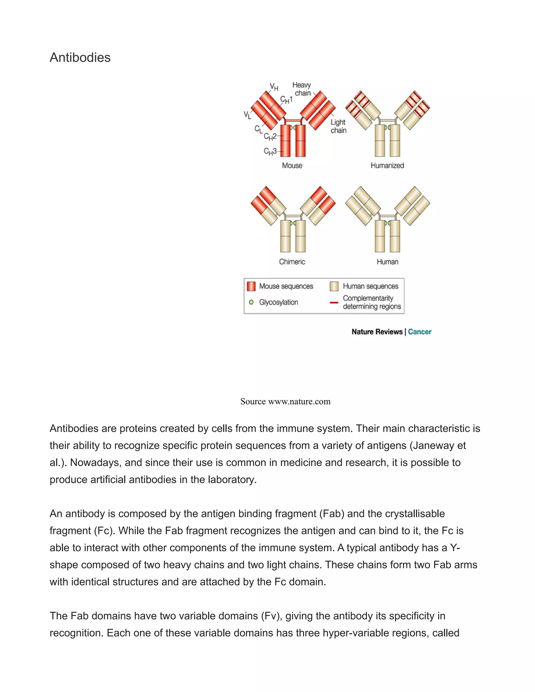
1. Antibody Humanization Techniques
OCTOBER 25, 2015 BY ADMIN (EDIT)
Overview
Antibodies are proteins with very important functions in the immune system. Thanks to their
ability to recognize specific sequences from pathogens and other threats, antibodies can
participate in the recognition and removal of foreign threats, such as virus and bacteria
(Janeway et al.). Every human body is able to create millions of slightly different antibodies;
every antibody has the ability to recognize one specific foreign antigen (Greger & Windhorst,
2013). Antibodies are, therefore, highly specific, which makes them very useful in the
biomedical research field and in applied medicine. Antibodies are crucial in several screening
and diagnosis methods, and in the last couple of decades they have emerged as treatment
option for several diseases, including arthritis, cardiovascular disease, transplant rejection
and cancer (Gura, 2002).
First generation antibodies come from mice, and since their use as therapeutic antibodies
requires for them to be injected into the human body, researchers encountered two major
problems: First, they elicit an immunogenic response, which, depending on its intensity, can
be dangerous for the patient. Second, they can also elicit an anti-antibody response. In this
case, the patient’s immune system destroys the therapeutic antibody, rendering them not
effective (Stern & Herrmann, 2005). These two obstacles to therapeutic antibody use come
from the fact that these antibodies, made in mice, are been recognized as foreign by the
patient’s immune system (Winter & Milstein, 991).
In order to surpass these obstacles, antibodies can be made more human, so the immune
system won’t attack them. Several strategies have been developed in order to achieve this
“humanization” of antibodies: The first approach consisted on the creation of chimeric
antibodies, which have a human part and a mice part. Afterward, antibodies were further
manipulated in order to increase their human composition, and these antibodies are known as
humanized antibodies (Carter, 2001). Currently, antibodies can also be fully human, thanks to
the existence of genetically engineered transgenic mice (Hudson & Souriau, 2003).
2. Antibodies
Antibodies are proteins created by cells from the immune system. Their main characteristic is
their ability to recognize specific protein sequences from a variety of antigens (Janeway et
al.). Nowadays, and since their use is common in medicine and research, it is possible to
produce artificial antibodies in the laboratory.
An antibody is composed by the antigen binding fragment (Fab) and the crystallisable
fragment (Fc). While the Fab fragment recognizes the antigen and can bind to it, the Fc is
able to interact with other components of the immune system. A typical antibody has a Y-
shape composed of two heavy chains and two light chains. These chains form two Fab arms
with identical structures and are attached by the Fc domain.
The Fab domains have two variable domains (Fv), giving the antibody its specificity in
recognition. Each one of these variable domains has three hyper-variable regions, called
Source www.nature.com
3. Complementary Determining Regions (CDRs). The CDRs are distributed in the Fv among
four less variable fragments (FRs). The CDRs are responsible for giving the antibody its
specific antigen recognition site (Sheriff et al, 1987).
An antibody from a non-human species (a mice or rat, for example) can be modified in order
to make it more similar to the antibodies produced by the human body. This is done by
antibody humanization techniques, and the result is antibodies that are useful for therapeutic
purpose (Hudson & Souriau, 2003). Antibodies created in mice have sequences that are
different to those naturally occurring in the human body, which causes them to be
immunogenic; the human body recognizes them as foreign. The subsequent immunological
reaction they cause in the human body is potentially dangerous and in the long term it means
they can be destroyed by the immune system before they exert their therapeutic effect.
Humanization of antibodies, therefore, makes them safer and fitter to use for therapy.
Chimeric Antibodies
Chimeric antibodies were envisioned as a solution to the immunogenic challenges posed by
mouse antibodies. Chimeric antibodies have a mixture of non-human (usually mouse) and
human components: around two thirds of their structure is human, while the remaining third
remains of animal composition. They are constructed by the fusion of the murine Fv region
(responsible for binding the antigen) and the Fc region of a human antibody (Hoogenboom et
al, 1996).
Chimeric antibodies started been developed in the 1980s, when recombinant technology
applied in genetic research started being available. Recombinant technology allows for
genetic material to be cut, spliced and put together from multiple sources. The creation of
chimeric antibodies is possible through the use of recombinant DNA and genetic engineering.
In live cells, mouse DNA encoding for the Fv region is merged with human DNA encoding for
the Fc region. This way, a fusion gene is created that will be translated into a recombinant
fusion protein (Fell & Folger-Bruce, 1993).
The final result is an antibody that has had the segments that made it recognizable as non-
human, replaced. By the genetic replacement of the murine constant domain for a human Fc
domain, the resulting antibody is recognized as human by the immune system and therefore
they have a reduced risk of eliciting an adverse immune response (Harding et al, 2010).
4. Chimeric antibodies have better binding affinity and reduced immunogenicity compared to
mouse antibodies; however, they are still immunogenic and can elicit an anti-immune and
anti-antigen response. Their therapeutic effectiveness is still not optimal, because this
immune response can eliminate them (Harding et al, 2010). Despite this, chimeric antibodies
have been successful in the treatment of several diseases, particularly cancer. There are,
currently, several of these chimeric antibodies available in the market for therapeutic use
(Elvin, Couston & van der Walle, 2013). These kinds of drugs are recognized by their suffix:
ximab.
Humanized Antibodies
The humanized antibodies were the next step after the creation and perfectioning of chimeric
antobodies. They are created by grafting the CDRs regions from a mouse antibody onto a
human Fv. A humanized antobody, then, contains CDR regions derived from the mouse that
have been engrafted into the human sequence-derived Fv. This way, they have around 90%
of human content (Winter & Harris, 1993).
These kinds of antibodies are designed and synthesized by the overlapping PCR method
(Oliphant et al, 2005). Besides, specific mutations can be done in order to generate the
desired antibody. The different chains of the antibody are generated and cloned into
expression vectors, and then co.expressed in another cell culture, like COS cells. This
process will generate the humanized version of the antibody, which will be found in the
supernatant and can be quantified by ELISA. An analysis of the resulting antibody is made
and in case it’s necessary changes can be done yu mutagenesis to make it more effective
(Oliphant et al, 2005).
Humanized antibodies have less immunogenicity than chimeric antibodies and mouse
antibodies. There are several of these types of antibodies in the market been used for
therapeutic purposes (Lahrtz, 2015). These drugs are known by their suffix, zumab.
Human Antibodies
Currently, it is possible to create fully human antibodies, with no murine sequence. They can
be produced in two ways: phage display technologies and transgenic mice (Winter, 1993).
Phage display is a technique that uses bacteriophages (viruses that infect bacteria) to
connect proteins. Antibodies will be displayed on the surface of phage by fusing the coding
5. sequence of the Fv regions to one of the phage’s coat proteins (Deantonio, 2014).
Human antibodies can also be created by the use of transgenic mice. In this technique, the
human immunoglobulin loci are introduced into the germ line of mice with inactivated antibody
machinery. This way, the mouse will be able to generate high-affinity and fully human
antibodies. The mice are presented with the antigen, the immunoglobulin transgenes undergo
joining and they end up creating high affinity monoclonal antibodies (Jakobovits, 1995).
In 2005 was aprooved the first fully human antibody for therapy. It was an antibody against
EGF. Since then, several other transgenic mice have been approved for the production of
other fully human antibodies, known by their suffix, umab (Elvin, 2013) The genetically
humanized mice are, therefore, a powerful tool for research and medical development.
6. Jones PT et al. Nature 321:523 (1986)
Concluding Remarks
Over the last years, the use of antibodies for therapy has increased. There are currently
several antibodies accepted for treatment against different conditions, including immune
disorders and cancer. There is potential for many more applications. In order for these
antibodies to be both safe and effective, they have to be non-immunogenic or elicit a small
immune response. In order to achieve this, antibodies can’t be completely animal in structure;
they have to be made more human. The humanization of antibodies can be achieved through
the creation of chimeric antibodies or humanized antibodies. Besides, currently, there is also
the possibility of creating fully human antibodies, thanks to the existence of humanized
transgenic mice and phage display technologies. However, no matter what method is used,
there’s still no antibody that’s completely immunogenically silent. Even if all antibodies still
elicit a small immune response, their therapeutic advantages have been demonstrated and
the use of antibodies is a reality in current medicine.
References
1. Janeway, C. A., Travers, P., Walport, M. J., & Shlomchik, M. J. (2001).Immunobiology:
the immune system in health and disease (Vol. 2). London: Churchill Livingstone.
2. Greger, R., & Windhorst, U. (Eds.). (2013). Comprehensive human physiology: from
cellular mechanisms to integration. Springer Science & Business Media.
3. Gura, T. (2002). Therapeutic antibodies: magic bullets hit the target. Nature,417(6889),
584-586.
4. Stern, M., & Herrmann, R. (2005). Overview of monoclonal antibodies in cancer therapy:
present and promise. Critical reviews in oncology/hematology, 54(1), 11-29.
5. Winter, G., & Milstein, C. (1991). Man-made antibodies. Nature, 349(6307), 293-299.
6. Carter, P. (2001). Improving the efficacy of antibody-based cancer therapies.Nature
Reviews Cancer, 1(2), 118-129.
7. 7. Sheriff, S., Silverton, E. W., Padlan, E. A., Cohen, G. H., Smith-Gill, S. J., Finzel, B. C.,
& Davies, D. R. (1987). Three-dimensional structure of an antibody-antigen complex.
Proceedings of the National Academy of Sciences,84(22), 8075-8079.
8. Hudson, P. J., & Souriau, C. (2003). Engineered antibodies. Nature medicine,9(1), 129-
134.
9. Hoogenboom, H. R., Baier, M., Jespers, L. S., & Winter, G. P. (1996). U.S. Patent No.
5,565,332. Washington, DC: U.S. Patent and Trademark Office.
10.Brüggemann, M., Winter, G. R. E. G., Waldmann, H. E. R. M. A. N., & Neuberger, M. S.
(1989). The immunogenicity of chimeric antibodies. The Journal of experimental
medicine, 170(6), 2153-2157.
11.Fell Jr, H. P., & Folger-Bruce, K. R. (1993). U.S. Patent No. 5,202,238. Washington, DC:
U.S. Patent and Trademark Office.
12.Harding, F. A., Stickler, M. M., Razo, J., & DuBridge, R. (2010, May). The
immunogenicity of humanized and fully human antibodies: residual immunogenicity
resides in the CDR regions. In MAbs (Vol. 2, No. 3, pp. 256-265). Taylor & Francis.
13.Elvin, J. G., Couston, R. G., & van der Walle, C. F. (2013). Therapeutic antibodies:
market considerations, disease targets and bioprocessing.International journal of
pharmaceutics, 440(1), 83-98.
14.Winter, G., & Harris, W. J. (1993). Humanized antibodies. Immunology today,14(6), 243-
246.
15.Oliphant, T., Engle, M., Nybakken, G. E., Doane, C., Johnson, S., Huang, L., … &
Diamond, M. S. (2005). Development of a humanized monoclonal antibody with
therapeutic potential against West Nile virus. Nature medicine, 11(5), 522-530.
16.Lahrtz, F. (2015). How to Successfully Patent Therapeutic Antibodies. Journal of
biomolecular screening, 1087057114567457.
8. 17.Deantonio, C., Cotella, D., Macor, P., Santoro, C., & Sblattero, D. (2014). Phage Display
Technology for Human Monoclonal Antibodies. In Human Monoclonal Antibodies (pp.
277-295). Humana Press.
18.Jakobovits, A. (1995). Production of fully human antibodies by transgenic mice.Current
Opinion in Biotechnology, 6(5), 561-566.
19.Elvin, J. G., Couston, R. G., & van der Walle, C. F. (2013). Therapeutic antibodies:
market considerations, disease targets and bioprocessing.International journal of
pharmaceutics, 440(1), 83-98.