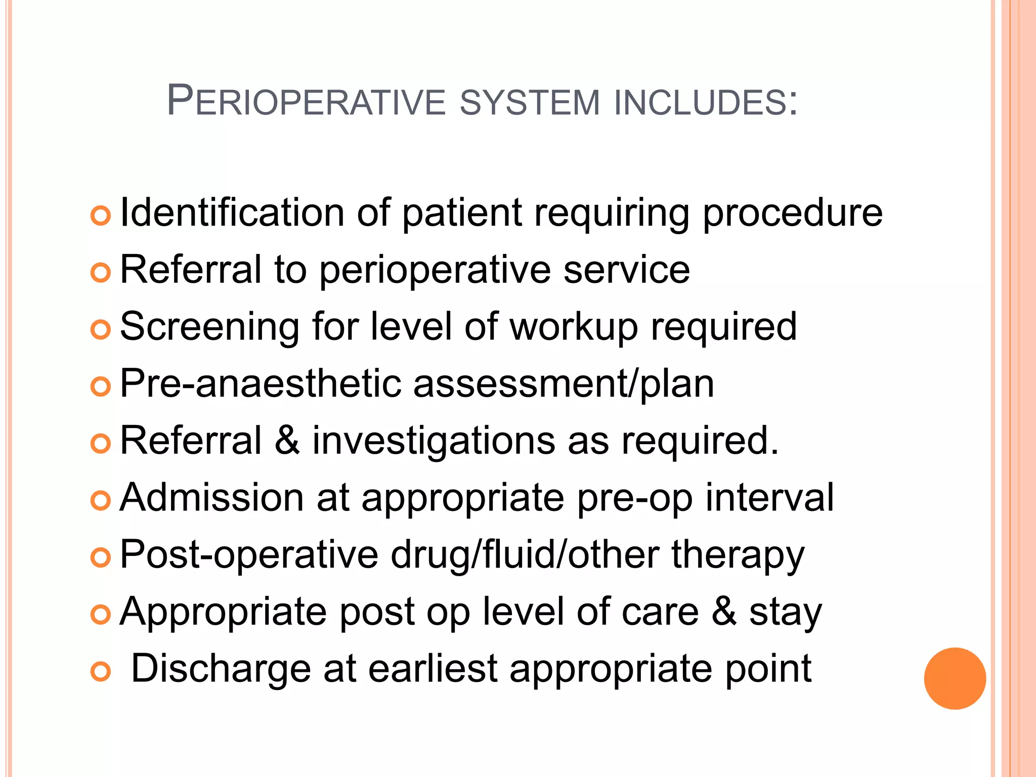
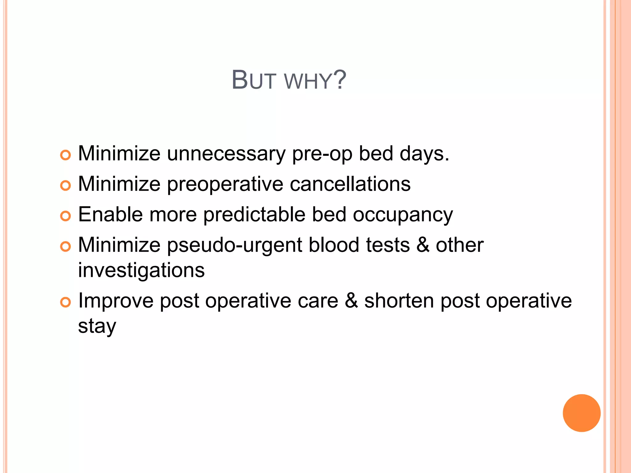
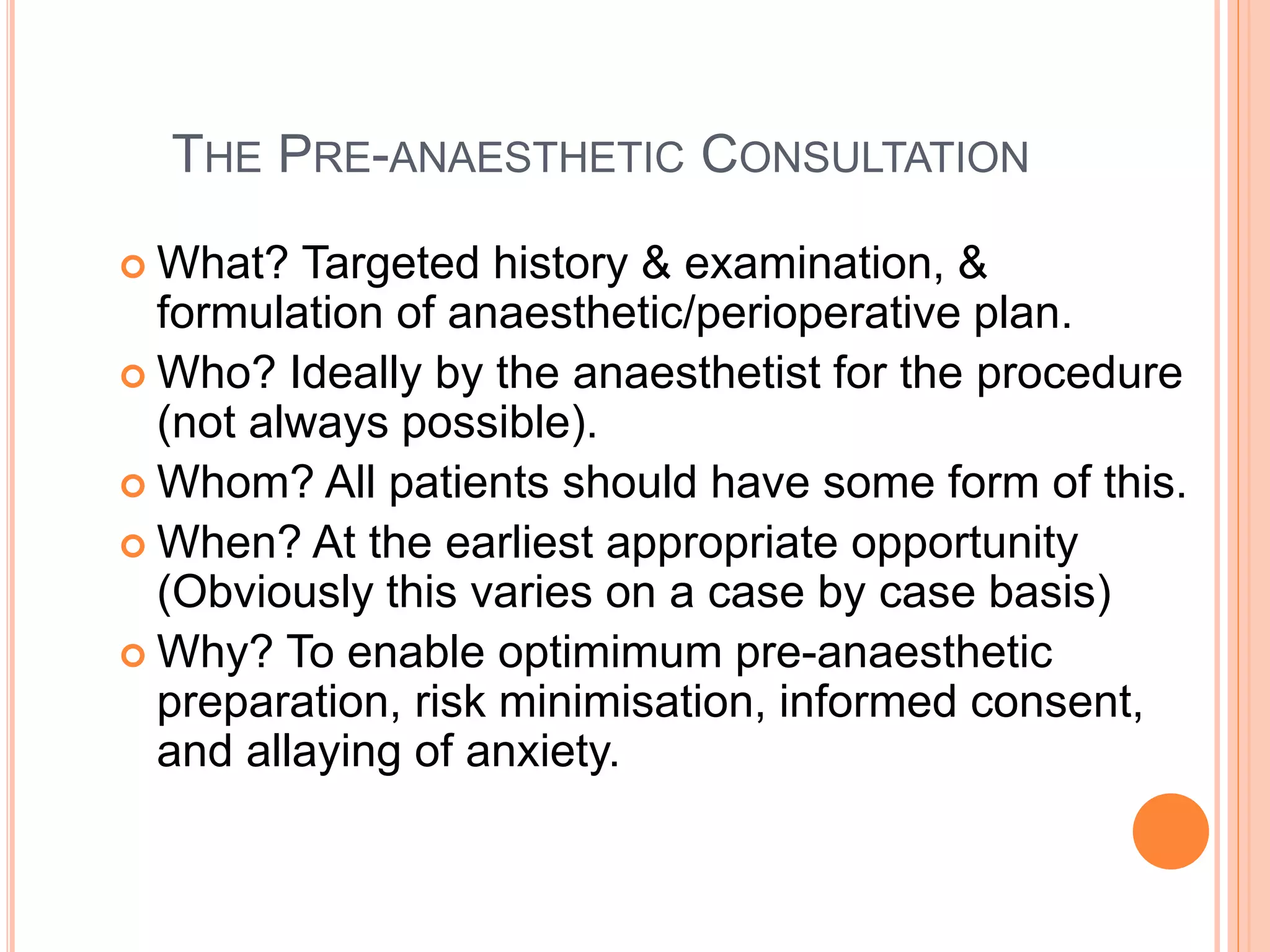
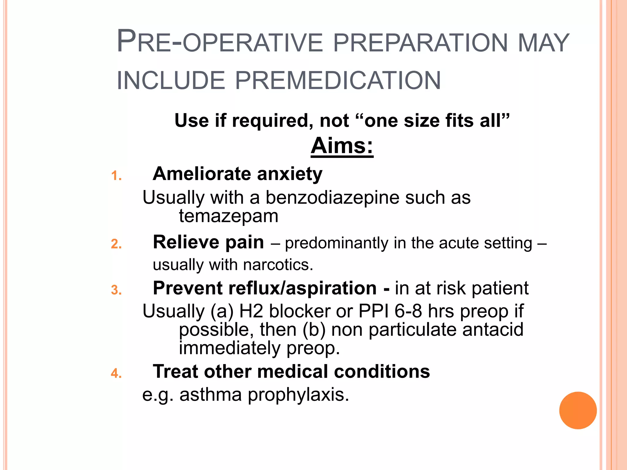
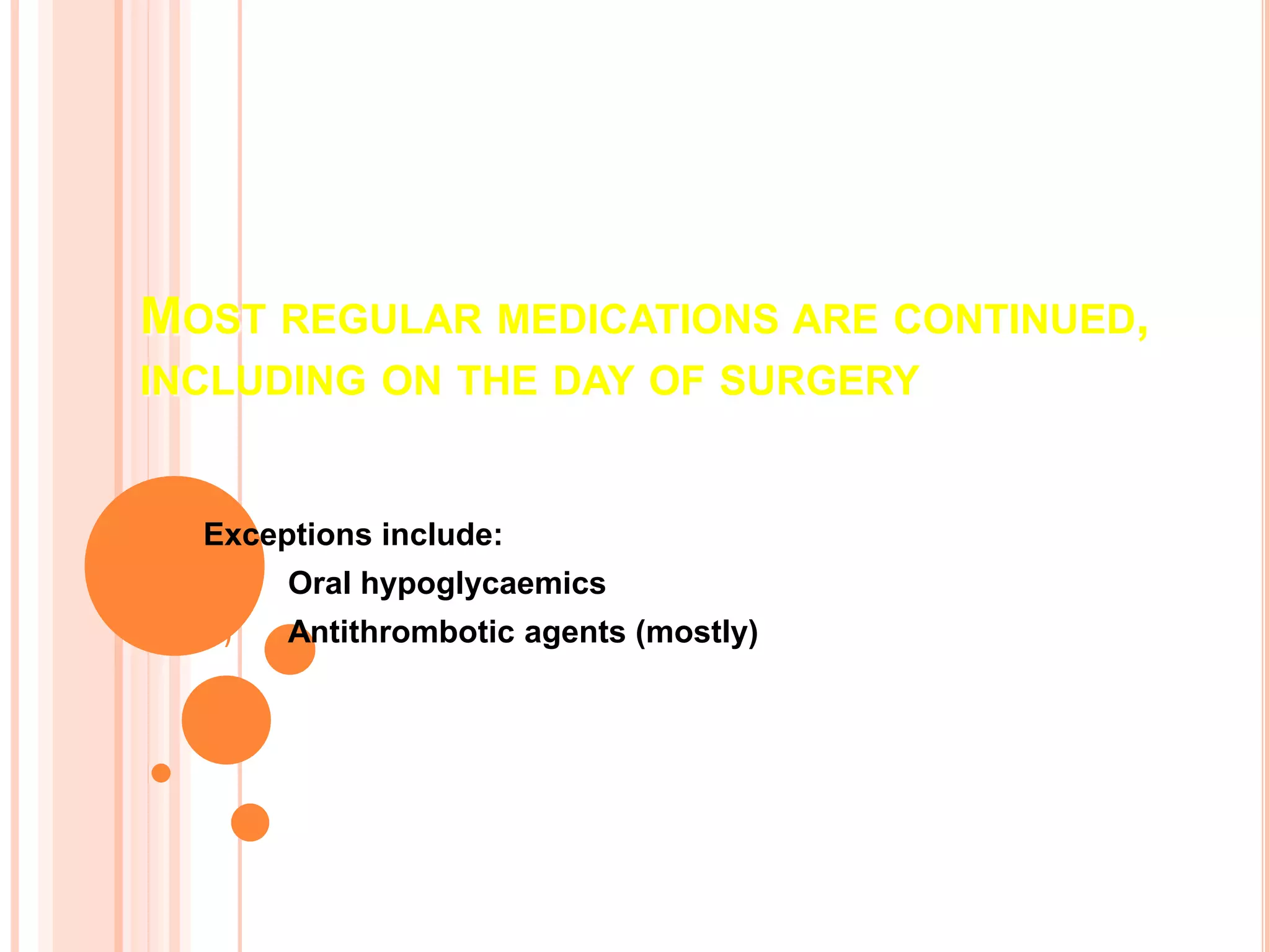
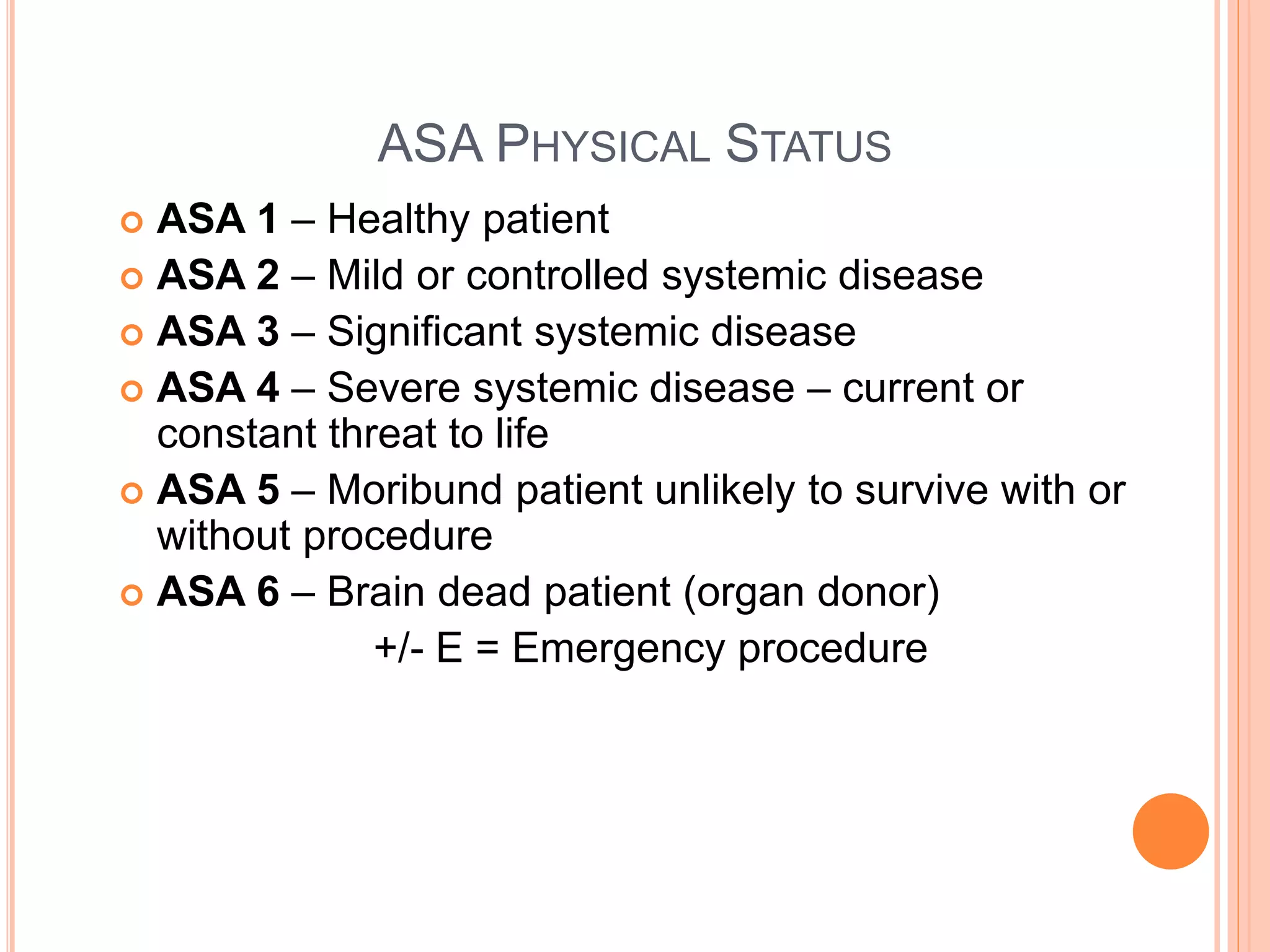
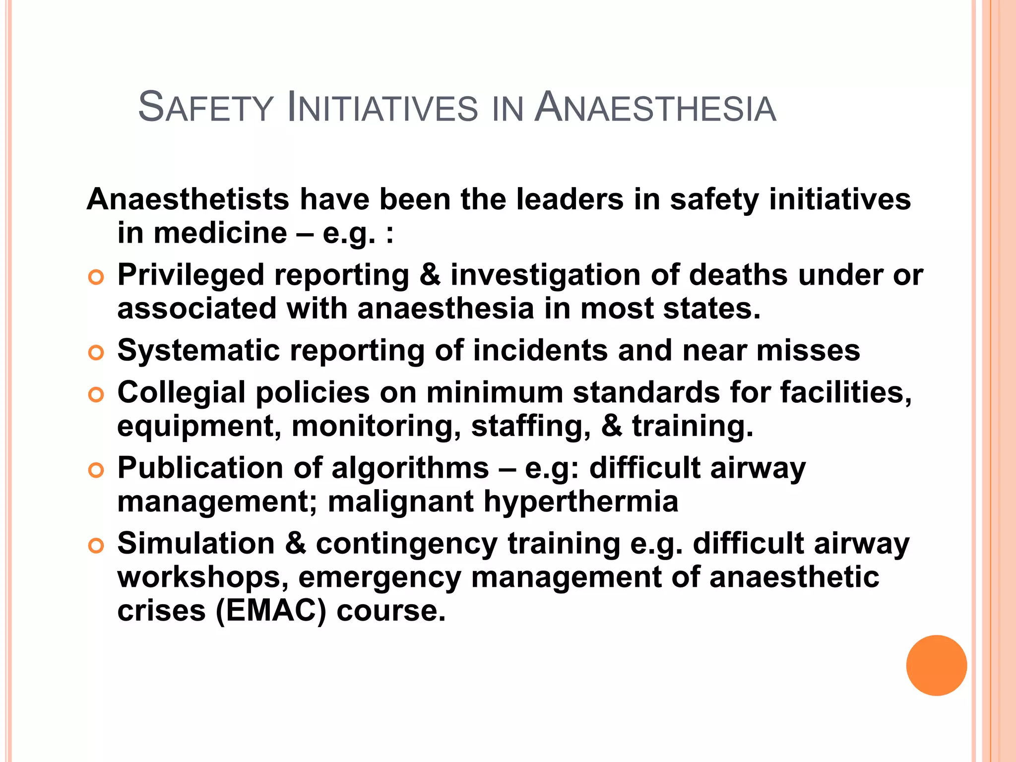
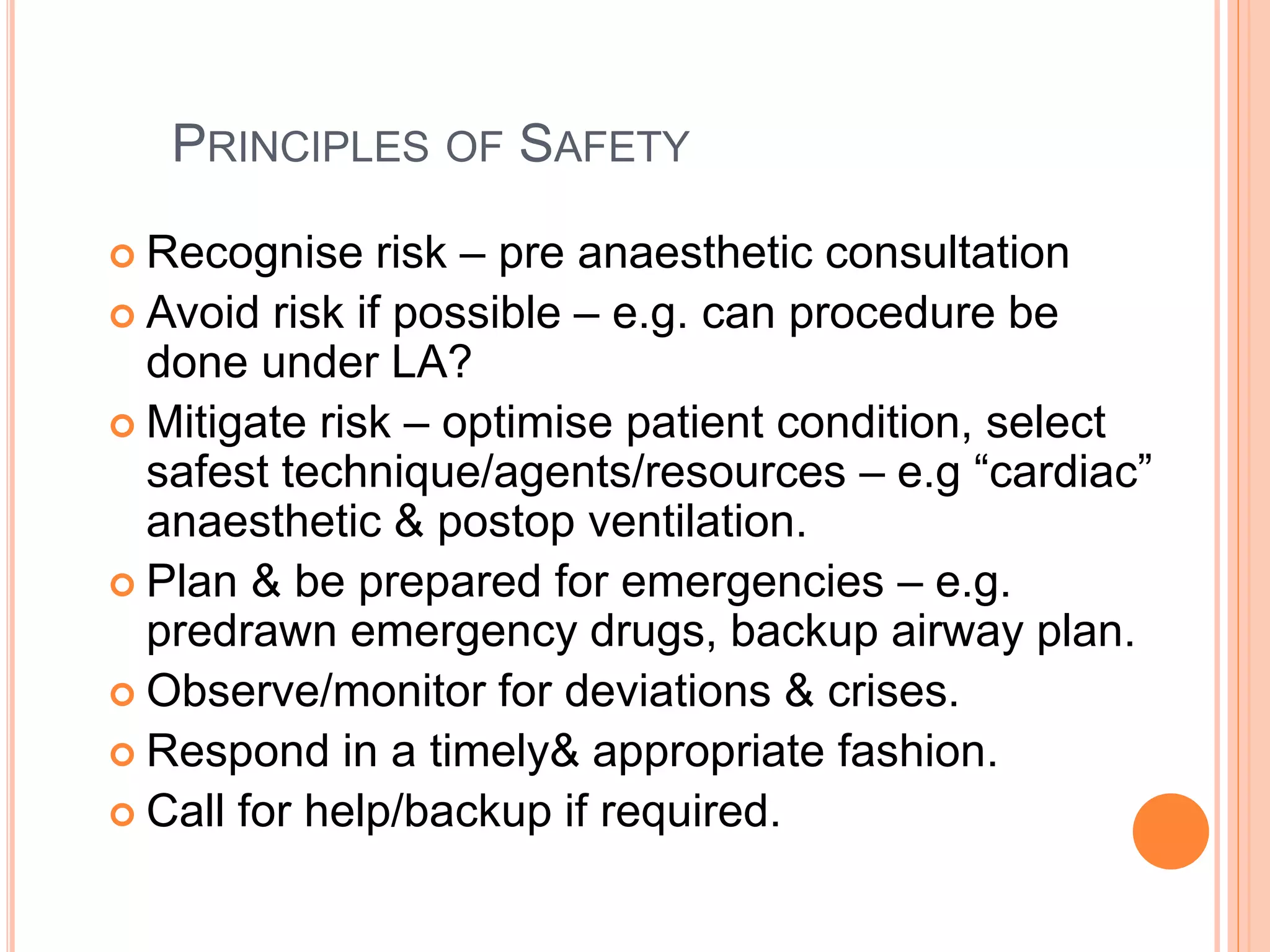
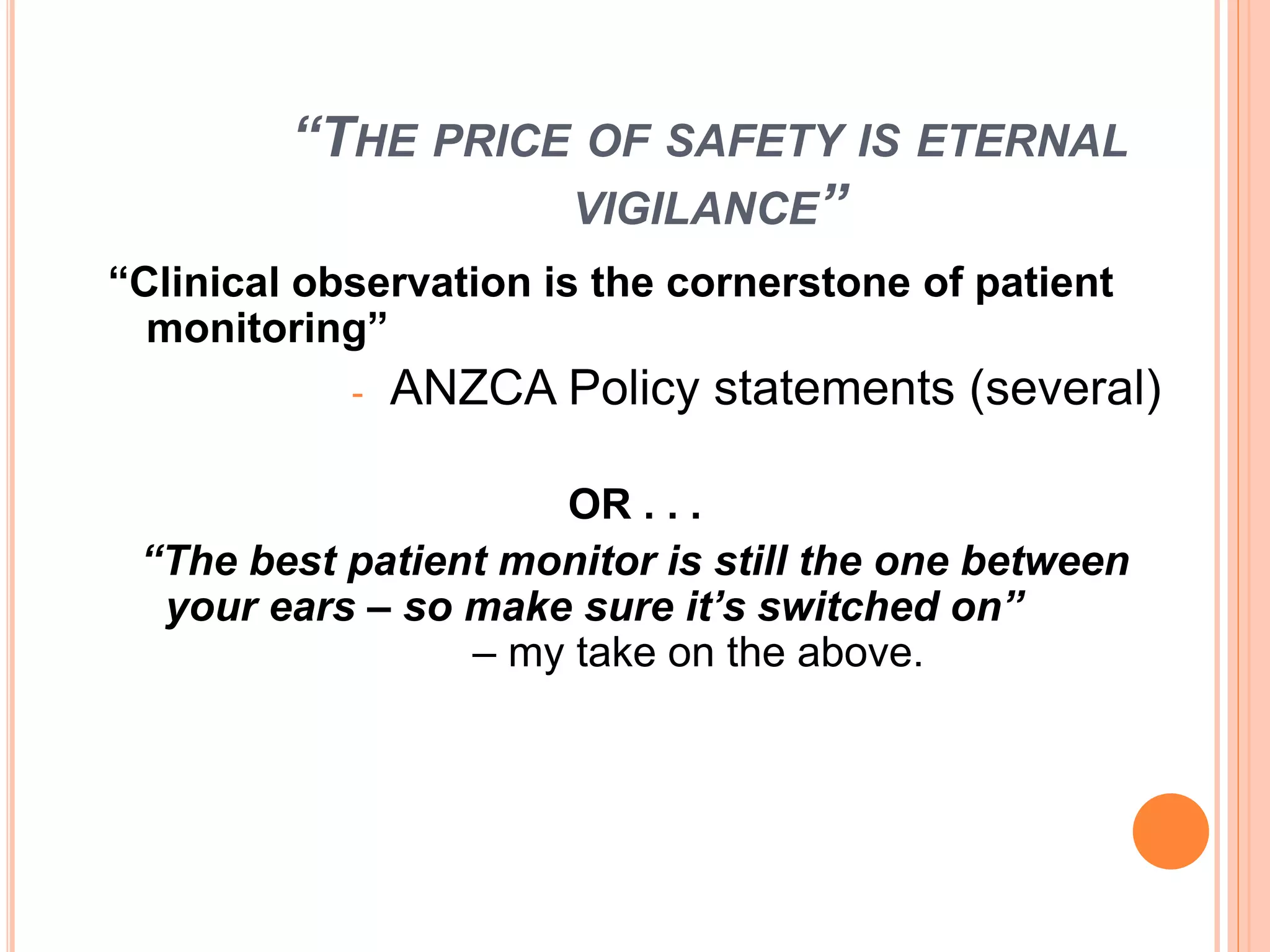
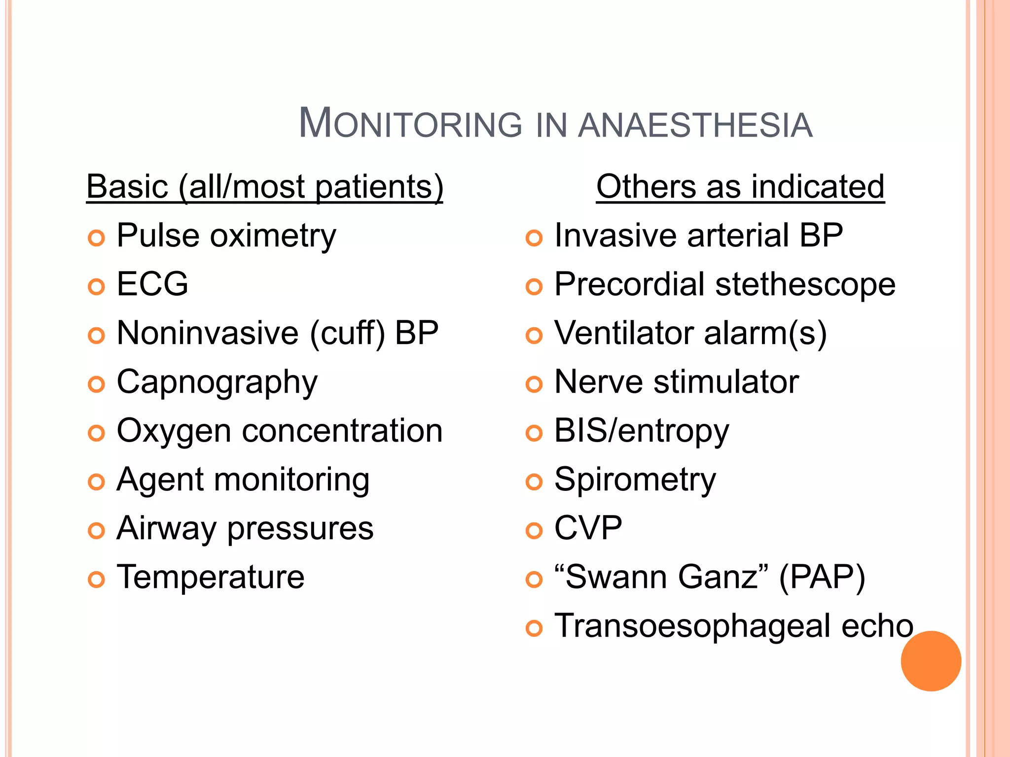
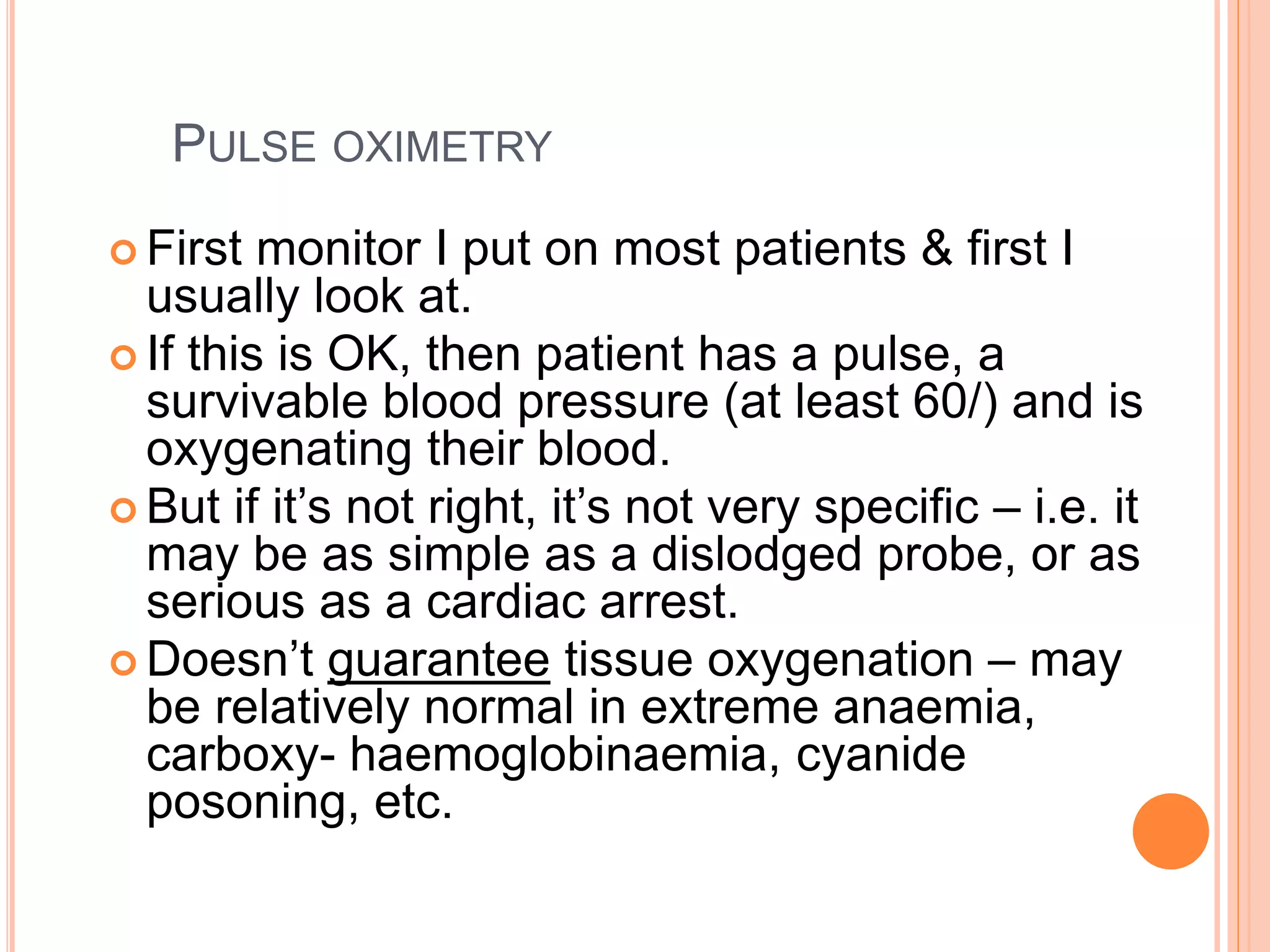
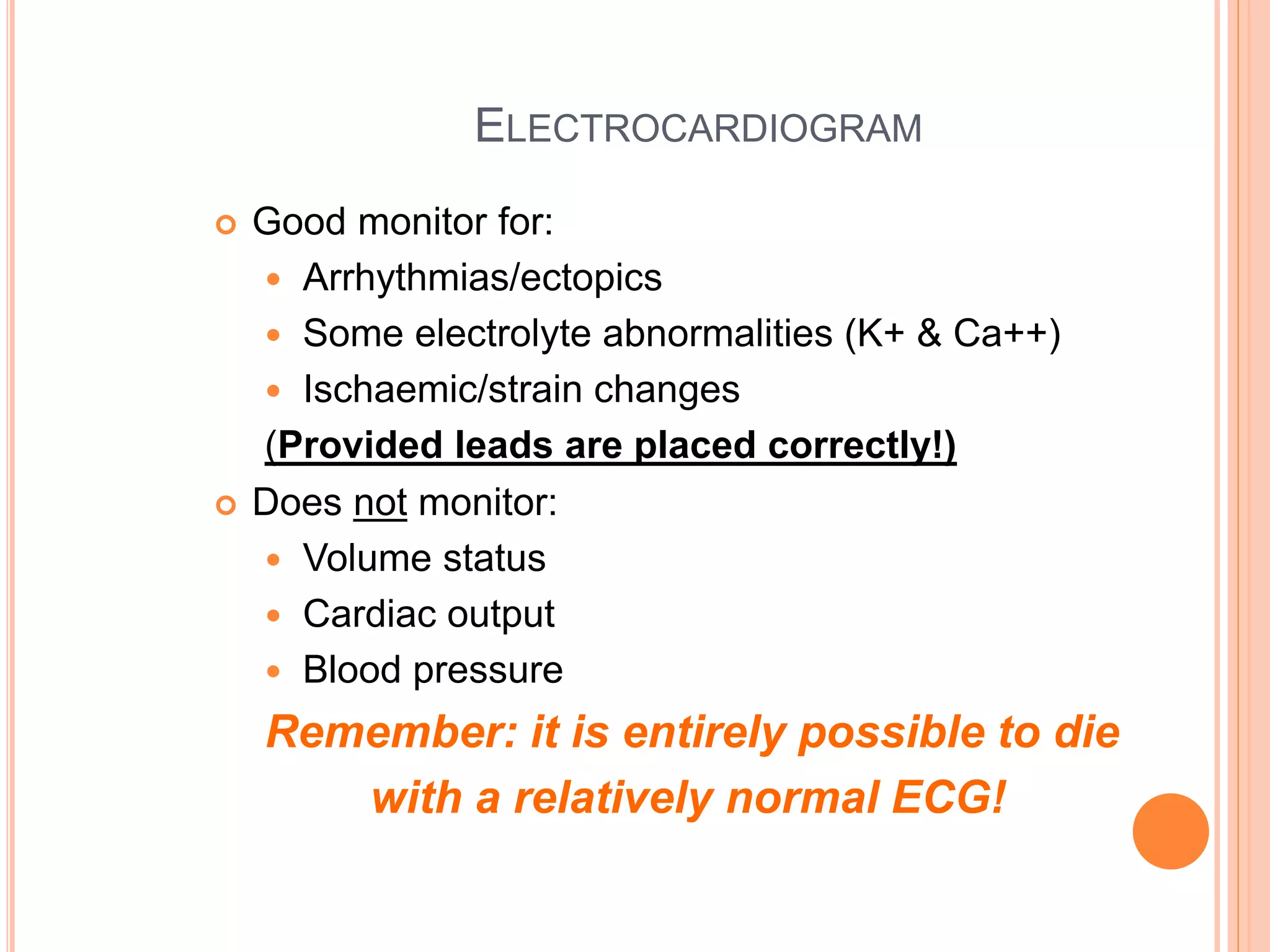
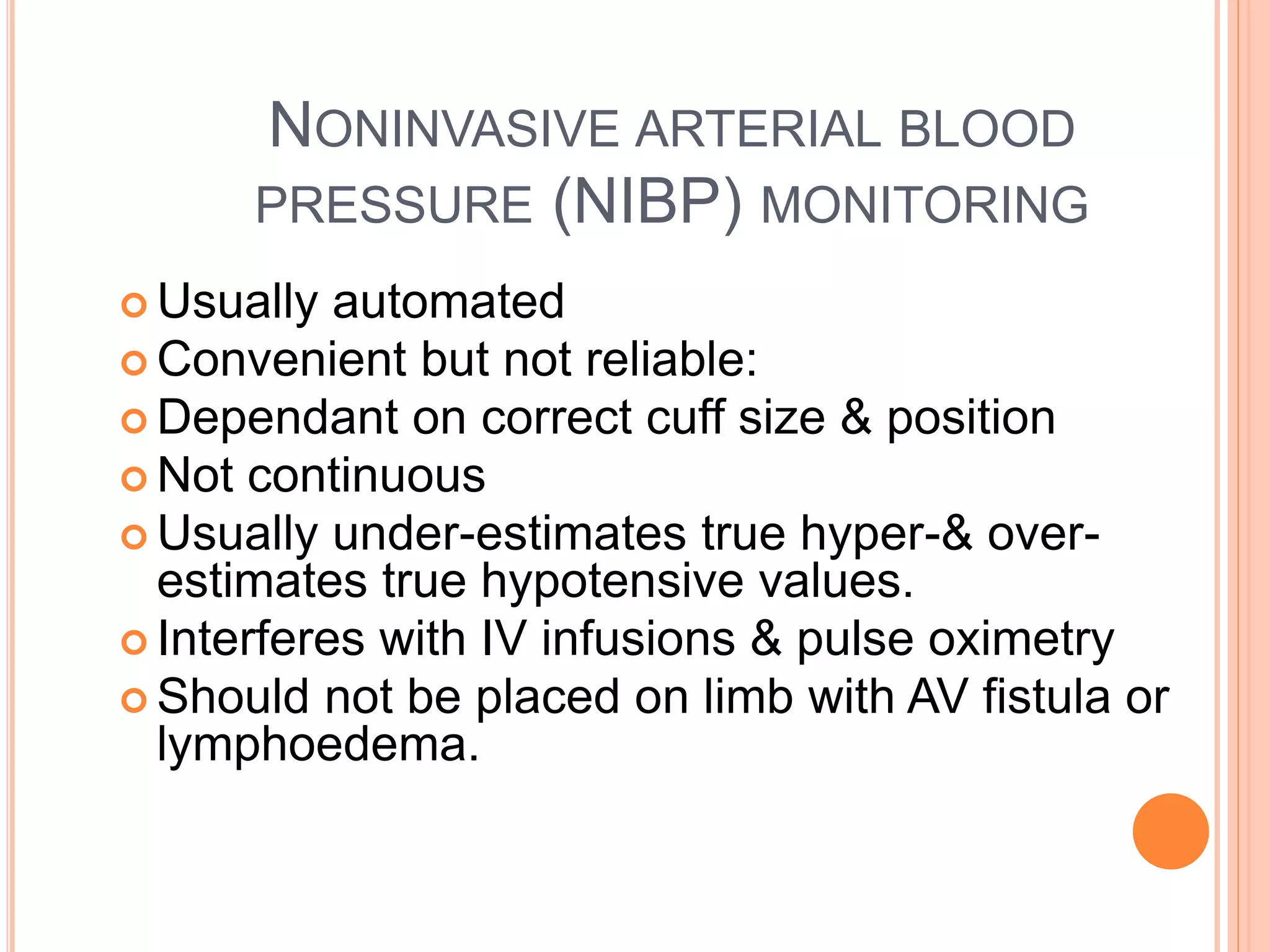
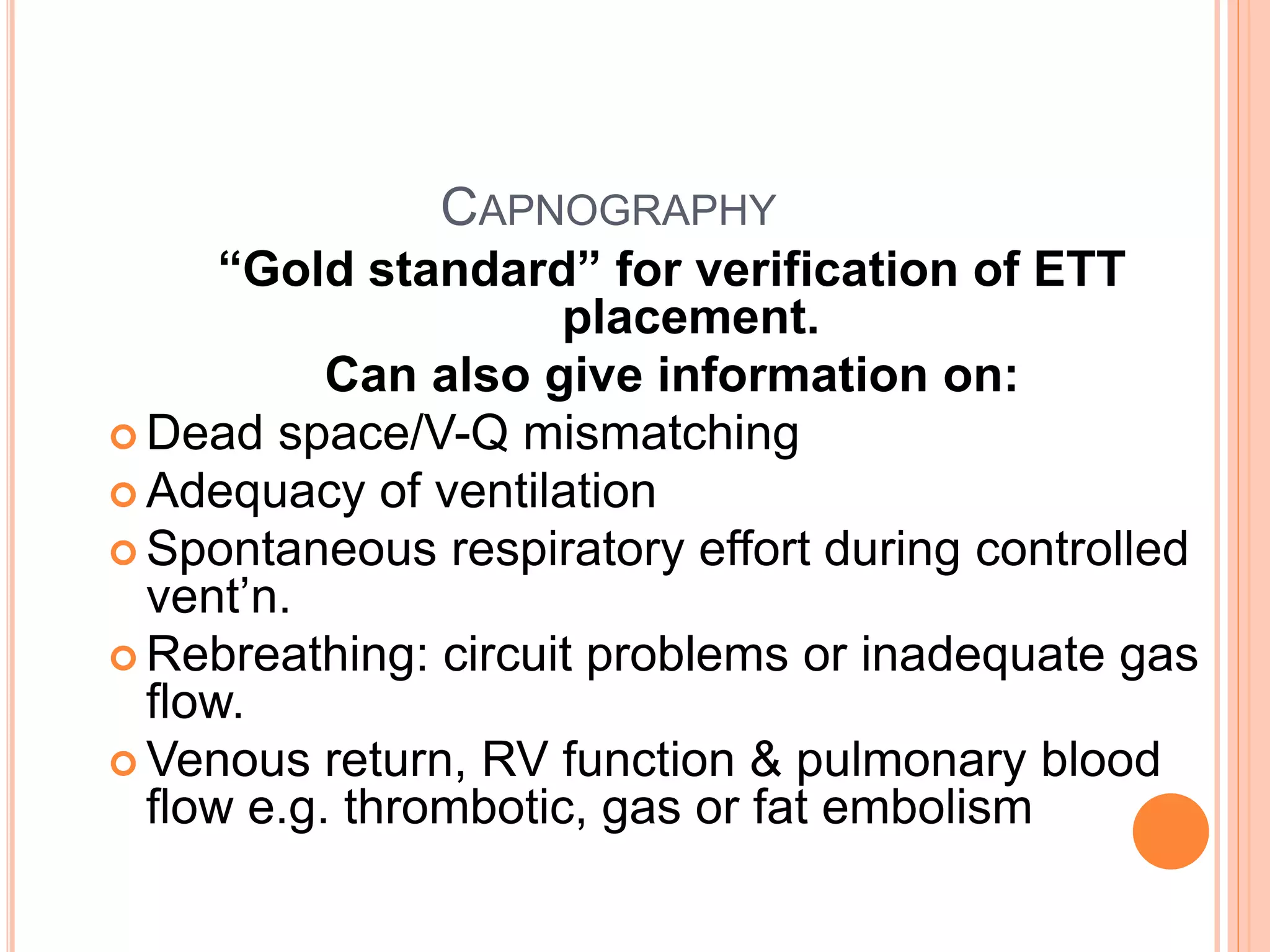
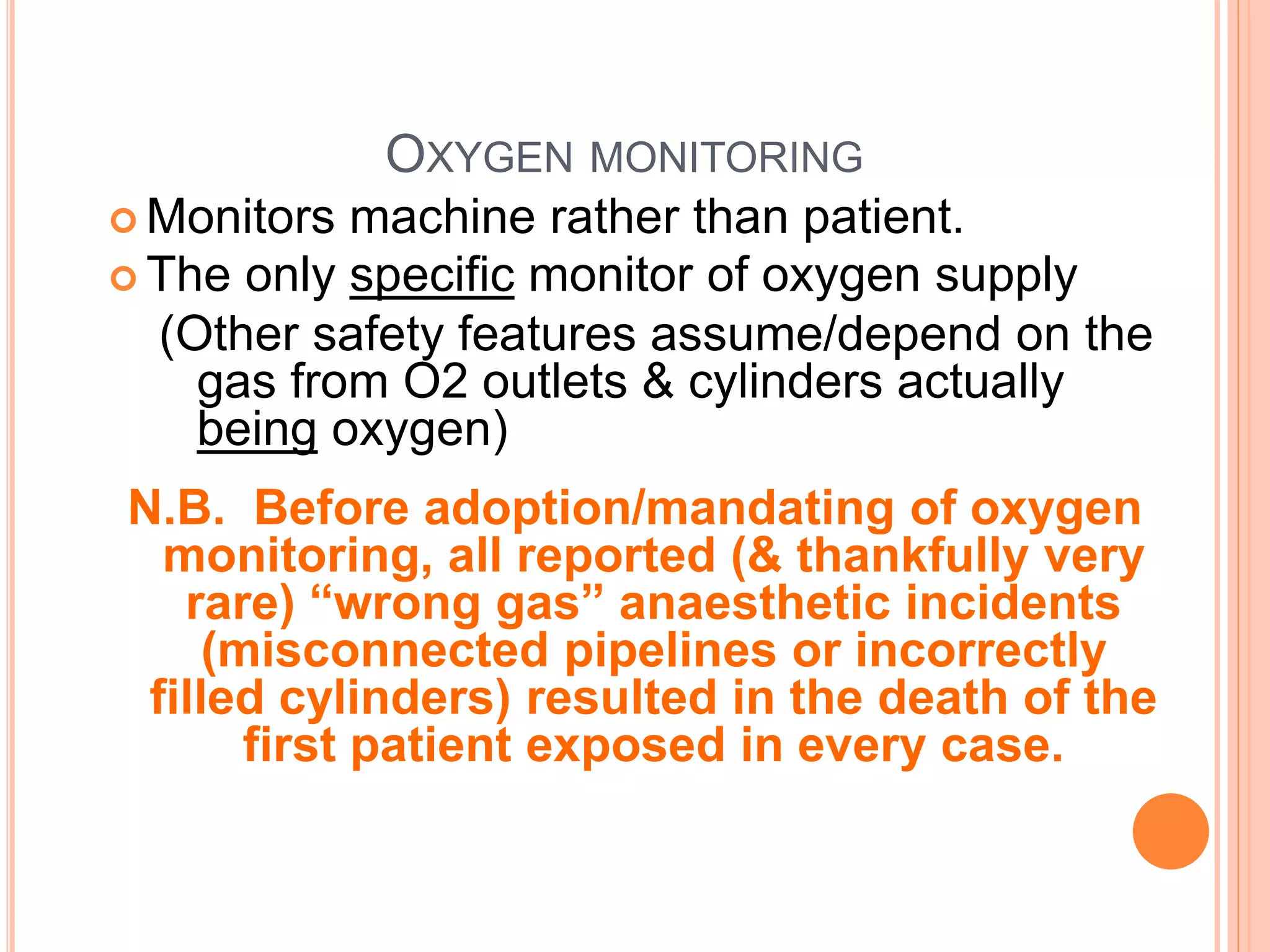
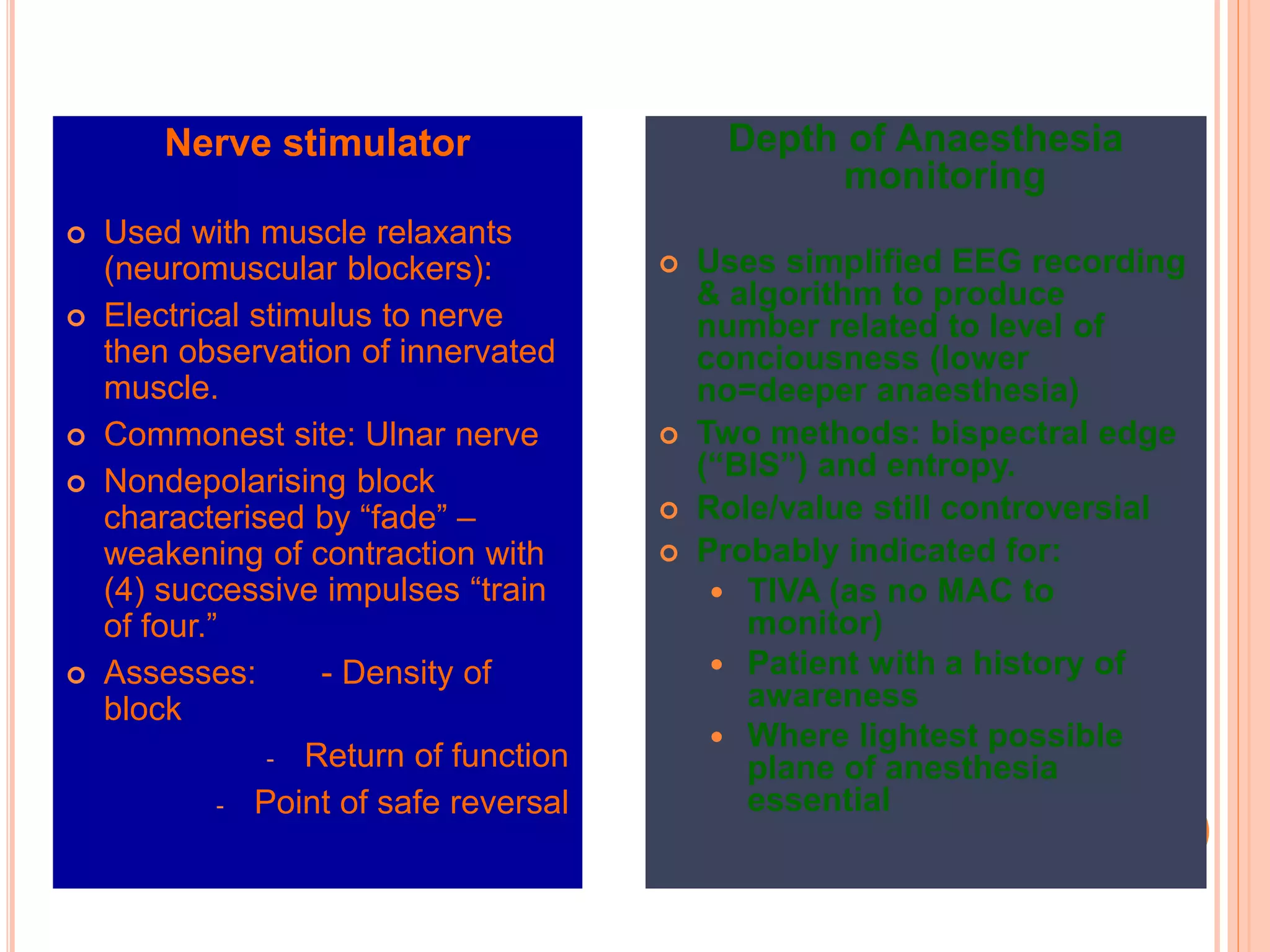
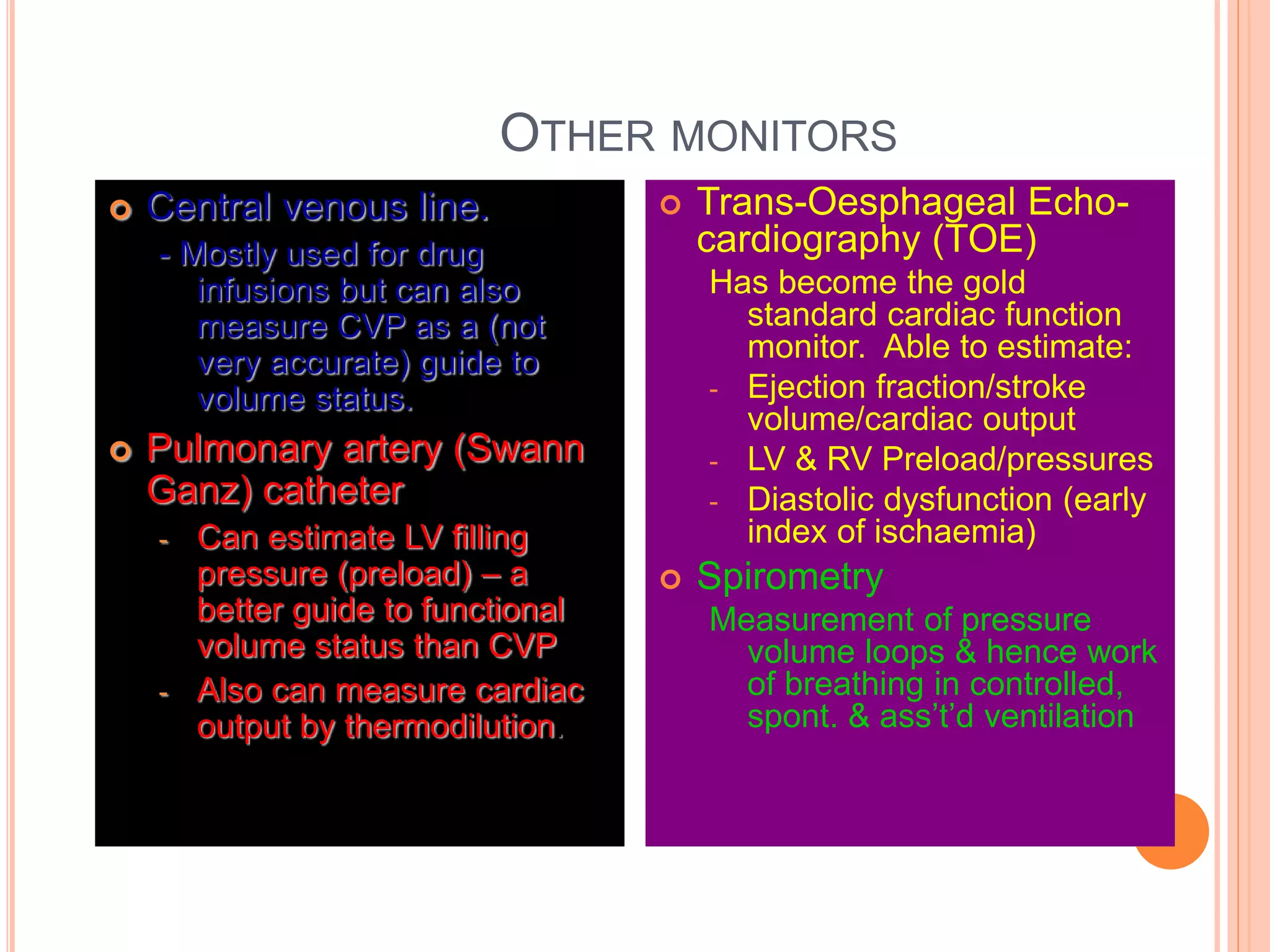
DR ROWAN MOLNAR PART II:
PERIOPERATIVE MEDICINE
“THE WAY OF THE FUTURE”
WHAT IS PERIOPERATIVE MEDICINE?
“Integrated multidisciplinary
management of the surgical
or procedural patient’s
hospital admission & stay.”
PERIOPERATIVE SYSTEM INCLUDES:
 Identification of patient requiring procedure
 Referral to perioperative service
 Screening for level of workup required
 Pre-anaesthetic assessment/plan
 Referral & investigations as required.
 Admission at appropriate pre-op interval
 Post-operative drug/fluid/other therapy
 Appropriate post op level of care & stay
 Discharge at earliest appropriate point
BUT WHY?
 Minimize unnecessary pre-op bed days.
 Minimize preoperative cancellations
 Enable more predictable bed occupancy
 Minimize pseudo-urgent blood tests & other
investigations
 Improve post operative care & shorten post operative
stay
THE PRE-ANAESTHETIC CONSULTATION
 What? Targeted history & examination, &
formulation of anaesthetic/perioperative plan.
 Who? Ideally by the anaesthetist for the procedure
(not always possible).
 Whom? All patients should have some form of this.
 When? At the earliest appropriate opportunity
(Obviously this varies on a case by case basis)
 Why? To enable optimimum pre-anaesthetic
preparation, risk minimisation, informed consent,
and allaying of anxiety.
PRE-OPERATIVE PREPARATION MAY
INCLUDE PREMEDICATION
Use if required, not “one size fits all”
Aims:
1. Ameliorate anxiety
Usually with a benzodiazepine such as
temazepam
2. Relieve pain – predominantly in the acute setting –
usually with narcotics.
3. Prevent reflux/aspiration - in at risk patient
Usually (a) H2 blocker or PPI 6-8 hrs preop if
possible, then (b) non particulate antacid
immediately preop.
4. Treat other medical conditions
e.g. asthma prophylaxis.
MOST REGULAR MEDICATIONS ARE CONTINUED,
INCLUDING ON THE DAY OF SURGERY
Exceptions include:
(a) Oral hypoglycaemics
(b) Antithrombotic agents (mostly)
ASA PHYSICAL STATUS
 ASA 1 – Healthy patient
 ASA 2 – Mild or controlled systemic disease
 ASA 3 – Significant systemic disease
 ASA 4 – Severe systemic disease – current or
constant threat to life
 ASA 5 – Moribund patient unlikely to survive with or
without procedure
 ASA 6 – Brain dead patient (organ donor)
+/- E = Emergency procedure
RELEVANCE OF THIS?
 Risk stratification
 Workload/resource utilisation planning
 Remuneration aspects
PERIOPERATIVE (PREANAESTHETIC)
CLINIC
Surgical clinic
Nurse Clinic
Checked up, satisfied
as fit & suitable
Decides to proceed
with planned time,
date & procedure
Not certain;sends
only case notes to
anaesthetist to review it
Satisfied with it;
decides to send
it back to her
for mx
Not quite satisfied;
takes over review & mx
Decides to further
investigate. May cancel,
postpone, refer case or
decide to do it
Surgeon refers case
Preanaesthetic Clinic
 The Doctor takes a quick history, leading
questions are allowed as major
diagnoses should already be known
 Asks for hypertension, diabetes,
asthma,epilepsy, previous anaesthetics,
allergies, complications, medications
being used
 A quick examination is done, Ix like Xray,
ECG, UES & Blood ix are done
 ASA categorised, anaesthesia decided
 Explained to patient about anaesthetics,
risks, PCA & possible complications
Preanaesthetic Clinic
Based on: History
Examination, Investigation . . .
Decision:
To do the
planned
procedure
To postpone the
procedure till fully
investigated
optimised
To cancel the
procedure
CASE STUDY II
Perioperative management
DIABETIC PATIENT FOR
VASCULAR SURGERY
HISTORY
 65 year old man, BMI 35
 Type II DM, 15 yrs, on OHGs, poor control
 Smoker 60+ pack years
 Hypertension
 Hypercholesterolaemia
 Ischaemic heart disease
 Diabetic nephropathy, (eGFR ~ 30mls/min)
For (R) femoro-popliteal bypass
What are the issues and risks here?
1.What are the issues and risks here?
2. How can we optimise him preoperatively?
1. What are the issues and risks here?
2. How can we optimise him preoperatively?
3. What are our anaesthetic options & problems?
1. What are the issues and risks here?
2. How can we optimise him preoperatively?
3. What are our anaesthetic options & problems?
4. How do we manage him postoperatively?
PART III:
SAFETY & MONITORING
IN ANAESTHESIA
SAFETY IN ANAESTHESIA IS PARAMOUNT
“When it goes right, no-one remembers. . . When it goes wrong,
no-one forgets”
. . . So the aim is to make anaesthesia as forgettable as possible!
SAFETY INITIATIVES IN ANAESTHESIA
Anaesthetists have been the leaders in safety initiatives
in medicine – e.g. :
 Privileged reporting & investigation of deaths under or
associated with anaesthesia in most states.
 Systematic reporting of incidents and near misses
 Collegial policies on minimum standards for facilities,
equipment, monitoring, staffing, & training.
 Publication of algorithms – e.g: difficult airway
management; malignant hyperthermia
 Simulation & contingency training e.g. difficult airway
workshops, emergency management of anaesthetic
crises (EMAC) course.
PRINCIPLES OF SAFETY
 Recognise risk – pre anaesthetic consultation
 Avoid risk if possible – e.g. can procedure be
done under LA?
 Mitigate risk – optimise patient condition, select
safest technique/agents/resources – e.g “cardiac”
anaesthetic & postop ventilation.
 Plan & be prepared for emergencies – e.g.
predrawn emergency drugs, backup airway plan.
 Observe/monitor for deviations & crises.
 Respond in a timely& appropriate fashion.
 Call for help/backup if required.
“THE PRICE OF SAFETY IS ETERNAL
VIGILANCE”
“Clinical observation is the cornerstone of patient
monitoring”
- ANZCA Policy statements (several)
OR . . .
“The best patient monitor is still the one between
your ears – so make sure it’s switched on”
– my take on the above.
MONITORING IN ANAESTHESIA
Basic (all/most patients)
 Pulse oximetry
 ECG
 Noninvasive (cuff) BP
 Capnography
 Oxygen concentration
 Agent monitoring
 Airway pressures
 Temperature
Others as indicated
 Invasive arterial BP
 Precordial stethescope
 Ventilator alarm(s)
 Nerve stimulator
 BIS/entropy
 Spirometry
 CVP
 “Swann Ganz” (PAP)
 Transoesophageal echo
PULSE OXIMETRY
 First monitor I put on most patients & first I
usually look at.
 If this is OK, then patient has a pulse, a
survivable blood pressure (at least 60/) and is
oxygenating their blood.
 But if it’s not right, it’s not very specific – i.e. it
may be as simple as a dislodged probe, or as
serious as a cardiac arrest.
 Doesn’t guarantee tissue oxygenation – may
be relatively normal in extreme anaemia,
carboxy- haemoglobinaemia, cyanide
posoning, etc.
ELECTROCARDIOGRAM
 Good monitor for:
 Arrhythmias/ectopics
 Some electrolyte abnormalities (K+ & Ca++)
 Ischaemic/strain changes
(Provided leads are placed correctly!)
 Does not monitor:
 Volume status
 Cardiac output
 Blood pressure
Remember: it is entirely possible to die
with a relatively normal ECG!
NONINVASIVE ARTERIAL BLOOD
PRESSURE (NIBP) MONITORING
 Usually automated
 Convenient but not reliable:
 Dependant on correct cuff size & position
 Not continuous
 Usually under-estimates true hyper-& over-
estimates true hypotensive values.
 Interferes with IV infusions & pulse oximetry
 Should not be placed on limb with AV fistula or
lymphoedema.
CAPNOGRAPHY
“Gold standard” for verification of ETT
placement.
Can also give information on:
 Dead space/V-Q mismatching
 Adequacy of ventilation
 Spontaneous respiratory effort during controlled
vent’n.
 Rebreathing: circuit problems or inadequate gas
flow.
 Venous return, RV function & pulmonary blood
flow e.g. thrombotic, gas or fat embolism
OXYGEN MONITORING
 Monitors machine rather than patient.
 The only specific monitor of oxygen supply
(Other safety features assume/depend on the
gas from O2 outlets & cylinders actually
being oxygen)
N.B. Before adoption/mandating of oxygen
monitoring, all reported (& thankfully very
rare) “wrong gas” anaesthetic incidents
(misconnected pipelines or incorrectly
filled cylinders) resulted in the death of the
first patient exposed in every case.
ANAESTHETIC AGENT MONITORING
 Identifies (hopefully confirms!) anaesthetic agent
being used
 Measures inspiratory & expiratory
concentrations
 Expiratory (alveolar) concentration enables
calculation of MAC fraction or multiple – i.e.
estimation of anaesthetic depth.
 Now mandatory when inhalational anaesthetic
agents are used.
TEMPERATURE MONITORING
 Anaesthesia promotes hypothermia by:
 Decreased metabolic rate -> decreased heat production
 Redistribution of blood flow -> increased heat loss
 Patients may need temperature support
 Passive (prevent heat loss)
 Active warming: forced air/ heated IV fluids
 What you support you must monitor
 Ideally monitor core temperature:
Nasopharyngeal/oesophageal/bladder/PV
Better than
Skin/axillary/oral/rectal
Airway manometry
 Usually analogue gauge
on circle circuit
 Monitors inflation
pressure
 With IPPV can help
identify:
Airway obstruction
Bronchospasm
Circuit leaks/faults
Ventilator monitor
 Mandatory when
mechanical IPPV
employed.
 Usually integrated into
ventilator w/automatic
activation.
 High (overpressure) &
low (disconnect)
functions
Precordial stethescope
 “Traditional” monitor
 Still used in some
paediatric cases
 Can monitors:
Heart & respiratory rate
Breath sounds presence &
quality.
Only as good as the
person listening to it!
Direct arterial pressure
monitoring
Invasive procedure, but:
 Gold standard for real
time haemodynamic
assessment
 Accurate, reliable.
 Immediate warning of
hypo/hypertension of
any aetiology.
Nerve stimulator
 Used with muscle relaxants
(neuromuscular blockers):
 Electrical stimulus to nerve
then observation of innervated
muscle.
 Commonest site: Ulnar nerve
 Nondepolarising block
characterised by “fade” –
weakening of contraction with
(4) successive impulses “train
of four.”
 Assesses: - Density of
block
- Return of function
- Point of safe reversal
Depth of Anaesthesia
monitoring
 Uses simplified EEG recording
& algorithm to produce
number related to level of
conciousness (lower
no=deeper anaesthesia)
 Two methods: bispectral edge
(“BIS”) and entropy.
 Role/value still controversial
 Probably indicated for:
 TIVA (as no MAC to
monitor)
 Patient with a history of
awareness
 Where lightest possible
plane of anesthesia
essential
OTHER MONITORS
 Central venous line.
- Mostly used for drug
infusions but can also
measure CVP as a (not
very accurate) guide to
volume status.
 Pulmonary artery (Swann
Ganz) catheter
- Can estimate LV filling
pressure (preload) – a
better guide to functional
volume status than CVP
- Also can measure cardiac
output by thermodilution.
 Trans-Oesphageal Echo-
cardiography (TOE)
Has become the gold
standard cardiac function
monitor. Able to estimate:
- Ejection fraction/stroke
volume/cardiac output
- LV & RV Preload/pressures
- Diastolic dysfunction (early
index of ischaemia)
 Spirometry
Measurement of pressure
volume loops & hence work
of breathing in controlled,
spont. & ass’t’d ventilation

Dr rowan molnar anaesthetics study guide part ii

  • 1.
    DR ROWAN MOLNARPART II: PERIOPERATIVE MEDICINE “THE WAY OF THE FUTURE”
  • 2.
    WHAT IS PERIOPERATIVEMEDICINE? “Integrated multidisciplinary management of the surgical or procedural patient’s hospital admission & stay.”
  • 3.
    PERIOPERATIVE SYSTEM INCLUDES: Identification of patient requiring procedure  Referral to perioperative service  Screening for level of workup required  Pre-anaesthetic assessment/plan  Referral & investigations as required.  Admission at appropriate pre-op interval  Post-operative drug/fluid/other therapy  Appropriate post op level of care & stay  Discharge at earliest appropriate point
  • 4.
    BUT WHY?  Minimizeunnecessary pre-op bed days.  Minimize preoperative cancellations  Enable more predictable bed occupancy  Minimize pseudo-urgent blood tests & other investigations  Improve post operative care & shorten post operative stay
  • 5.
    THE PRE-ANAESTHETIC CONSULTATION What? Targeted history & examination, & formulation of anaesthetic/perioperative plan.  Who? Ideally by the anaesthetist for the procedure (not always possible).  Whom? All patients should have some form of this.  When? At the earliest appropriate opportunity (Obviously this varies on a case by case basis)  Why? To enable optimimum pre-anaesthetic preparation, risk minimisation, informed consent, and allaying of anxiety.
  • 6.
    PRE-OPERATIVE PREPARATION MAY INCLUDEPREMEDICATION Use if required, not “one size fits all” Aims: 1. Ameliorate anxiety Usually with a benzodiazepine such as temazepam 2. Relieve pain – predominantly in the acute setting – usually with narcotics. 3. Prevent reflux/aspiration - in at risk patient Usually (a) H2 blocker or PPI 6-8 hrs preop if possible, then (b) non particulate antacid immediately preop. 4. Treat other medical conditions e.g. asthma prophylaxis.
  • 7.
    MOST REGULAR MEDICATIONSARE CONTINUED, INCLUDING ON THE DAY OF SURGERY Exceptions include: (a) Oral hypoglycaemics (b) Antithrombotic agents (mostly)
  • 8.
    ASA PHYSICAL STATUS ASA 1 – Healthy patient  ASA 2 – Mild or controlled systemic disease  ASA 3 – Significant systemic disease  ASA 4 – Severe systemic disease – current or constant threat to life  ASA 5 – Moribund patient unlikely to survive with or without procedure  ASA 6 – Brain dead patient (organ donor) +/- E = Emergency procedure
  • 9.
    RELEVANCE OF THIS? Risk stratification  Workload/resource utilisation planning  Remuneration aspects
  • 10.
    PERIOPERATIVE (PREANAESTHETIC) CLINIC Surgical clinic NurseClinic Checked up, satisfied as fit & suitable Decides to proceed with planned time, date & procedure Not certain;sends only case notes to anaesthetist to review it Satisfied with it; decides to send it back to her for mx Not quite satisfied; takes over review & mx Decides to further investigate. May cancel, postpone, refer case or decide to do it Surgeon refers case
  • 11.
    Preanaesthetic Clinic  TheDoctor takes a quick history, leading questions are allowed as major diagnoses should already be known  Asks for hypertension, diabetes, asthma,epilepsy, previous anaesthetics, allergies, complications, medications being used  A quick examination is done, Ix like Xray, ECG, UES & Blood ix are done  ASA categorised, anaesthesia decided  Explained to patient about anaesthetics, risks, PCA & possible complications
  • 12.
    Preanaesthetic Clinic Based on:History Examination, Investigation . . . Decision: To do the planned procedure To postpone the procedure till fully investigated optimised To cancel the procedure
  • 13.
  • 14.
  • 15.
    HISTORY  65 yearold man, BMI 35  Type II DM, 15 yrs, on OHGs, poor control  Smoker 60+ pack years  Hypertension  Hypercholesterolaemia  Ischaemic heart disease  Diabetic nephropathy, (eGFR ~ 30mls/min) For (R) femoro-popliteal bypass
  • 16.
    What are theissues and risks here?
  • 17.
    1.What are theissues and risks here? 2. How can we optimise him preoperatively?
  • 18.
    1. What arethe issues and risks here? 2. How can we optimise him preoperatively? 3. What are our anaesthetic options & problems?
  • 19.
    1. What arethe issues and risks here? 2. How can we optimise him preoperatively? 3. What are our anaesthetic options & problems? 4. How do we manage him postoperatively?
  • 20.
    PART III: SAFETY &MONITORING IN ANAESTHESIA
  • 21.
    SAFETY IN ANAESTHESIAIS PARAMOUNT “When it goes right, no-one remembers. . . When it goes wrong, no-one forgets” . . . So the aim is to make anaesthesia as forgettable as possible!
  • 22.
    SAFETY INITIATIVES INANAESTHESIA Anaesthetists have been the leaders in safety initiatives in medicine – e.g. :  Privileged reporting & investigation of deaths under or associated with anaesthesia in most states.  Systematic reporting of incidents and near misses  Collegial policies on minimum standards for facilities, equipment, monitoring, staffing, & training.  Publication of algorithms – e.g: difficult airway management; malignant hyperthermia  Simulation & contingency training e.g. difficult airway workshops, emergency management of anaesthetic crises (EMAC) course.
  • 23.
    PRINCIPLES OF SAFETY Recognise risk – pre anaesthetic consultation  Avoid risk if possible – e.g. can procedure be done under LA?  Mitigate risk – optimise patient condition, select safest technique/agents/resources – e.g “cardiac” anaesthetic & postop ventilation.  Plan & be prepared for emergencies – e.g. predrawn emergency drugs, backup airway plan.  Observe/monitor for deviations & crises.  Respond in a timely& appropriate fashion.  Call for help/backup if required.
  • 24.
    “THE PRICE OFSAFETY IS ETERNAL VIGILANCE” “Clinical observation is the cornerstone of patient monitoring” - ANZCA Policy statements (several) OR . . . “The best patient monitor is still the one between your ears – so make sure it’s switched on” – my take on the above.
  • 25.
    MONITORING IN ANAESTHESIA Basic(all/most patients)  Pulse oximetry  ECG  Noninvasive (cuff) BP  Capnography  Oxygen concentration  Agent monitoring  Airway pressures  Temperature Others as indicated  Invasive arterial BP  Precordial stethescope  Ventilator alarm(s)  Nerve stimulator  BIS/entropy  Spirometry  CVP  “Swann Ganz” (PAP)  Transoesophageal echo
  • 26.
    PULSE OXIMETRY  Firstmonitor I put on most patients & first I usually look at.  If this is OK, then patient has a pulse, a survivable blood pressure (at least 60/) and is oxygenating their blood.  But if it’s not right, it’s not very specific – i.e. it may be as simple as a dislodged probe, or as serious as a cardiac arrest.  Doesn’t guarantee tissue oxygenation – may be relatively normal in extreme anaemia, carboxy- haemoglobinaemia, cyanide posoning, etc.
  • 27.
    ELECTROCARDIOGRAM  Good monitorfor:  Arrhythmias/ectopics  Some electrolyte abnormalities (K+ & Ca++)  Ischaemic/strain changes (Provided leads are placed correctly!)  Does not monitor:  Volume status  Cardiac output  Blood pressure Remember: it is entirely possible to die with a relatively normal ECG!
  • 28.
    NONINVASIVE ARTERIAL BLOOD PRESSURE(NIBP) MONITORING  Usually automated  Convenient but not reliable:  Dependant on correct cuff size & position  Not continuous  Usually under-estimates true hyper-& over- estimates true hypotensive values.  Interferes with IV infusions & pulse oximetry  Should not be placed on limb with AV fistula or lymphoedema.
  • 29.
    CAPNOGRAPHY “Gold standard” forverification of ETT placement. Can also give information on:  Dead space/V-Q mismatching  Adequacy of ventilation  Spontaneous respiratory effort during controlled vent’n.  Rebreathing: circuit problems or inadequate gas flow.  Venous return, RV function & pulmonary blood flow e.g. thrombotic, gas or fat embolism
  • 30.
    OXYGEN MONITORING  Monitorsmachine rather than patient.  The only specific monitor of oxygen supply (Other safety features assume/depend on the gas from O2 outlets & cylinders actually being oxygen) N.B. Before adoption/mandating of oxygen monitoring, all reported (& thankfully very rare) “wrong gas” anaesthetic incidents (misconnected pipelines or incorrectly filled cylinders) resulted in the death of the first patient exposed in every case.
  • 31.
    ANAESTHETIC AGENT MONITORING Identifies (hopefully confirms!) anaesthetic agent being used  Measures inspiratory & expiratory concentrations  Expiratory (alveolar) concentration enables calculation of MAC fraction or multiple – i.e. estimation of anaesthetic depth.  Now mandatory when inhalational anaesthetic agents are used.
  • 32.
    TEMPERATURE MONITORING  Anaesthesiapromotes hypothermia by:  Decreased metabolic rate -> decreased heat production  Redistribution of blood flow -> increased heat loss  Patients may need temperature support  Passive (prevent heat loss)  Active warming: forced air/ heated IV fluids  What you support you must monitor  Ideally monitor core temperature: Nasopharyngeal/oesophageal/bladder/PV Better than Skin/axillary/oral/rectal
  • 33.
    Airway manometry  Usuallyanalogue gauge on circle circuit  Monitors inflation pressure  With IPPV can help identify: Airway obstruction Bronchospasm Circuit leaks/faults Ventilator monitor  Mandatory when mechanical IPPV employed.  Usually integrated into ventilator w/automatic activation.  High (overpressure) & low (disconnect) functions
  • 34.
    Precordial stethescope  “Traditional”monitor  Still used in some paediatric cases  Can monitors: Heart & respiratory rate Breath sounds presence & quality. Only as good as the person listening to it! Direct arterial pressure monitoring Invasive procedure, but:  Gold standard for real time haemodynamic assessment  Accurate, reliable.  Immediate warning of hypo/hypertension of any aetiology.
  • 35.
    Nerve stimulator  Usedwith muscle relaxants (neuromuscular blockers):  Electrical stimulus to nerve then observation of innervated muscle.  Commonest site: Ulnar nerve  Nondepolarising block characterised by “fade” – weakening of contraction with (4) successive impulses “train of four.”  Assesses: - Density of block - Return of function - Point of safe reversal Depth of Anaesthesia monitoring  Uses simplified EEG recording & algorithm to produce number related to level of conciousness (lower no=deeper anaesthesia)  Two methods: bispectral edge (“BIS”) and entropy.  Role/value still controversial  Probably indicated for:  TIVA (as no MAC to monitor)  Patient with a history of awareness  Where lightest possible plane of anesthesia essential
  • 36.
    OTHER MONITORS  Centralvenous line. - Mostly used for drug infusions but can also measure CVP as a (not very accurate) guide to volume status.  Pulmonary artery (Swann Ganz) catheter - Can estimate LV filling pressure (preload) – a better guide to functional volume status than CVP - Also can measure cardiac output by thermodilution.  Trans-Oesphageal Echo- cardiography (TOE) Has become the gold standard cardiac function monitor. Able to estimate: - Ejection fraction/stroke volume/cardiac output - LV & RV Preload/pressures - Diastolic dysfunction (early index of ischaemia)  Spirometry Measurement of pressure volume loops & hence work of breathing in controlled, spont. & ass’t’d ventilation