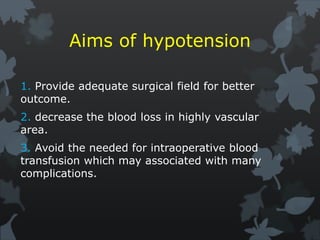
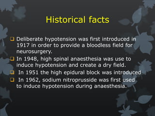
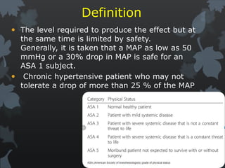
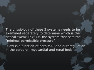
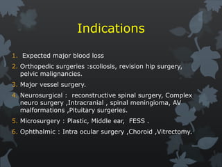
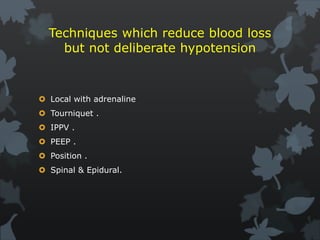
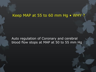
Hypotensive techniques
Dr. Radhwan Hazem Alkhashab
Consultant anesthesia & ICU
2020
Introduction
 Deliberate hypotensive technique is important
during many surgical procedures. One of them
are ENT surgery
Aims of hypotension
1. Provide adequate surgical field for better
outcome.
2. decrease the blood loss in highly vascular
area.
3. Avoid the needed for intraoperative blood
transfusion which may associated with many
complications.
Historical facts
 Deliberate hypotension was first introduced in
1917 in order to provide a bloodless field for
neurosurgery.
 In 1948, high spinal anaesthesia was use to
induce hypotension and create a dry field.
 In 1951 the high epidural block was introduced
 In 1962, sodium nitroprusside was first used
to induce hypotension during anaesthesia.
Definition
 The level required to produce the effect but at
the same time is limited by safety.
Generally, it is taken that a MAP as low as 50
mmHg or a 30% drop in MAP is safe for an
ASA 1 subject.
 Chronic hypertensive patient who may not
tolerate a drop of more than 25 % of the MAP
 It is recommended that hypotensive
anesthesia be adjusted in relation to the
patient's preoperative blood pressure rather
than to a specific target pressure and be
limited to that level necessary to reduce
bleeding in the surgical field and in duration
to that part of the surgical procedure
deemed to benefit by it.
 A mean arterial blood pressure (MAP)
30% below a patient's usual MAP, with a
minimum MAP of 50 mm Hg in ASA Class I
patients and a MAP not less than 80 mm
Hg in the elderly, is suggested to be
clinically acceptable.
The physiology of these 3 systems needs to be
examined separately to determine which is the
critical “weak link” i.e. the system that sets the
“minimal permissible pressure”.
Flow is a function of both MAP and autoregulation
in the cerebral, myocardial and renal beds
Indications
1. Expected major blood loss
2. Orthopedic surgeries :scoliosis, revision hip surgery,
pelvic malignancies.
3. Major vessel surgery.
4. Neurosurgical : reconstructive spinal surgery, Complex
neuro surgery ,Intracranial , spinal meningioma, AV
malformations ,Pituitary surgeries.
5. Microsurgery : Plastic, Middle ear, FESS .
6. Ophthalmic : Intra ocular surgery ,Choroid ,Vitrectomy.
Indications shortly (SPINE)
 S = SPINAL .
 P= PITUITARY, PELVIC, PLASTIC .
 I = INTRACRANIAL, INTRAOCULAR .
 N =NEURO, NEUROVASCULAR .
 E = EAR, ENDOSCOPIC SINUS.
Techniques which reduce blood loss
but not deliberate hypotension
 Local with adrenaline
 Tourniquet .
 IPPV .
 PEEP .
 Position .
 Spinal & Epidural.
Keep MAP at 55 to 60 mm Hg • WHY ?
Auto regulation of Coronary and cerebral
blood flow stops at MAP at 50 to 55 mm Hg
Organs capable of autoregulation are able to
maintain their perfusion over a wide range of
pressure changes.
This critical pressure varies from vessel to vessel,
organ to organ, and probably from individual to
individual.
CEREBRAL CIRCULATION
Autoregulation
Normal cerebral blood flow is maintained at
45-50mls/100g/min
MAP range of 50-150mmHg
Autoregulation & CBF
Factors influencing CBF
1. PaCO2:-
For every 1mmHg increase in PaCO2 there is an
increase in CBF in the order of 1ml/100g/min
2. PaO2-
1. Changes in pao2 also alters CBF
2. 100% O2 administration in hyperbaric
produce toxic effects on cerebral function
and
3. Reduces CBF by 1/5
4. Administration 100% O2 during induced
hypotension not beneficial
3. Volatiles agents:
 Volatile anaesthetics attenuate or abolish the
autoregulation of cerebral blood flow in a dose
dependent manner in the following order :
 Halothane > enflurane > isoflurane
4. TEMPERATURE:
 Cerebral blood flow changes 5-7% per degree
celcius change in temperature.
 Hypothermia causes cerebral vasoconstriction
whereas an increase in body temperature causes
cerebral vasodilation.
5. VASODILATORS
Vasodilators such as nitroprusside and
nitroglycerine attenuate the autoregulation of
CBF in a similar manner to that of volatile
agents.
6. POSITIONING:
 Elevation of the head during hypotensive
anaesthesia can aggrevate the decrease in
cerebral perfusion pressure.
 The perfusion pressure decreases by 2mmHg
for every 2.5cm the head is raised above the
point of monitoring
CORONARY CIRCULATION
 Coronary blood flow is dependent upon the
aortic diastolic blood pressure and the coronary
vascular resistance.
 Control of coronary blood flow is autoregulated
predominantly by means of alteration in coronary
vascular resistance that are made to meet
myocardial oxygen demand.
 Any increase in myocardial oxygen demand
requires a parallel increase in coronary artery
blood flow.
Hypotensive anaesthesia may substantially:
1. Decrease coronary blood flow.
2. Decreases myocardial oxygen demand
3. Due to reduction in afterload or preload
4. Patients with CAD may have areas of
myocardium that are entirely dependent
upon
pressure to supply adequate blood flow.
5. Use of vasodilators in these patients may
induce
a steal phenomenon.
6. Significant intraoperative risk of myocardial
infarction.
RENAL CIRCULATION
 Autoregulation over the range 80-180
mmHg
 MAP less than 75 mmHg leads to
decrease in GFR
 Opioids and inhalational agents stimulate
ADH
HEPATIC CIRCULATION
Liver is not an autoregulated organ. decrease in
arterial pressure will lead to a decrease in liver
blood flow.
An increase in PaCO2 or a decrease in PaO2 will
lead to a catecholamine response which causes
splanchnic vasoconstriction and therefore a
decrease in liver blood flow.
Hypocapnia produced incidentally by
hyperventialtion during IPPV leads to a decrease
in liver blood flow as a result of the mechanical
effects.
Liver blood flow may be altered directly by the
effects of anaesthetic agents on splanchnic blood
flow.
RESPIRATORY SYSTEM
• During controlled hypotensive anaesthesia the
following occurs:
• Pulmonary blood flow gravitates to the dependent
areas of the lungs.
• The use of vasodilators to induce hypotension
inhibits the hypoxic pulmonary vasoconstriction
response thereby increasing intra-pulmonary shunt.
• All these factors result in hypercarbia, an
increase in arterial-end tidal CO2 gradient and
hypoxaemia
Contraindications of
techniques
1. Anaethetist factors:
 Lack of understanding of the technique.
 Lack of technical experience.
 Inability to monitor the patient adequately
2. Patient factors
 Cardiac disease .
 Diabetes .
 Severe Anemia.
 Hepatic disease.
 Cerebrovascular disease.
 Renal disease.
 Respiratory insufficiency.
 Severe systemic hypertension.
 Intolerance to drugs used for
hypotensive anaesthesia
 Pregnancy.
 Glaucoma.
Techniques
MAP = CO x SYSTEMIC VASCULAR RESISTANCE
The key equation in the provision of hypotensive
anaesthesia .
MAP can be manipulated by reducing either SVR
or Cardiac output or both.
METHODS TO REDUCE CARDIAC OUTPUT
METHODS TO REDUCE PERIPHERAL
VASCULAR RESISTANCE
Pharmacologic technique
 Ideal agent
 Ease of administration
 Predictable & dose-dependent effect
 Rapid onset/offset
 Quick elimination without the production
of toxic metabolites
 Minimal effects on blood flow to vital
organs
Inhalational anesthetics
negative inotropic effect vasodilation
Advantage:
1. Provides surgical
anesthesia
2. Rapid onset/offset.
3. Easy to titrate
4. Cerebral protection
Disadvantage:
1. Decreases CO
2. Cerebral vasodilation
Isoflurane is ideal • 2 MAC • SVR decrease without myocardial
depression • Inh. of baroreceptor reflexes thro anaesthetic action
• Halo OK but think of brady and myocardial depression.
Nitrates drugs
Which includes :
1. Sodium nitroprusside (SNP).
2. Nitroglycerin (GTN).
Mechanism of action of
nitrate
Sodium nitroprusside and other nitrovasodilators
relax both arteriolar and venous smooth muscle.
Its primary mechanism of action is shared with
other nitrates (eg, hydralazine and nitroglycerin).
As these drugs are metabolized, they release
nitric oxide
Mechanism of action Cont.
 Nitric oxide, a naturally occurring potent
vasodilator released by endothelial cells
(endothelium-derived relaxing factor), plays an
important role in regulating vascular tone
throughout the body. Its ultrashort half-life (<5
s) provides sensitive endogenous control of
regional blood flow.
 Inhaled nitric oxide is a selective pulmonary
vasodilator that is beneficial and routinely used
in the treatment of reversible pulmonary
hypertension.
Clinical Uses
Sodium nitroprusside is a potent and reliable
antihypertensive. It is usually diluted to a
concentration of 100 mcg/mL and administered as
a continuous intravenous infusion (0.5–10
mcg/kg/min). Its extremely rapid onset of action
(1–2 min) and fleeting duration of action allow
precise titration of arterial blood pressure. A bolus
of 1–2 mcg/kg minimizes blood pressure elevation
during laryngoscopy but can cause transient
hypotension in some patients.
CLINICAL USES CONT.
potency of this drug requires frequent blood
pressure measurements—or, preferably,
intraarterial monitoring—and the use of mechanical
infusion pumps. Solutions of sodium nitroprusside
must be protected from light because of
photodegradation
Sodium nitroprusside
Direct vasodilator (nitric oxide release)
Advantages
 Rapid onset/offset
 East to titrate
 Increases CO
Disadvantages
1) Cyanide/thiocyanate
toxicity
2) Increased ICP
3) Increased pulm. Shunt
4) Sympathetic stimulation
5) Rebound hypertension
6) Coronary steal
7) Tachycardia
Onset 30 sec, peak 2 min. • 1 to 1.5 mic /Kg /min • Toxicity due
to cyanide as metabolite – common after 8 mic /Kg /min •
Systemic and pulmonary vasodilation .
Nitroglycerin
Direct vasodilator (nitric oxide release)
Advantages
 Rapid onset/offset
 East to titrate
 Limited increase in heart rate
 No coronary steal
Disadvantages
 Lack of efficacy depending on
anesthetic technique
 Increased ICP
 Increased pulm. shunt
 Methemoglobinemia
 Inhibition of plt. aggregation
More effect on capacitance vessels • Better in IHD patients •
Action a little delayed : 2 -10 min. • Reflex tachycardia
Beta adrenergic antagonist
Advantage
 Rapid onset/offset
 Decreased myocardial O2
consumption
 No increase in ICP
 No increase in pulm. shunt
Disadvantage
 Decreased CO
 Heart block
 Bronchospasm
 Limited efficacy when used
alone
β-Blocker therapy should be maintained perioperatively in
patients who are being treated with β-blockers as a part of
their routine medical regimen
Calcium channel blocker -
vasodilation
Advantage
 Rapid onset
 Limited increase in HR
 Increase CO
 No effect on airway reactivity
 Increased GFR/urine output
Disadvantage
 Prolonged duration of action
 Increased ICP
 Increased pulm. shunt
DELIBERATE HYPOTENSION: NEW TECHNIQUES
 Use the natural hypotensive effects of anaesthetic drugs
with regard to the definition of the ideal hypotensive
agent.
 Remifentanil( 0.05-2 µg/kg/min) .
 Propofol (2-3 mg/kg)
 Sevoflurane(2-2.5 %)
 Clonidine IV (α2 agonist).
MECHANICAL MANOEUVERS TO
POTENTIATE
THE ACTION OF HYPOTENSIVE AGENTS
1. Positioning:
 Position of the patient is criticalto ensure success
of the controlled hypotensive technique.
 Elevation of the site of operation allows easy
venous drainage from the site of surgery. This is
critical to ensure a bloodless field
 change in blood pressure is at a rate of
0.77mmHg per cm change in vertical height from
the heart.
2. Positive airway pressure
 An attractive adjunct to hypotensive
anaesthesia is the use of positive pressure
ventilation
1. with high tidal volumes,
2. prolonged inspiratory times and
3. raising positive end expiratory pressure.
Anaesthetic management
Preoperative management
 Thorough knowledge by the anaesthetist.
 Proper patient evaluation and selection.
 HB of 10 g/dl.
 Arterial blood gas analysis sampling.
 Good level of anxiolytics ,analgesics .
 Vagolytic drugs should be avoided.
Intraoperative management
 Stress free induction.
 Enough peripheral venous access.
Monitoring
 HR,NIBP,SPO2,ETCO2
 Invasive blood pressure .
 ECG V5 lead with ST segment analysis.
 Central venous pressure.
 Urine output.
 Temperature.
Fluid therapy
 Proper fluid therapy is essential during hypotensive
anaesthesia.
 Preoperative fluid status must be assessed and corrected.
 At the same time maintenance volumes need to be
infused.
 Blood loss must be replaced with an equal amount of
colloid or three to four times the amount of crystalloid.
 If the blood loss exceeds a predetermined level (eg. 20-
25% of thepatient’s total blood volume ), a blood
transfusion is warranted
Postoperative management
 Rebound hypertension.
 Reactionary hemorrhage.
CONCLUSION
 advantage of miminimisng blood loss during
surgery thereby reducing blood transfusion
requirements.
 an improved surgical field results thereby
improving surgical technique and dissection and
reducing the need for electrocauterization.
 reduce post operative pain and sepsis.
 It is also a safe technique provided appropriate
patient evaluation and selection, proper
positioning and monitoring and adequate fluid
therapy .
Hypotensive techniques

Hypotensive techniques

  • 1.
    Hypotensive techniques Dr. RadhwanHazem Alkhashab Consultant anesthesia & ICU 2020
  • 2.
    Introduction  Deliberate hypotensivetechnique is important during many surgical procedures. One of them are ENT surgery
  • 3.
    Aims of hypotension 1.Provide adequate surgical field for better outcome. 2. decrease the blood loss in highly vascular area. 3. Avoid the needed for intraoperative blood transfusion which may associated with many complications.
  • 4.
    Historical facts  Deliberatehypotension was first introduced in 1917 in order to provide a bloodless field for neurosurgery.  In 1948, high spinal anaesthesia was use to induce hypotension and create a dry field.  In 1951 the high epidural block was introduced  In 1962, sodium nitroprusside was first used to induce hypotension during anaesthesia.
  • 5.
    Definition  The levelrequired to produce the effect but at the same time is limited by safety. Generally, it is taken that a MAP as low as 50 mmHg or a 30% drop in MAP is safe for an ASA 1 subject.  Chronic hypertensive patient who may not tolerate a drop of more than 25 % of the MAP
  • 6.
     It isrecommended that hypotensive anesthesia be adjusted in relation to the patient's preoperative blood pressure rather than to a specific target pressure and be limited to that level necessary to reduce bleeding in the surgical field and in duration to that part of the surgical procedure deemed to benefit by it.
  • 7.
     A meanarterial blood pressure (MAP) 30% below a patient's usual MAP, with a minimum MAP of 50 mm Hg in ASA Class I patients and a MAP not less than 80 mm Hg in the elderly, is suggested to be clinically acceptable.
  • 8.
    The physiology ofthese 3 systems needs to be examined separately to determine which is the critical “weak link” i.e. the system that sets the “minimal permissible pressure”. Flow is a function of both MAP and autoregulation in the cerebral, myocardial and renal beds
  • 9.
    Indications 1. Expected majorblood loss 2. Orthopedic surgeries :scoliosis, revision hip surgery, pelvic malignancies. 3. Major vessel surgery. 4. Neurosurgical : reconstructive spinal surgery, Complex neuro surgery ,Intracranial , spinal meningioma, AV malformations ,Pituitary surgeries. 5. Microsurgery : Plastic, Middle ear, FESS . 6. Ophthalmic : Intra ocular surgery ,Choroid ,Vitrectomy.
  • 10.
    Indications shortly (SPINE) S = SPINAL .  P= PITUITARY, PELVIC, PLASTIC .  I = INTRACRANIAL, INTRAOCULAR .  N =NEURO, NEUROVASCULAR .  E = EAR, ENDOSCOPIC SINUS.
  • 11.
    Techniques which reduceblood loss but not deliberate hypotension  Local with adrenaline  Tourniquet .  IPPV .  PEEP .  Position .  Spinal & Epidural.
  • 12.
    Keep MAP at55 to 60 mm Hg • WHY ? Auto regulation of Coronary and cerebral blood flow stops at MAP at 50 to 55 mm Hg
  • 13.
    Organs capable ofautoregulation are able to maintain their perfusion over a wide range of pressure changes. This critical pressure varies from vessel to vessel, organ to organ, and probably from individual to individual.
  • 14.
    CEREBRAL CIRCULATION Autoregulation Normal cerebralblood flow is maintained at 45-50mls/100g/min MAP range of 50-150mmHg
  • 15.
  • 16.
    Factors influencing CBF 1.PaCO2:- For every 1mmHg increase in PaCO2 there is an increase in CBF in the order of 1ml/100g/min
  • 17.
    2. PaO2- 1. Changesin pao2 also alters CBF 2. 100% O2 administration in hyperbaric produce toxic effects on cerebral function and 3. Reduces CBF by 1/5 4. Administration 100% O2 during induced hypotension not beneficial
  • 18.
    3. Volatiles agents: Volatile anaesthetics attenuate or abolish the autoregulation of cerebral blood flow in a dose dependent manner in the following order :  Halothane > enflurane > isoflurane
  • 19.
    4. TEMPERATURE:  Cerebralblood flow changes 5-7% per degree celcius change in temperature.  Hypothermia causes cerebral vasoconstriction whereas an increase in body temperature causes cerebral vasodilation. 5. VASODILATORS Vasodilators such as nitroprusside and nitroglycerine attenuate the autoregulation of CBF in a similar manner to that of volatile agents.
  • 20.
    6. POSITIONING:  Elevationof the head during hypotensive anaesthesia can aggrevate the decrease in cerebral perfusion pressure.  The perfusion pressure decreases by 2mmHg for every 2.5cm the head is raised above the point of monitoring
  • 21.
    CORONARY CIRCULATION  Coronaryblood flow is dependent upon the aortic diastolic blood pressure and the coronary vascular resistance.  Control of coronary blood flow is autoregulated predominantly by means of alteration in coronary vascular resistance that are made to meet myocardial oxygen demand.  Any increase in myocardial oxygen demand requires a parallel increase in coronary artery blood flow.
  • 22.
    Hypotensive anaesthesia maysubstantially: 1. Decrease coronary blood flow. 2. Decreases myocardial oxygen demand 3. Due to reduction in afterload or preload 4. Patients with CAD may have areas of myocardium that are entirely dependent upon pressure to supply adequate blood flow. 5. Use of vasodilators in these patients may induce a steal phenomenon. 6. Significant intraoperative risk of myocardial infarction.
  • 23.
    RENAL CIRCULATION  Autoregulationover the range 80-180 mmHg  MAP less than 75 mmHg leads to decrease in GFR  Opioids and inhalational agents stimulate ADH
  • 24.
    HEPATIC CIRCULATION Liver isnot an autoregulated organ. decrease in arterial pressure will lead to a decrease in liver blood flow. An increase in PaCO2 or a decrease in PaO2 will lead to a catecholamine response which causes splanchnic vasoconstriction and therefore a decrease in liver blood flow. Hypocapnia produced incidentally by hyperventialtion during IPPV leads to a decrease in liver blood flow as a result of the mechanical effects. Liver blood flow may be altered directly by the effects of anaesthetic agents on splanchnic blood flow.
  • 25.
    RESPIRATORY SYSTEM • Duringcontrolled hypotensive anaesthesia the following occurs: • Pulmonary blood flow gravitates to the dependent areas of the lungs. • The use of vasodilators to induce hypotension inhibits the hypoxic pulmonary vasoconstriction response thereby increasing intra-pulmonary shunt. • All these factors result in hypercarbia, an increase in arterial-end tidal CO2 gradient and hypoxaemia
  • 26.
  • 27.
    1. Anaethetist factors: Lack of understanding of the technique.  Lack of technical experience.  Inability to monitor the patient adequately
  • 28.
    2. Patient factors Cardiac disease .  Diabetes .  Severe Anemia.  Hepatic disease.  Cerebrovascular disease.  Renal disease.  Respiratory insufficiency.  Severe systemic hypertension.  Intolerance to drugs used for hypotensive anaesthesia  Pregnancy.  Glaucoma.
  • 29.
    Techniques MAP = COx SYSTEMIC VASCULAR RESISTANCE The key equation in the provision of hypotensive anaesthesia . MAP can be manipulated by reducing either SVR or Cardiac output or both.
  • 30.
    METHODS TO REDUCECARDIAC OUTPUT
  • 31.
    METHODS TO REDUCEPERIPHERAL VASCULAR RESISTANCE
  • 32.
    Pharmacologic technique  Idealagent  Ease of administration  Predictable & dose-dependent effect  Rapid onset/offset  Quick elimination without the production of toxic metabolites  Minimal effects on blood flow to vital organs
  • 33.
    Inhalational anesthetics negative inotropiceffect vasodilation Advantage: 1. Provides surgical anesthesia 2. Rapid onset/offset. 3. Easy to titrate 4. Cerebral protection Disadvantage: 1. Decreases CO 2. Cerebral vasodilation Isoflurane is ideal • 2 MAC • SVR decrease without myocardial depression • Inh. of baroreceptor reflexes thro anaesthetic action • Halo OK but think of brady and myocardial depression.
  • 34.
    Nitrates drugs Which includes: 1. Sodium nitroprusside (SNP). 2. Nitroglycerin (GTN).
  • 35.
    Mechanism of actionof nitrate Sodium nitroprusside and other nitrovasodilators relax both arteriolar and venous smooth muscle. Its primary mechanism of action is shared with other nitrates (eg, hydralazine and nitroglycerin). As these drugs are metabolized, they release nitric oxide
  • 36.
    Mechanism of actionCont.  Nitric oxide, a naturally occurring potent vasodilator released by endothelial cells (endothelium-derived relaxing factor), plays an important role in regulating vascular tone throughout the body. Its ultrashort half-life (<5 s) provides sensitive endogenous control of regional blood flow.  Inhaled nitric oxide is a selective pulmonary vasodilator that is beneficial and routinely used in the treatment of reversible pulmonary hypertension.
  • 37.
    Clinical Uses Sodium nitroprussideis a potent and reliable antihypertensive. It is usually diluted to a concentration of 100 mcg/mL and administered as a continuous intravenous infusion (0.5–10 mcg/kg/min). Its extremely rapid onset of action (1–2 min) and fleeting duration of action allow precise titration of arterial blood pressure. A bolus of 1–2 mcg/kg minimizes blood pressure elevation during laryngoscopy but can cause transient hypotension in some patients.
  • 38.
    CLINICAL USES CONT. potencyof this drug requires frequent blood pressure measurements—or, preferably, intraarterial monitoring—and the use of mechanical infusion pumps. Solutions of sodium nitroprusside must be protected from light because of photodegradation
  • 39.
    Sodium nitroprusside Direct vasodilator(nitric oxide release) Advantages  Rapid onset/offset  East to titrate  Increases CO Disadvantages 1) Cyanide/thiocyanate toxicity 2) Increased ICP 3) Increased pulm. Shunt 4) Sympathetic stimulation 5) Rebound hypertension 6) Coronary steal 7) Tachycardia Onset 30 sec, peak 2 min. • 1 to 1.5 mic /Kg /min • Toxicity due to cyanide as metabolite – common after 8 mic /Kg /min • Systemic and pulmonary vasodilation .
  • 40.
    Nitroglycerin Direct vasodilator (nitricoxide release) Advantages  Rapid onset/offset  East to titrate  Limited increase in heart rate  No coronary steal Disadvantages  Lack of efficacy depending on anesthetic technique  Increased ICP  Increased pulm. shunt  Methemoglobinemia  Inhibition of plt. aggregation More effect on capacitance vessels • Better in IHD patients • Action a little delayed : 2 -10 min. • Reflex tachycardia
  • 41.
    Beta adrenergic antagonist Advantage Rapid onset/offset  Decreased myocardial O2 consumption  No increase in ICP  No increase in pulm. shunt Disadvantage  Decreased CO  Heart block  Bronchospasm  Limited efficacy when used alone β-Blocker therapy should be maintained perioperatively in patients who are being treated with β-blockers as a part of their routine medical regimen
  • 42.
    Calcium channel blocker- vasodilation Advantage  Rapid onset  Limited increase in HR  Increase CO  No effect on airway reactivity  Increased GFR/urine output Disadvantage  Prolonged duration of action  Increased ICP  Increased pulm. shunt
  • 43.
    DELIBERATE HYPOTENSION: NEWTECHNIQUES  Use the natural hypotensive effects of anaesthetic drugs with regard to the definition of the ideal hypotensive agent.  Remifentanil( 0.05-2 µg/kg/min) .  Propofol (2-3 mg/kg)  Sevoflurane(2-2.5 %)  Clonidine IV (α2 agonist).
  • 44.
    MECHANICAL MANOEUVERS TO POTENTIATE THEACTION OF HYPOTENSIVE AGENTS
  • 45.
    1. Positioning:  Positionof the patient is criticalto ensure success of the controlled hypotensive technique.  Elevation of the site of operation allows easy venous drainage from the site of surgery. This is critical to ensure a bloodless field  change in blood pressure is at a rate of 0.77mmHg per cm change in vertical height from the heart.
  • 46.
    2. Positive airwaypressure  An attractive adjunct to hypotensive anaesthesia is the use of positive pressure ventilation 1. with high tidal volumes, 2. prolonged inspiratory times and 3. raising positive end expiratory pressure.
  • 47.
  • 48.
    Preoperative management  Thoroughknowledge by the anaesthetist.  Proper patient evaluation and selection.  HB of 10 g/dl.  Arterial blood gas analysis sampling.  Good level of anxiolytics ,analgesics .  Vagolytic drugs should be avoided.
  • 49.
    Intraoperative management  Stressfree induction.  Enough peripheral venous access.
  • 50.
    Monitoring  HR,NIBP,SPO2,ETCO2  Invasiveblood pressure .  ECG V5 lead with ST segment analysis.  Central venous pressure.  Urine output.  Temperature.
  • 51.
    Fluid therapy  Properfluid therapy is essential during hypotensive anaesthesia.  Preoperative fluid status must be assessed and corrected.  At the same time maintenance volumes need to be infused.  Blood loss must be replaced with an equal amount of colloid or three to four times the amount of crystalloid.  If the blood loss exceeds a predetermined level (eg. 20- 25% of thepatient’s total blood volume ), a blood transfusion is warranted
  • 52.
    Postoperative management  Reboundhypertension.  Reactionary hemorrhage.
  • 53.
    CONCLUSION  advantage ofmiminimisng blood loss during surgery thereby reducing blood transfusion requirements.  an improved surgical field results thereby improving surgical technique and dissection and reducing the need for electrocauterization.  reduce post operative pain and sepsis.  It is also a safe technique provided appropriate patient evaluation and selection, proper positioning and monitoring and adequate fluid therapy .