SlideShare a Scribd company logo
THE HEART, COR
Synonym ‘cardia’ gives rise to
‘cardiology’, ‘electrocardiography’ and
other medical terms.
The heart, cor is the central
circulatory organ.
It occupies the middle division of inferior
mediastinum yet it is dislocated leftwards
so that the two thirds are found to the
left from the median line and the rest — to
the right.
The axis of heart runs from
 superior to inferior,
 posterior to anterior
 right to left.
The mart sizes 14*10*7 cm and weighs
about 250-300 grams.
-
The parts, surfaces and borders of heart:
The heart appears as flattened conic body.
The external features distinguishable are the
 Base
 Apex
• the base of heart, basis cordis is the upper wider portion of
heart. It is directed superiorly, posteriorly and rightwards.
The base attaches to large blood vessels;
• the apex of heart, apex cordis is the lower narrowed and
rounded portion directed inferiorly and leftwards. To the
right from the apex one can distinguish a small notch of
cardiac apex, incisura apicis cordis;
• the anterior (sternocostal) surface, facies anterior
(sternocostalis) slightly convex, it is directed anteriorly,
superiorly and leftwards;
• the diaphragmatic (inferior) surface, facies diaphragmatica
(inferior)1 is directed inferiorly, posteriorly and rightwards;
this surface adheres to the diaphragm;
• the left and light pulmonary surfaces, facies pulmonalis
dextra et sinistra face the respective lungs.
• the posterior surface, facies vertebralis (posterior).
• the right border, margo dexter a
sharp border that delimits the sur-
faces on the right; the border is
directed inferiorly and
rightwards;
• the left border, margo sinister.
• the inferior border, margo
inferior.
The sulci of heart
The surface of heart features one
transverse and two longitudinal sulci that delimit
the cardiac chambers
from outside. The sulci contain the blood vessels
and fat tissue:
• the coronary sulcus, sulcus coronarius is a
deep circular groove that delimits the atriums
and the ventricles. The anterior deeper portion
of the groove is covered with the auricles;
• the anterior interventricular sulcus, sulcus
interventricularis anterior runs along the
anterior surface of heart. The sulcus delimits
the ventricles anteriorly;
• the posterior interventricular sulcus, sulcus
interventricularis posterior runs along the
inferior surface of heart. It delimits the
ventricles posteriorly.
The sulci join at the notch of cardiac apex.
The cardiac chambers
The heart features four cham-
bers —
• two atriums (left and right) and
• two ventricles (also left and right).
The atriums are separated by the
interatrial septum, septum interatriale
and the ventricles — by the
interventricular septum, septum in-
terventriculare.
The right atrium and the right
ventricle constitute the right (venous)
part of heart;
the left atrium and the left ventricle
constitute the left (arterial) part.
The right atrium is a cavity of cubic shape with a well
distinguishable evagination directed anteromedially — this is the
right auricle, auricular dextra that neighbors the aorta on the
right. The auricle is delimited from the venae cavae inlet by the
sulcus terminalis cordis.
The veins that run into the right atrium
These veins are both venae cavae and the coronary sinus:
• the superior vena cava, opens into the atrium with the
opening (ostium) of superior vena cava; the opening is found
between the superior and the inferior atrium walls;
• the inferior vena cava, opens on the posterior wall with the
opening of inferior vena cava; the opening neighbors the
sulcus terminalis. A crescent-shaped fold that expands between
the lower border of opening and the interatrial septum is the
valve of inferior vena cava. The venous openings are delimited
by a small tuberculum intervenosum. The area that accepts the
venae cavae is called the sinus of venae cavae;
• sinus coronarius is the venous collector that drains the cardiac
walls. Its opening found in between the atrioventricular orifice
and the opening of inferior vena cava is rimmed with a small
fold — the valve of coronary sinus.
The right atrium, atrium dextrum
The internal surface of the right atrium has the features as
follows:
• the fossa ovalis is a central excavation of the interatrial
septum (1.5-2 cm wide). The floor of the fossa is made up of
thin endocardial duplication. The fossa is rimmed with the
limbus fossae ovalis. In embryo, the area features the wide
opening — the foramen ovale, that communicates the
atriums. Shortly after delivery the foramen closes;
• the musculi pectinati are the small muscles situated next to
right auricle and the right wall of atrium. The rest of the
internal surface of right atrium is smooth.
The right ventricle features a cavity of
irregular pyramidal shape with anterior, inferior and
medial walls distinguishable. A cross-sectioned cavity
resembles crescent.
Superiorly, the ventricular cavity
communicates with the right atrium via wide ostium
atrioventriculare dexter; anteriorly, the ventricle is
continuous with the pulmonary trunk.
The tricuspid valve (the right atrioventricular
valve), resides within the right atrioventricular orifice.
Its name arises from the number of cusps featured. The
valve features the anterior, the posterior and the
septal cusps (the valve cusps are irregular in number):
• the anterior cusp, cuspis anterior resides anteriorly;
• the posterior cusp, cuspis posterior resides
posteriorly
• the septal cusp, cuspis septalis is the smallest medial
cusp.
The cusps fix firmly to the right fibrous ring that
encircles the orifice. The chordae tendineae arise from
the papillary muscles and the ventricular walls (the
latter are the false chordae tendineae) and attach to free
margins of cusps.
The right ventricle, ventriculus dexter
The papillary muscles, are the conic muscular
projections on the internal surface of ventricle. The right
ventricle features the muscles as follows:
• the anterior papillary muscle, the greatest muscle of the
group. It attaches to the anterior and the posterior valve cusps
(by means of chordae tendineae);
• the posterior papillary muscle, musculus papillaris posterior
some-what smaller, it attaches to the anterior and the posterior
cusps;
• the septal papillary muscle, appears as a group of small
muscular projections that attach to the septal and the anterior
cusps.
The internal surface of the ventricle features numerous
trabeculae carneae that form the networks.
The conus arteriosus is the antero-superior portion of
the ventricle with smooth walls. The conus narrows and becomes
continuous with the pulmonary trunk. Its opening — the
opening of pulmonary trunk, features the valve.
The right ventricle, ventriculus dexter
The pulmonary valve situated within
the opening of pulmonary trunk features
three cusps; they appear as three small
pouches:
• valvula semilunaris anterior;
• valvula semilunaris sinistra;
• valvula semilunaris dextra;
The spaces bounded by the
cusps and the trunk wall are the sinuses
of pulmonary trunk. Each free margin
of the cusp features small nodules of
semilunar cusp, for better cusp contact.
The left atrium, atrium sinistrum
The space of left atrium is of
irregular cubic shape. The anterior wall
projection is the auricula sisnistra. The
auricle neighbors the pulmonary trunk. The
internal surface of auricle features well
visible ridges formed of musculi pectinati.
The rest of internal surface is smooth. The
interatrial septum features the thinner area
related to the fossa ovalis.
The pulmonary veins
The left atrium accepts four pulmonary
veins (two from each lung) that carry
oxygenated blood:
• vena pulmonalis superior dextra:
• vena pulmonalis inferior dextra;
• vena pulmonalis superior sinistra;
• vena pulmonalis inferior sinistra;
The openings of pulmonary
veins, ostia venarum pulmonalium
reside on the posterior wall of atrium;
the veins lack valves. Sometimes any
pair of veins merges to open into the
atrium with an unpaired trunk. In this
case, the atrium features fewer
openings.
The left ventricle, ventriculus sinister
The left ventricle features a cone-shaped
cavity. Cross-sectioned cavity is of oval shape. The
upper portion of the ventricle features two openings —
the ostium atrioventriculare sinistrum and the ostium
aortae (found superiorly and to the right from the left
atrioventricular orifice).
The mitral (left) atrioventricular valve (valva
mitralis) occupies the respective opening. Because of
two cusps present, the valve sometimes is called the
bicuspid valve.The mitral valve features the anterior and
the posterior cusps:
• the anterior cusp, resides anteriorly and on the right;
• the posterior cusp, somewhat smaller, it is found
posteriorly and on the left;
The cusps fix firmly to the left fibrous ring that
encircles the orifice. The chordae tendineae given by the
papillary muscles attach to the free margins of cusps.
The papillary muscles
The left ventricle features two greater papillary
muscles — the anterior and the posterior:
• musculus papillaris anterior resides on the
anterior ventricular wall;
• musculus papillaris posterior resides on the
posterior ventricular wall;
The muscles attach to both cusps by means of
chordae tendineae. The internal surface of the
ventricle features numerous trabeculae carneae
that run in all directions and give a reticular
look to the inner ventricular surface.
The left ventricle, ventriculus sinister
The aortic valve:
The ventricular cavity becomes continuous
with the aorta; the aortic orifice, ostium aortae
features the aortic valve, valva aortae. The valve
comprises three semilunar cusps :
• valvula semilunaris posterior (valvula non
coronaria);
• valvula semilunaris (coronaria) sinistra;
• valvula semilunaris (coronaria) dextra.
The spaces bounded by the cusps and the
aortic wall are the aortic sinuses, sinus aortae. The
right and the left sinuses house the openings of the
respective coronary arteries. The free margins of
cusps feature nodules of semilunar cusp. Laterally
from the nodules, one can distinguish thickened areas
called the lunulae valvularum semilunarium.
Clinical applications
The heart valves often undergo pathological
alterations. The damaged cusps may fuse or
become dense and inflexible. The effected
valves produce two common states — valve
insufficiency and stenosis. Insufficiency
constitutes incomplete closing of valve cusps
followed by blood regurgitation (backflow).
Stenosis (narrowing) stands for failure of valve
to open completely, which slows the blood flow.
Combined pathologies are also of frequent
occurrence. The mitral valve is most frequently
affected (two thirds of acquired pathologies);
the aortic valve occupies the second place in the
list. Nowadays, the pathologies are successfully
treated by replacement of the valves affected
with the artificial valve prostheses (the xeno -
grafted valves are also in use).
The interventricular septum (IVS), (or ventricular
septum, or during development septum inferius), is the stout
wall separating the lower chambers (the ventricles) of the
heart from one another.
The ventricular septum is directed obliquely
backward to the right, and curved with the convexity toward
the right ventricle; its margins correspond with the anterior
and posterior longitudinal sulci.
Portions:
• The greater portion of it is thick and muscular and
constitutes the muscular interventricular septum.
• Its upper and posterior part, which separates the aortic
vestibule from the lower part of the right atrium and upper
part of the right ventricle, is thin and fibrous, and is
termed the membranous ventricular septum (septum
membranaceum)..
The interventricular septum
The wall layers
The cardiac wall consists of three layers — the
epicardium, the myocardium and the
endocardium:
• the epicardium is the external layer
represented with visceral plate of cardiac
serous coating (the pericardium), immediately
below it, one can see fat tissue best developed
in the sulci;
• the myocardium is the thickest middle layer
of heart. The myocardium contains specific
muscular bundles of the conductive system of
heart;
• the endocardium is the internal investment
of cardiac chambers. The valves are the
endocardial duplications with dense fibrous
tissue featured.
.
WALL STRUCTURE OF HEART
The fibrous rings, anuli fibrosi
The muscular fibers and the valves attach to the
fibrous rings that constitute a dense fibrous
skeleton of heart. The heart features four fibrous
rings:
1) anulus fibrosus sinister rims the left
atrioventricular orifice;
2) anulus fibrosus dexter rims the right
atrioventricular orifice;
3) the fibrous ring around the opening of
pulmonary trunk;
4) the fibrous ring around the aortic opening.
The left fibrous ring fuses with the aortic
ring to give rise to two left and right trigonum
fibrosum dexter et sinister situated posterior to
the aorta. The rings and the trigones separate the
atrial myocardium from the ventricular
WALL STRUCTURE OF HEART
The atrial myocardium is a thinner (2-3
mm) muscular layer. It arises from the fibrous rings
and features two layers —
•the superficial and
•the deep.
The superficial layer shared by atriums features
transverse muscular fibers.
In the deep layer, the fibers are longitudinal and
circular. The longitudinal fibers prevail; they enfold
each atrium separately and form sphincters around
venous openings. The longitudinal fibers give rise to
muscili pectinati.
The ventricular myocardium is thicker than
that of atriums. The left ventricle has the thickest
muscular layer — about 1.5 cm. The right ventricle
wall is somewhat thinner — about 0.7-0.8 cm.
The ventricular myocardium comprises complex
system of interlaced and looping fibers arranged in
three layers:
•the external (oblique) layer arises from anterior
portion of each fibrous ring; it runs slantwise inferiorly
and leftwards to reach the apex. On reaching the apex,
the fibers loop to form the vortex of heart, vortex
cordis and become continuous with the
•internal longitudinal layer. The longitudinal layer
gives rise to the trabeculae carneae and the papillary
muscles. Both layers are shared by the ventricles;
•the middle layer comprises transverse fibers that
enfold each ventricle separately. This layer is the
strongest one.
Nowadays, the cardiac muscular fibers are
believed to form the three dimensional network.
Continuous changing of fibers direction provides more
efficient force application.
THE CONDUCTING SYSTEM OF HEART
The conducting system of heart comprises specific
muscular fibers (light, poor in myofilaments and reach in
sarcoplasm) with featured automation i.e. with possibility of
generation and transmission of rhythmic impulses to the
working myocardium. The system ensures successive
contractions (systole) of the atriums and the ventricles. The
conducting system features nodes and bundles (the latter
conduct the impulses) .
• The sinu-atrial node, (node of Keith-Flack) resides
within the right atrial wall immediately below the
epicardium between the superior vena cava and the right
auricle. The node sizes 2X2 mm. the node accepts numerous
nerve fibers. The node generates impulses at rate of 60-80
bpm (resting rate); the impulses expand along the bundles to
reach the atrial myocardium and the atrioventricular node.
The node is the principal pacemaker (all components of
conductive system have pacemaking properties).
The atrioventricular node, (node of Aschoff-
Tawara) resides right below the endocardium
within the upper portion of the interventricular
septum on the right. It sizes 5 X 3 mm. The
atrioventricular node is associated with the sinu-
atrial node. The atrioventricular node gives rise
to the atrioventricular bundle.
THE CONDUCTING SYSTEM OF HEART
The atrioventricular bundle,
fasciculus atrioventricularis (the bundle of His)
arises from the atrioventricular node, traverses
the right fibrous triangle and becomes evident
within the interventricular septum as 1 cm long
trunk. Within the interventricular septum, the
bundle splits into the left and the right bundles,
crus dextrum et crus sinistrum. The crura
descend below the endocardium on each side of
the interventricular septum. The crura split into
numerous subendocardial branches, rami
subendocardiales (the fibers of Purkinje) that
penetrate the trabeculae carneae, the papillary
muscles and working myocardium. The
atrioventricular bundle conducts the impulses
from the atriums to the ventricular myocardium.
Clinical applications
Impulses generated by the conducting
system of heart can easily be registered by
means of ECG. As far as many cardiac
diseases are complicated with conductivity
disorders, ECG is a very important
diagnostic procedure. Myocardial infarction
of the interventricular septum (where the
atrioventricular bundle passes) is quite
dangerous because it causes conductivity
block.

More Related Content

What's hot

Anatomy of the thorax
Anatomy of the thoraxAnatomy of the thorax
Anatomy of the thorax
نصار ايوب
 
Muscles and nerves in the thoracic wall
Muscles and nerves in the thoracic wallMuscles and nerves in the thoracic wall
Muscles and nerves in the thoracic wall
Dr. Mohammad Mahmoud
 
Adductor canal
Adductor canalAdductor canal
Adductor canal
Dr. sana yaseen
 
Internal feature of right and left atria
Internal feature of right and left atriaInternal feature of right and left atria
Internal feature of right and left atria
farranajwa
 
Anatomy of respiratory system
Anatomy of respiratory systemAnatomy of respiratory system
Anatomy of respiratory system
DrJagadish Jena
 
Coverings of brain & spinal cord
Coverings of brain & spinal cordCoverings of brain & spinal cord
Coverings of brain & spinal cord
Abdul Ansari
 
ANATOMY AND PHYSIOLOGY OF LIVER.pptx
ANATOMY  AND  PHYSIOLOGY   OF LIVER.pptxANATOMY  AND  PHYSIOLOGY   OF LIVER.pptx
ANATOMY AND PHYSIOLOGY OF LIVER.pptx
Saili Gaude
 
Spinal cord Anatomy
Spinal cord AnatomySpinal cord Anatomy
Spinal cord Anatomy
CIMS
 
Intercostal muscles
Intercostal musclesIntercostal muscles
Intercostal muscles
Idris Siddiqui
 
abdomen
abdomenabdomen
abdomen
deoechok
 
Anatomy of heart presentation
Anatomy of heart presentationAnatomy of heart presentation
Anatomy of heart presentation
Rajeshkumarm16
 
Lymphatic system ppt
Lymphatic system pptLymphatic system ppt
Lymphatic system ppt
Rikkideogade
 
Blood supply of heart
Blood supply of heartBlood supply of heart
Blood supply of heart
Nikhil Vaishnav
 
Intercostal muscles
Intercostal musclesIntercostal muscles
Intercostal muscles
Dr Asma Lashari
 
Arm and cubital fossa
Arm and cubital fossaArm and cubital fossa
Arm and cubital fossa
Dr Laxman Khanal
 
Anatomy of Thorax
Anatomy of Thorax         Anatomy of Thorax
Anatomy of Thorax
DonBenny2
 
Tongue
TongueTongue
Neck
NeckNeck
Gross anatomy & histology of ileum, jejunum
Gross anatomy & histology of ileum, jejunumGross anatomy & histology of ileum, jejunum
Gross anatomy & histology of ileum, jejunum
Abdul Ansari
 
Ascending pathways
Ascending pathwaysAscending pathways
Ascending pathways
Dr ABU SURAIH SAKHRI
 

What's hot (20)

Anatomy of the thorax
Anatomy of the thoraxAnatomy of the thorax
Anatomy of the thorax
 
Muscles and nerves in the thoracic wall
Muscles and nerves in the thoracic wallMuscles and nerves in the thoracic wall
Muscles and nerves in the thoracic wall
 
Adductor canal
Adductor canalAdductor canal
Adductor canal
 
Internal feature of right and left atria
Internal feature of right and left atriaInternal feature of right and left atria
Internal feature of right and left atria
 
Anatomy of respiratory system
Anatomy of respiratory systemAnatomy of respiratory system
Anatomy of respiratory system
 
Coverings of brain & spinal cord
Coverings of brain & spinal cordCoverings of brain & spinal cord
Coverings of brain & spinal cord
 
ANATOMY AND PHYSIOLOGY OF LIVER.pptx
ANATOMY  AND  PHYSIOLOGY   OF LIVER.pptxANATOMY  AND  PHYSIOLOGY   OF LIVER.pptx
ANATOMY AND PHYSIOLOGY OF LIVER.pptx
 
Spinal cord Anatomy
Spinal cord AnatomySpinal cord Anatomy
Spinal cord Anatomy
 
Intercostal muscles
Intercostal musclesIntercostal muscles
Intercostal muscles
 
abdomen
abdomenabdomen
abdomen
 
Anatomy of heart presentation
Anatomy of heart presentationAnatomy of heart presentation
Anatomy of heart presentation
 
Lymphatic system ppt
Lymphatic system pptLymphatic system ppt
Lymphatic system ppt
 
Blood supply of heart
Blood supply of heartBlood supply of heart
Blood supply of heart
 
Intercostal muscles
Intercostal musclesIntercostal muscles
Intercostal muscles
 
Arm and cubital fossa
Arm and cubital fossaArm and cubital fossa
Arm and cubital fossa
 
Anatomy of Thorax
Anatomy of Thorax         Anatomy of Thorax
Anatomy of Thorax
 
Tongue
TongueTongue
Tongue
 
Neck
NeckNeck
Neck
 
Gross anatomy & histology of ileum, jejunum
Gross anatomy & histology of ileum, jejunumGross anatomy & histology of ileum, jejunum
Gross anatomy & histology of ileum, jejunum
 
Ascending pathways
Ascending pathwaysAscending pathways
Ascending pathways
 

Similar to Heart 10-2 (1).ppt

Heart
HeartHeart
Heart.pptx
Heart.pptxHeart.pptx
Heart.pptx
MuhammadAdnan693
 
Cardiovascular anatomy and imaging Techniques
Cardiovascular anatomy and   imaging TechniquesCardiovascular anatomy and   imaging Techniques
Cardiovascular anatomy and imaging Techniques
Milan Silwal
 
Heart.pptx
Heart.pptxHeart.pptx
Heart.pptx
SpandanaRavikanti
 
Heart wps office (4)
Heart wps office (4)Heart wps office (4)
Heart wps office (4)
drhitesh7
 
Lecture 4 Heart Anatomy
Lecture 4 Heart AnatomyLecture 4 Heart Anatomy
Lecture 4 Heart Anatomy
Dr. Noura El Tahawy
 
Anatomy of heart .pptx
Anatomy of heart .pptxAnatomy of heart .pptx
Anatomy of heart .pptx
sonipranshu810
 
Anatomy of heart
Anatomy of heartAnatomy of heart
Anatomy of heart
Dr Amrit Parihar
 
The heart chambers
The heart chambersThe heart chambers
The heart chambers
Idris Siddiqui
 
Heart
HeartHeart
3-180318193312.pdf
3-180318193312.pdf3-180318193312.pdf
3-180318193312.pdf
snithiyuvarajayuvara
 
3. internal structure of the heart
3. internal structure of the heart3. internal structure of the heart
3. internal structure of the heart
Dr. Mohammad Mahmoud
 
3-pericardium & Heart.pptx
3-pericardium & Heart.pptx3-pericardium & Heart.pptx
3-pericardium & Heart.pptx
GulAndama2
 
Anatomy of Human Heart along with internal and external features.
Anatomy of Human Heart along with internal and external features.Anatomy of Human Heart along with internal and external features.
Anatomy of Human Heart along with internal and external features.
ssuser7bf0a2
 
3-pericardium & Heart.pptx
3-pericardium & Heart.pptx3-pericardium & Heart.pptx
3-pericardium & Heart.pptx
muhammadsumair19
 
Anatomy of heart presenter dr.smita valani
Anatomy of heart  presenter dr.smita valaniAnatomy of heart  presenter dr.smita valani
Anatomy of heart presenter dr.smita valani
Smita Valani
 
anatomy and physiology of human heart BY DEEPIKA.R
anatomy and physiology of human heart BY DEEPIKA.Ranatomy and physiology of human heart BY DEEPIKA.R
anatomy and physiology of human heart BY DEEPIKA.R
DeepikaLingam2
 
heart.pptx
heart.pptxheart.pptx
heart.pptx
ssuser65b0c5
 
Anatomy of the heart and cardiovascular system
Anatomy of the heart and cardiovascular systemAnatomy of the heart and cardiovascular system
Anatomy of the heart and cardiovascular system
TeshaleTekle1
 
Cardiovascular system ppt
Cardiovascular system pptCardiovascular system ppt

Similar to Heart 10-2 (1).ppt (20)

Heart
HeartHeart
Heart
 
Heart.pptx
Heart.pptxHeart.pptx
Heart.pptx
 
Cardiovascular anatomy and imaging Techniques
Cardiovascular anatomy and   imaging TechniquesCardiovascular anatomy and   imaging Techniques
Cardiovascular anatomy and imaging Techniques
 
Heart.pptx
Heart.pptxHeart.pptx
Heart.pptx
 
Heart wps office (4)
Heart wps office (4)Heart wps office (4)
Heart wps office (4)
 
Lecture 4 Heart Anatomy
Lecture 4 Heart AnatomyLecture 4 Heart Anatomy
Lecture 4 Heart Anatomy
 
Anatomy of heart .pptx
Anatomy of heart .pptxAnatomy of heart .pptx
Anatomy of heart .pptx
 
Anatomy of heart
Anatomy of heartAnatomy of heart
Anatomy of heart
 
The heart chambers
The heart chambersThe heart chambers
The heart chambers
 
Heart
HeartHeart
Heart
 
3-180318193312.pdf
3-180318193312.pdf3-180318193312.pdf
3-180318193312.pdf
 
3. internal structure of the heart
3. internal structure of the heart3. internal structure of the heart
3. internal structure of the heart
 
3-pericardium & Heart.pptx
3-pericardium & Heart.pptx3-pericardium & Heart.pptx
3-pericardium & Heart.pptx
 
Anatomy of Human Heart along with internal and external features.
Anatomy of Human Heart along with internal and external features.Anatomy of Human Heart along with internal and external features.
Anatomy of Human Heart along with internal and external features.
 
3-pericardium & Heart.pptx
3-pericardium & Heart.pptx3-pericardium & Heart.pptx
3-pericardium & Heart.pptx
 
Anatomy of heart presenter dr.smita valani
Anatomy of heart  presenter dr.smita valaniAnatomy of heart  presenter dr.smita valani
Anatomy of heart presenter dr.smita valani
 
anatomy and physiology of human heart BY DEEPIKA.R
anatomy and physiology of human heart BY DEEPIKA.Ranatomy and physiology of human heart BY DEEPIKA.R
anatomy and physiology of human heart BY DEEPIKA.R
 
heart.pptx
heart.pptxheart.pptx
heart.pptx
 
Anatomy of the heart and cardiovascular system
Anatomy of the heart and cardiovascular systemAnatomy of the heart and cardiovascular system
Anatomy of the heart and cardiovascular system
 
Cardiovascular system ppt
Cardiovascular system pptCardiovascular system ppt
Cardiovascular system ppt
 

Recently uploaded

Artificial Intelligence Symposium (THAIS)
Artificial Intelligence Symposium (THAIS)Artificial Intelligence Symposium (THAIS)
Artificial Intelligence Symposium (THAIS)
Josep Vidal-Alaball
 
Top 10 Best Ayurvedic Kidney Stone Syrups in India
Top 10 Best Ayurvedic Kidney Stone Syrups in IndiaTop 10 Best Ayurvedic Kidney Stone Syrups in India
Top 10 Best Ayurvedic Kidney Stone Syrups in India
Swastik Ayurveda
 
The Best Ayurvedic Antacid Tablets in India
The Best Ayurvedic Antacid Tablets in IndiaThe Best Ayurvedic Antacid Tablets in India
The Best Ayurvedic Antacid Tablets in India
Swastik Ayurveda
 
K CỔ TỬ CUNG.pdf tự ghi chép, chữ hơi xấu
K CỔ TỬ CUNG.pdf tự ghi chép, chữ hơi xấuK CỔ TỬ CUNG.pdf tự ghi chép, chữ hơi xấu
K CỔ TỬ CUNG.pdf tự ghi chép, chữ hơi xấu
HongBiThi1
 
Hemodialysis: Chapter 4, Dialysate Circuit - Dr.Gawad
Hemodialysis: Chapter 4, Dialysate Circuit - Dr.GawadHemodialysis: Chapter 4, Dialysate Circuit - Dr.Gawad
Hemodialysis: Chapter 4, Dialysate Circuit - Dr.Gawad
NephroTube - Dr.Gawad
 
Ear and its clinical correlations By Dr. Rabia Inam Gandapore.pptx
Ear and its clinical correlations By Dr. Rabia Inam Gandapore.pptxEar and its clinical correlations By Dr. Rabia Inam Gandapore.pptx
Ear and its clinical correlations By Dr. Rabia Inam Gandapore.pptx
Dr. Rabia Inam Gandapore
 
Aortic Association CBL Pilot April 19 – 20 Bern
Aortic Association CBL Pilot April 19 – 20 BernAortic Association CBL Pilot April 19 – 20 Bern
Aortic Association CBL Pilot April 19 – 20 Bern
suvadeepdas911
 
A Classical Text Review on Basavarajeeyam
A Classical Text Review on BasavarajeeyamA Classical Text Review on Basavarajeeyam
A Classical Text Review on Basavarajeeyam
Dr. Jyothirmai Paindla
 
8 Surprising Reasons To Meditate 40 Minutes A Day That Can Change Your Life.pptx
8 Surprising Reasons To Meditate 40 Minutes A Day That Can Change Your Life.pptx8 Surprising Reasons To Meditate 40 Minutes A Day That Can Change Your Life.pptx
8 Surprising Reasons To Meditate 40 Minutes A Day That Can Change Your Life.pptx
Holistified Wellness
 
Complementary feeding in infant IAP PROTOCOLS
Complementary feeding in infant IAP PROTOCOLSComplementary feeding in infant IAP PROTOCOLS
Complementary feeding in infant IAP PROTOCOLS
chiranthgowda16
 
Does Over-Masturbation Contribute to Chronic Prostatitis.pptx
Does Over-Masturbation Contribute to Chronic Prostatitis.pptxDoes Over-Masturbation Contribute to Chronic Prostatitis.pptx
Does Over-Masturbation Contribute to Chronic Prostatitis.pptx
walterHu5
 
The Electrocardiogram - Physiologic Principles
The Electrocardiogram - Physiologic PrinciplesThe Electrocardiogram - Physiologic Principles
The Electrocardiogram - Physiologic Principles
MedicoseAcademics
 
Histololgy of Female Reproductive System.pptx
Histololgy of Female Reproductive System.pptxHistololgy of Female Reproductive System.pptx
Histololgy of Female Reproductive System.pptx
AyeshaZaid1
 
Part II - Body Grief: Losing parts of ourselves and our identity before, duri...
Part II - Body Grief: Losing parts of ourselves and our identity before, duri...Part II - Body Grief: Losing parts of ourselves and our identity before, duri...
Part II - Body Grief: Losing parts of ourselves and our identity before, duri...
bkling
 
Chapter 11 Nutrition and Chronic Diseases.pptx
Chapter 11 Nutrition and Chronic Diseases.pptxChapter 11 Nutrition and Chronic Diseases.pptx
Chapter 11 Nutrition and Chronic Diseases.pptx
Earlene McNair
 
Diabetic nephropathy diagnosis treatment
Diabetic nephropathy diagnosis treatmentDiabetic nephropathy diagnosis treatment
Diabetic nephropathy diagnosis treatment
arahmanzai5
 
Clinic ^%[+27633867063*Abortion Pills For Sale In Tembisa Central
Clinic ^%[+27633867063*Abortion Pills For Sale In Tembisa CentralClinic ^%[+27633867063*Abortion Pills For Sale In Tembisa Central
Clinic ^%[+27633867063*Abortion Pills For Sale In Tembisa Central
19various
 
CHEMOTHERAPY_RDP_CHAPTER 3_ANTIFUNGAL AGENT.pdf
CHEMOTHERAPY_RDP_CHAPTER 3_ANTIFUNGAL AGENT.pdfCHEMOTHERAPY_RDP_CHAPTER 3_ANTIFUNGAL AGENT.pdf
CHEMOTHERAPY_RDP_CHAPTER 3_ANTIFUNGAL AGENT.pdf
rishi2789
 
Post-Menstrual Smell- When to Suspect Vaginitis.pptx
Post-Menstrual Smell- When to Suspect Vaginitis.pptxPost-Menstrual Smell- When to Suspect Vaginitis.pptx
Post-Menstrual Smell- When to Suspect Vaginitis.pptx
FFragrant
 
CHEMOTHERAPY_RDP_CHAPTER 6_Anti Malarial Drugs.pdf
CHEMOTHERAPY_RDP_CHAPTER 6_Anti Malarial Drugs.pdfCHEMOTHERAPY_RDP_CHAPTER 6_Anti Malarial Drugs.pdf
CHEMOTHERAPY_RDP_CHAPTER 6_Anti Malarial Drugs.pdf
rishi2789
 

Recently uploaded (20)

Artificial Intelligence Symposium (THAIS)
Artificial Intelligence Symposium (THAIS)Artificial Intelligence Symposium (THAIS)
Artificial Intelligence Symposium (THAIS)
 
Top 10 Best Ayurvedic Kidney Stone Syrups in India
Top 10 Best Ayurvedic Kidney Stone Syrups in IndiaTop 10 Best Ayurvedic Kidney Stone Syrups in India
Top 10 Best Ayurvedic Kidney Stone Syrups in India
 
The Best Ayurvedic Antacid Tablets in India
The Best Ayurvedic Antacid Tablets in IndiaThe Best Ayurvedic Antacid Tablets in India
The Best Ayurvedic Antacid Tablets in India
 
K CỔ TỬ CUNG.pdf tự ghi chép, chữ hơi xấu
K CỔ TỬ CUNG.pdf tự ghi chép, chữ hơi xấuK CỔ TỬ CUNG.pdf tự ghi chép, chữ hơi xấu
K CỔ TỬ CUNG.pdf tự ghi chép, chữ hơi xấu
 
Hemodialysis: Chapter 4, Dialysate Circuit - Dr.Gawad
Hemodialysis: Chapter 4, Dialysate Circuit - Dr.GawadHemodialysis: Chapter 4, Dialysate Circuit - Dr.Gawad
Hemodialysis: Chapter 4, Dialysate Circuit - Dr.Gawad
 
Ear and its clinical correlations By Dr. Rabia Inam Gandapore.pptx
Ear and its clinical correlations By Dr. Rabia Inam Gandapore.pptxEar and its clinical correlations By Dr. Rabia Inam Gandapore.pptx
Ear and its clinical correlations By Dr. Rabia Inam Gandapore.pptx
 
Aortic Association CBL Pilot April 19 – 20 Bern
Aortic Association CBL Pilot April 19 – 20 BernAortic Association CBL Pilot April 19 – 20 Bern
Aortic Association CBL Pilot April 19 – 20 Bern
 
A Classical Text Review on Basavarajeeyam
A Classical Text Review on BasavarajeeyamA Classical Text Review on Basavarajeeyam
A Classical Text Review on Basavarajeeyam
 
8 Surprising Reasons To Meditate 40 Minutes A Day That Can Change Your Life.pptx
8 Surprising Reasons To Meditate 40 Minutes A Day That Can Change Your Life.pptx8 Surprising Reasons To Meditate 40 Minutes A Day That Can Change Your Life.pptx
8 Surprising Reasons To Meditate 40 Minutes A Day That Can Change Your Life.pptx
 
Complementary feeding in infant IAP PROTOCOLS
Complementary feeding in infant IAP PROTOCOLSComplementary feeding in infant IAP PROTOCOLS
Complementary feeding in infant IAP PROTOCOLS
 
Does Over-Masturbation Contribute to Chronic Prostatitis.pptx
Does Over-Masturbation Contribute to Chronic Prostatitis.pptxDoes Over-Masturbation Contribute to Chronic Prostatitis.pptx
Does Over-Masturbation Contribute to Chronic Prostatitis.pptx
 
The Electrocardiogram - Physiologic Principles
The Electrocardiogram - Physiologic PrinciplesThe Electrocardiogram - Physiologic Principles
The Electrocardiogram - Physiologic Principles
 
Histololgy of Female Reproductive System.pptx
Histololgy of Female Reproductive System.pptxHistololgy of Female Reproductive System.pptx
Histololgy of Female Reproductive System.pptx
 
Part II - Body Grief: Losing parts of ourselves and our identity before, duri...
Part II - Body Grief: Losing parts of ourselves and our identity before, duri...Part II - Body Grief: Losing parts of ourselves and our identity before, duri...
Part II - Body Grief: Losing parts of ourselves and our identity before, duri...
 
Chapter 11 Nutrition and Chronic Diseases.pptx
Chapter 11 Nutrition and Chronic Diseases.pptxChapter 11 Nutrition and Chronic Diseases.pptx
Chapter 11 Nutrition and Chronic Diseases.pptx
 
Diabetic nephropathy diagnosis treatment
Diabetic nephropathy diagnosis treatmentDiabetic nephropathy diagnosis treatment
Diabetic nephropathy diagnosis treatment
 
Clinic ^%[+27633867063*Abortion Pills For Sale In Tembisa Central
Clinic ^%[+27633867063*Abortion Pills For Sale In Tembisa CentralClinic ^%[+27633867063*Abortion Pills For Sale In Tembisa Central
Clinic ^%[+27633867063*Abortion Pills For Sale In Tembisa Central
 
CHEMOTHERAPY_RDP_CHAPTER 3_ANTIFUNGAL AGENT.pdf
CHEMOTHERAPY_RDP_CHAPTER 3_ANTIFUNGAL AGENT.pdfCHEMOTHERAPY_RDP_CHAPTER 3_ANTIFUNGAL AGENT.pdf
CHEMOTHERAPY_RDP_CHAPTER 3_ANTIFUNGAL AGENT.pdf
 
Post-Menstrual Smell- When to Suspect Vaginitis.pptx
Post-Menstrual Smell- When to Suspect Vaginitis.pptxPost-Menstrual Smell- When to Suspect Vaginitis.pptx
Post-Menstrual Smell- When to Suspect Vaginitis.pptx
 
CHEMOTHERAPY_RDP_CHAPTER 6_Anti Malarial Drugs.pdf
CHEMOTHERAPY_RDP_CHAPTER 6_Anti Malarial Drugs.pdfCHEMOTHERAPY_RDP_CHAPTER 6_Anti Malarial Drugs.pdf
CHEMOTHERAPY_RDP_CHAPTER 6_Anti Malarial Drugs.pdf
 

Heart 10-2 (1).ppt

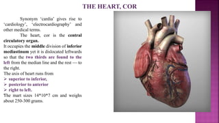
  • 1. THE HEART, COR Synonym ‘cardia’ gives rise to ‘cardiology’, ‘electrocardiography’ and other medical terms. The heart, cor is the central circulatory organ. It occupies the middle division of inferior mediastinum yet it is dislocated leftwards so that the two thirds are found to the left from the median line and the rest — to the right. The axis of heart runs from  superior to inferior,  posterior to anterior  right to left. The mart sizes 14*10*7 cm and weighs about 250-300 grams. -
  • 2. The parts, surfaces and borders of heart: The heart appears as flattened conic body. The external features distinguishable are the  Base  Apex • the base of heart, basis cordis is the upper wider portion of heart. It is directed superiorly, posteriorly and rightwards. The base attaches to large blood vessels; • the apex of heart, apex cordis is the lower narrowed and rounded portion directed inferiorly and leftwards. To the right from the apex one can distinguish a small notch of cardiac apex, incisura apicis cordis; • the anterior (sternocostal) surface, facies anterior (sternocostalis) slightly convex, it is directed anteriorly, superiorly and leftwards; • the diaphragmatic (inferior) surface, facies diaphragmatica (inferior)1 is directed inferiorly, posteriorly and rightwards; this surface adheres to the diaphragm; • the left and light pulmonary surfaces, facies pulmonalis dextra et sinistra face the respective lungs. • the posterior surface, facies vertebralis (posterior).
  • 3. • the right border, margo dexter a sharp border that delimits the sur- faces on the right; the border is directed inferiorly and rightwards; • the left border, margo sinister. • the inferior border, margo inferior.
  • 4. The sulci of heart The surface of heart features one transverse and two longitudinal sulci that delimit the cardiac chambers from outside. The sulci contain the blood vessels and fat tissue: • the coronary sulcus, sulcus coronarius is a deep circular groove that delimits the atriums and the ventricles. The anterior deeper portion of the groove is covered with the auricles; • the anterior interventricular sulcus, sulcus interventricularis anterior runs along the anterior surface of heart. The sulcus delimits the ventricles anteriorly; • the posterior interventricular sulcus, sulcus interventricularis posterior runs along the inferior surface of heart. It delimits the ventricles posteriorly. The sulci join at the notch of cardiac apex.
  • 5.
  • 6. The cardiac chambers The heart features four cham- bers — • two atriums (left and right) and • two ventricles (also left and right). The atriums are separated by the interatrial septum, septum interatriale and the ventricles — by the interventricular septum, septum in- terventriculare. The right atrium and the right ventricle constitute the right (venous) part of heart; the left atrium and the left ventricle constitute the left (arterial) part.
  • 7. The right atrium is a cavity of cubic shape with a well distinguishable evagination directed anteromedially — this is the right auricle, auricular dextra that neighbors the aorta on the right. The auricle is delimited from the venae cavae inlet by the sulcus terminalis cordis. The veins that run into the right atrium These veins are both venae cavae and the coronary sinus: • the superior vena cava, opens into the atrium with the opening (ostium) of superior vena cava; the opening is found between the superior and the inferior atrium walls; • the inferior vena cava, opens on the posterior wall with the opening of inferior vena cava; the opening neighbors the sulcus terminalis. A crescent-shaped fold that expands between the lower border of opening and the interatrial septum is the valve of inferior vena cava. The venous openings are delimited by a small tuberculum intervenosum. The area that accepts the venae cavae is called the sinus of venae cavae; • sinus coronarius is the venous collector that drains the cardiac walls. Its opening found in between the atrioventricular orifice and the opening of inferior vena cava is rimmed with a small fold — the valve of coronary sinus. The right atrium, atrium dextrum
  • 8. The internal surface of the right atrium has the features as follows: • the fossa ovalis is a central excavation of the interatrial septum (1.5-2 cm wide). The floor of the fossa is made up of thin endocardial duplication. The fossa is rimmed with the limbus fossae ovalis. In embryo, the area features the wide opening — the foramen ovale, that communicates the atriums. Shortly after delivery the foramen closes; • the musculi pectinati are the small muscles situated next to right auricle and the right wall of atrium. The rest of the internal surface of right atrium is smooth.
  • 9. The right ventricle features a cavity of irregular pyramidal shape with anterior, inferior and medial walls distinguishable. A cross-sectioned cavity resembles crescent. Superiorly, the ventricular cavity communicates with the right atrium via wide ostium atrioventriculare dexter; anteriorly, the ventricle is continuous with the pulmonary trunk. The tricuspid valve (the right atrioventricular valve), resides within the right atrioventricular orifice. Its name arises from the number of cusps featured. The valve features the anterior, the posterior and the septal cusps (the valve cusps are irregular in number): • the anterior cusp, cuspis anterior resides anteriorly; • the posterior cusp, cuspis posterior resides posteriorly • the septal cusp, cuspis septalis is the smallest medial cusp. The cusps fix firmly to the right fibrous ring that encircles the orifice. The chordae tendineae arise from the papillary muscles and the ventricular walls (the latter are the false chordae tendineae) and attach to free margins of cusps. The right ventricle, ventriculus dexter
  • 10. The papillary muscles, are the conic muscular projections on the internal surface of ventricle. The right ventricle features the muscles as follows: • the anterior papillary muscle, the greatest muscle of the group. It attaches to the anterior and the posterior valve cusps (by means of chordae tendineae); • the posterior papillary muscle, musculus papillaris posterior some-what smaller, it attaches to the anterior and the posterior cusps; • the septal papillary muscle, appears as a group of small muscular projections that attach to the septal and the anterior cusps. The internal surface of the ventricle features numerous trabeculae carneae that form the networks. The conus arteriosus is the antero-superior portion of the ventricle with smooth walls. The conus narrows and becomes continuous with the pulmonary trunk. Its opening — the opening of pulmonary trunk, features the valve. The right ventricle, ventriculus dexter
  • 11. The pulmonary valve situated within the opening of pulmonary trunk features three cusps; they appear as three small pouches: • valvula semilunaris anterior; • valvula semilunaris sinistra; • valvula semilunaris dextra; The spaces bounded by the cusps and the trunk wall are the sinuses of pulmonary trunk. Each free margin of the cusp features small nodules of semilunar cusp, for better cusp contact.
  • 12.
  • 13. The left atrium, atrium sinistrum The space of left atrium is of irregular cubic shape. The anterior wall projection is the auricula sisnistra. The auricle neighbors the pulmonary trunk. The internal surface of auricle features well visible ridges formed of musculi pectinati. The rest of internal surface is smooth. The interatrial septum features the thinner area related to the fossa ovalis.
  • 14. The pulmonary veins The left atrium accepts four pulmonary veins (two from each lung) that carry oxygenated blood: • vena pulmonalis superior dextra: • vena pulmonalis inferior dextra; • vena pulmonalis superior sinistra; • vena pulmonalis inferior sinistra; The openings of pulmonary veins, ostia venarum pulmonalium reside on the posterior wall of atrium; the veins lack valves. Sometimes any pair of veins merges to open into the atrium with an unpaired trunk. In this case, the atrium features fewer openings.
  • 15. The left ventricle, ventriculus sinister The left ventricle features a cone-shaped cavity. Cross-sectioned cavity is of oval shape. The upper portion of the ventricle features two openings — the ostium atrioventriculare sinistrum and the ostium aortae (found superiorly and to the right from the left atrioventricular orifice). The mitral (left) atrioventricular valve (valva mitralis) occupies the respective opening. Because of two cusps present, the valve sometimes is called the bicuspid valve.The mitral valve features the anterior and the posterior cusps: • the anterior cusp, resides anteriorly and on the right; • the posterior cusp, somewhat smaller, it is found posteriorly and on the left; The cusps fix firmly to the left fibrous ring that encircles the orifice. The chordae tendineae given by the papillary muscles attach to the free margins of cusps.
  • 16. The papillary muscles The left ventricle features two greater papillary muscles — the anterior and the posterior: • musculus papillaris anterior resides on the anterior ventricular wall; • musculus papillaris posterior resides on the posterior ventricular wall; The muscles attach to both cusps by means of chordae tendineae. The internal surface of the ventricle features numerous trabeculae carneae that run in all directions and give a reticular look to the inner ventricular surface.
  • 17. The left ventricle, ventriculus sinister The aortic valve: The ventricular cavity becomes continuous with the aorta; the aortic orifice, ostium aortae features the aortic valve, valva aortae. The valve comprises three semilunar cusps : • valvula semilunaris posterior (valvula non coronaria); • valvula semilunaris (coronaria) sinistra; • valvula semilunaris (coronaria) dextra. The spaces bounded by the cusps and the aortic wall are the aortic sinuses, sinus aortae. The right and the left sinuses house the openings of the respective coronary arteries. The free margins of cusps feature nodules of semilunar cusp. Laterally from the nodules, one can distinguish thickened areas called the lunulae valvularum semilunarium.
  • 18. Clinical applications The heart valves often undergo pathological alterations. The damaged cusps may fuse or become dense and inflexible. The effected valves produce two common states — valve insufficiency and stenosis. Insufficiency constitutes incomplete closing of valve cusps followed by blood regurgitation (backflow). Stenosis (narrowing) stands for failure of valve to open completely, which slows the blood flow. Combined pathologies are also of frequent occurrence. The mitral valve is most frequently affected (two thirds of acquired pathologies); the aortic valve occupies the second place in the list. Nowadays, the pathologies are successfully treated by replacement of the valves affected with the artificial valve prostheses (the xeno - grafted valves are also in use).
  • 19. The interventricular septum (IVS), (or ventricular septum, or during development septum inferius), is the stout wall separating the lower chambers (the ventricles) of the heart from one another. The ventricular septum is directed obliquely backward to the right, and curved with the convexity toward the right ventricle; its margins correspond with the anterior and posterior longitudinal sulci. Portions: • The greater portion of it is thick and muscular and constitutes the muscular interventricular septum. • Its upper and posterior part, which separates the aortic vestibule from the lower part of the right atrium and upper part of the right ventricle, is thin and fibrous, and is termed the membranous ventricular septum (septum membranaceum).. The interventricular septum
  • 20. The wall layers The cardiac wall consists of three layers — the epicardium, the myocardium and the endocardium: • the epicardium is the external layer represented with visceral plate of cardiac serous coating (the pericardium), immediately below it, one can see fat tissue best developed in the sulci; • the myocardium is the thickest middle layer of heart. The myocardium contains specific muscular bundles of the conductive system of heart; • the endocardium is the internal investment of cardiac chambers. The valves are the endocardial duplications with dense fibrous tissue featured. . WALL STRUCTURE OF HEART
  • 21. The fibrous rings, anuli fibrosi The muscular fibers and the valves attach to the fibrous rings that constitute a dense fibrous skeleton of heart. The heart features four fibrous rings: 1) anulus fibrosus sinister rims the left atrioventricular orifice; 2) anulus fibrosus dexter rims the right atrioventricular orifice; 3) the fibrous ring around the opening of pulmonary trunk; 4) the fibrous ring around the aortic opening. The left fibrous ring fuses with the aortic ring to give rise to two left and right trigonum fibrosum dexter et sinister situated posterior to the aorta. The rings and the trigones separate the atrial myocardium from the ventricular
  • 22. WALL STRUCTURE OF HEART The atrial myocardium is a thinner (2-3 mm) muscular layer. It arises from the fibrous rings and features two layers — •the superficial and •the deep. The superficial layer shared by atriums features transverse muscular fibers. In the deep layer, the fibers are longitudinal and circular. The longitudinal fibers prevail; they enfold each atrium separately and form sphincters around venous openings. The longitudinal fibers give rise to muscili pectinati. The ventricular myocardium is thicker than that of atriums. The left ventricle has the thickest muscular layer — about 1.5 cm. The right ventricle wall is somewhat thinner — about 0.7-0.8 cm.
  • 23. The ventricular myocardium comprises complex system of interlaced and looping fibers arranged in three layers: •the external (oblique) layer arises from anterior portion of each fibrous ring; it runs slantwise inferiorly and leftwards to reach the apex. On reaching the apex, the fibers loop to form the vortex of heart, vortex cordis and become continuous with the •internal longitudinal layer. The longitudinal layer gives rise to the trabeculae carneae and the papillary muscles. Both layers are shared by the ventricles; •the middle layer comprises transverse fibers that enfold each ventricle separately. This layer is the strongest one. Nowadays, the cardiac muscular fibers are believed to form the three dimensional network. Continuous changing of fibers direction provides more efficient force application.
  • 24.
  • 25. THE CONDUCTING SYSTEM OF HEART The conducting system of heart comprises specific muscular fibers (light, poor in myofilaments and reach in sarcoplasm) with featured automation i.e. with possibility of generation and transmission of rhythmic impulses to the working myocardium. The system ensures successive contractions (systole) of the atriums and the ventricles. The conducting system features nodes and bundles (the latter conduct the impulses) . • The sinu-atrial node, (node of Keith-Flack) resides within the right atrial wall immediately below the epicardium between the superior vena cava and the right auricle. The node sizes 2X2 mm. the node accepts numerous nerve fibers. The node generates impulses at rate of 60-80 bpm (resting rate); the impulses expand along the bundles to reach the atrial myocardium and the atrioventricular node. The node is the principal pacemaker (all components of conductive system have pacemaking properties).
  • 26. The atrioventricular node, (node of Aschoff- Tawara) resides right below the endocardium within the upper portion of the interventricular septum on the right. It sizes 5 X 3 mm. The atrioventricular node is associated with the sinu- atrial node. The atrioventricular node gives rise to the atrioventricular bundle.
  • 27. THE CONDUCTING SYSTEM OF HEART The atrioventricular bundle, fasciculus atrioventricularis (the bundle of His) arises from the atrioventricular node, traverses the right fibrous triangle and becomes evident within the interventricular septum as 1 cm long trunk. Within the interventricular septum, the bundle splits into the left and the right bundles, crus dextrum et crus sinistrum. The crura descend below the endocardium on each side of the interventricular septum. The crura split into numerous subendocardial branches, rami subendocardiales (the fibers of Purkinje) that penetrate the trabeculae carneae, the papillary muscles and working myocardium. The atrioventricular bundle conducts the impulses from the atriums to the ventricular myocardium.
  • 28. Clinical applications Impulses generated by the conducting system of heart can easily be registered by means of ECG. As far as many cardiac diseases are complicated with conductivity disorders, ECG is a very important diagnostic procedure. Myocardial infarction of the interventricular septum (where the atrioventricular bundle passes) is quite dangerous because it causes conductivity block.