
Recommended
More Related Content
What's hot
What's hot (20)
Similar to Heart anatomy
Similar to Heart anatomy (20)
More from RobbinsHobbin
Recently uploaded
Recently uploaded (20)
Heart anatomy
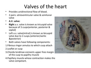
- 1. Valves of the heart • Provides unidirectional flow of blood. • 2 pairs: atrioventricular valve & semilunar valve • A.V. valve: • Right a.v. valve is known as tricuspid valve because of 3 cusps(anterior, posterior & septal) • Left a.v. valve(mitral) is known as bicuspid valve due to 2 cusps.(anterior/aortic &posterior) • Both valves have following components: 1.Fibrous ringor annulus to which cusp attach 2.Leaflet or cusp 3.Chorda tendinae-connects upper free margin of the cusp to papillry muscle 4.Papillary muscle-whose contraction makes the valve competant.
- 2. Semilunar valve • Aortic & pulmonary valve are semilunar valves-cusps are semilunar in shape. • Each valve has 3 cusps which are attached to the fibrous scallop of the orifice by convex margin. • Free margin shows fibrous nodule (nodule of Arantius ) at the centre with 2 thin lunule at the side. • Opposite the cusp, vessel walls are dilated to form aortic & pulmonary sinuses. • Applied anatomy: stenosis, valvar incompetence leading to regurgitation (murmur)
- 3. Skeleton of the heart • It consists of 4 fibrous rings around A.v. orifice, pulmonary & aortic orifices • Pulmonary & aortic rings are • connected by fibrous septum called tendon of infundibulum. • Right & left A.V. rings are united by trigonum fibrosum dextrum. • Aortic & left A.V. rings are connected by trigonum fibrosum sinistrum. • These interconnected fibrous rings provide attachment to cardiac muscle & supports the valve • It disturbs the continuity between atrial & ventricular muscle. So impulses are propagated through conductng system of the heart
- 4. Conducting system of the Heart • They are the specialized myocytes for initiation & conduction of cardiac impulses. It includes: • Sinuatrial node(S.A node) • Atrioventricular Bundle & its 2 limbs • Subendocardial plexus of purkinje fibers. • Heart beat is myogenic.
- 5. Conducting system of the Heart
- 6. Blood supply of the heart • Heart is supplied by 2 coronary arteries- rt.<. arising from ascending aorta. • coronary is derived from the word `crown’which encircles the base of ventricle as a crown • Each coronary artery is vasvasorum • Right coronary artery: • Arises from ant. Aortic sinus & passes forward to the right betn pulmonary trunk & rt auricle. • Then runs in the rt. Coronary sulcus to the junction of rt. & inferior border of the heart. • It winds around the inferior border to reach diaphragmatic surface of the heart. Here it runs backward & to the left to reach posterior iv groove & terminates anastomosing with left coronary artery.
- 7. Blood supply of the heart
- 8. Contd.. • Left coronary artery: Larger than right coronary artery. • Arises from left posterior aortic sinus . • Pecularities: • Sometimes left coronary artery may arise from pulmonary trunk. • Sympathetic stimulation constricts the epicardial artery & dilate intramuscular arteries. • It is the only vessel where blood flows in diastole. • Cardiac & extracardiac anastomosis • 85% of Inter- atrial anastomosis takes place in precapillary level &15% in capillary level. So they are not typical end arteries but functionally behave like end arteries.
- 9. Inter-atrial anastomosis • Sites of anastomosis are: • Kugel’s artery: artterial branch of both coronary arteries communicates at ant. atrial wall. • Anastomosis between anterior & posterior interventricular artery near to the apex. • Annulus of Vieussens- anastomosis betn rt. & left conal artery around the infundibulum. • In the intervenricular septum • Near the crux of the heart • Coronary perdominance: • Most case right coronary predominance where posterior interventricular artery is derived from right coronary artery(70%). • Minority of population has left coronary predominance-from left coronary artery. • In balanced pattern branches of both run in or near the sulcus.
- 10. Venous drainage of the heart • Great cardiac vein • Middle cardiac vein • Small cardiac vein • Oblique vein of left atrium(of Marshal) • Left marginal vein • Anterior cardiac vein • Venae cordis minimae(Thebesian vein) • All veins excep last two drain into coronary sinus which open into rt. Atrium.
- 11. Contd..
- 12. Nerve supply of the heart • Derived from cardiac plexus formed by sympathetic (T1-T4/T5) & parasympathetic(vagus) nerves. • Cardiac plexus suprficial-below the arch of aorta deep-infront of bifurcation of trachea
- 13. Applied anatomy • Common cause of death in developed countries-ischaemic heart disease • Sudden obstruction of major branch of coronary vessels lesd to myocrdial infarction. • Common branches for occlusion are: • Anterior interventricular artery • Rt. coronary artery • Circuflex branch of left coronary artery