Downloaded 106 times
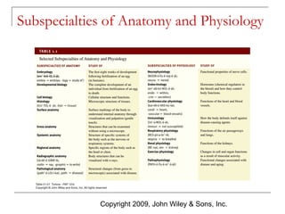
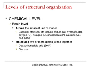
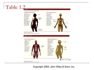
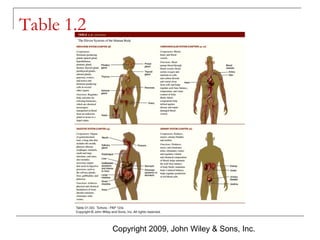
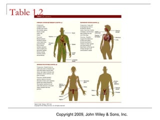
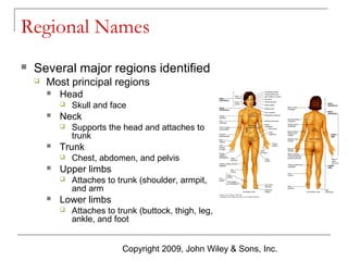
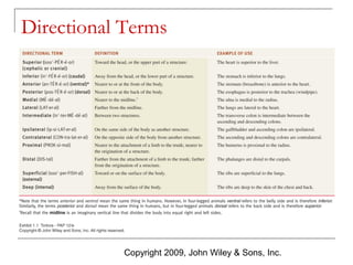
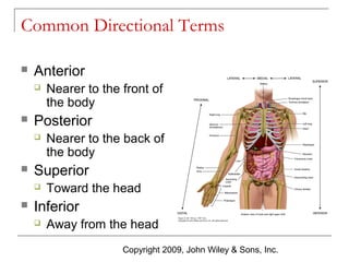
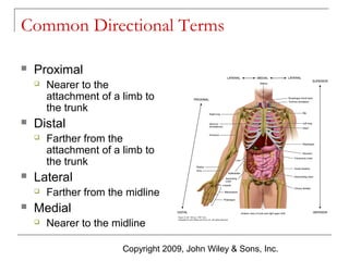
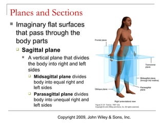
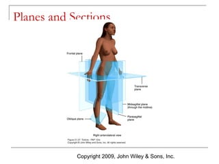

The document provides an overview of human anatomy and physiology. It defines anatomy and physiology as the study of body structures and functions. It describes the six levels of structural organization from chemical to organismal. It also discusses homeostasis and how feedback systems help regulate the internal environment. Key concepts covered include anatomical position and directional terms used to describe body structures in relation to one another.


![08 [chapter 8 the skeletal system appendicular skeleton]](https://cdn.slidesharecdn.com/ss_thumbnails/08chapter8theskeletalsystem-appendicularskeleton-170828041008-thumbnail.jpg?width=640&height=640&fit=bounds)


![28 [chapter 28 the reproductive system]](https://cdn.slidesharecdn.com/ss_thumbnails/28chapter28thereproductivesystem-170828134050-thumbnail.jpg?width=640&height=640&fit=bounds)

![22 [chapter 22 the lymphatic system and immunity]](https://cdn.slidesharecdn.com/ss_thumbnails/22chapter22thelymphaticsystemandimmunity-170828153258-thumbnail.jpg?width=640&height=640&fit=bounds)

![20 [chapter 20 the cardiovascular system the heart]](https://cdn.slidesharecdn.com/ss_thumbnails/20chapter20thecardiovascularsystem-theheart-170828133506-thumbnail.jpg?width=640&height=640&fit=bounds)


![PERI-PROSTHETIC FRACTURE NAIL-PLATE CONSTRUCT [NPC].pptx](https://cdn.slidesharecdn.com/ss_thumbnails/drarunkumardrmohamedashrafperiprostheticfrasturenail-plateconstructnpc-260209164459-7e9d15a1-thumbnail.jpg?width=640&height=640&fit=bounds)


![ONFH[AVN HIP] -TRIPLE REGIME -A NOVAL SURGICAL CONCEPT .pptx](https://cdn.slidesharecdn.com/ss_thumbnails/onfhavnhip2026koaconcalicutdrgokuldevdrmashraf-260210064517-213ec005-thumbnail.jpg?width=640&height=640&fit=bounds)


