Case record...Hypoparathyroidism
•
9 likes•3,053 views

Case record...Hypoparathyroidism

Recommended
More Related Content
What's hot
What's hot (20)
Viewers also liked
Viewers also liked (20)
Similar to Case record...Hypoparathyroidism
Similar to Case record...Hypoparathyroidism (20)
More from Professor Yasser Metwally
More from Professor Yasser Metwally (20)
Recently uploaded
Recently uploaded (20)
Case record...Hypoparathyroidism
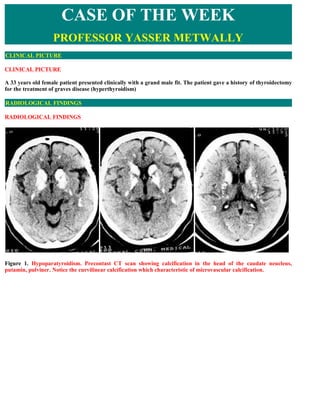
- 1. CASE OF THE WEEK PROFESSOR YASSER METWALLY CLINICAL PICTURE CLINICAL PICTURE A 33 years old female patient presented clinically with a grand male fit. The patient gave a history of thyroidectomy for the treatment of graves disease (hyperthyroidism) RADIOLOGICAL FINDINGS RADIOLOGICAL FINDINGS Figure 1. Hypoparatyroidism. Precontast CT scan showing calcification in the head of the caudate neucleus, putamin, pulviner. Notice the curvilinear calcification which characteristic of microvascular calcification.
- 2. Figure 2. Hypoparathyroidism. Notice the curvilinear, periventricular calcification which is characteristic of microvascular calcification. Figure 3. Hypoparathyroidism. Precontrast CT scan showing calcification in the dentate nucleus. Figure 4. Precontrast CT scan showing calcification in the caudate nucleus, putamen, thalamus and choroid plexus (left image), the middle image shows cerebellar calcification, right image is a bone window of figure (4a)
- 3. Figure 5. CT scan precontrast showing calcification of the head of the caudate nucleus, notice the curvilinear calcification radiating from the frontal horn of the lateral ventricle, which characteristic of microvascular calcification Figure 6. CT scan bone window showing the perivascular curvilinear calcification that characterizes hypoparathyroidism DIAGNOSIS: DIAGNOSIS: BRAIN CALCIFICATION SECONDARY TO HYPOPARATHYROIDISM DISCUSSION DISCUSSION The disturbance in the calcium and phosphorus metabolism that results from deficiency in the activity of the parathyroid gland produces a train of neurological problems that includes epilepsy, tetany, mental confusion, dementia, chorea, athetosis, torticollis, papilledema and others. Biochemically patients with hypoparathyroidism have reduced serum calcium level and elevated serum phosphorus level with inability to mobilize calcium from tissues in general and bone in particular. Background: Hypoparathyroidism is a condition of parathyroid hormone (PTH) deficiency. Primary hypoparathyroidism is a state of inadequate PTH activity. In the absence of adequate PTH activity, the ionized calcium concentration in the extracellular fluid falls below the reference range. Primary hypoparathyroidism, the subject of this article, is a syndrome resulting from many rare diseases. Secondary hypoparathyroidism is a physiologic state in which PTH levels are low in response to a primary process, which causes hypercalcemia. Pathophysiology: The ionized calcium concentration in the extracellular fluid (ECF) remains nearly constant at a level of approximately 1 mm. Ionized calcium in the ECF is in equilibrium with ionized calcium in storage pools such as bone, proteins in the circulation, and within the intracellular fluid. The intracellular fluid concentration of calcium is more than 10,000-fold lower than in the ECF. The maintenance of ionized calcium concentrations in both the intracellular and extracellular fluids is highly regulated and modulates the functions of bone, renal tubular cells, clotting factors, adhesion molecules, excitable tissues, and a myriad of intracellular processes. An extracellular calcium-sensing receptor has been isolated from parathyroid, kidney, and brain cells. The extracellular calcium-sensing receptor is G protein coupled. Mutations in the extracellular calcium-sensing receptor have been demonstrated to result in hypercalcemic or hypocalcemic states. Normally, the extracellular calcium-
- 4. sensing receptor is extremely sensitive and responds to changes in the ECF calcium ion concentration as small as 2%. In parathyroid cells, the extracellular calcium-sensing receptor regulates the secretion of PTH. Inactivating mutations of the extracellular calcium-sensing receptor lead to hypercalcemia, as observed in familial hypocalciuric hypercalcemia (heterozygous mutation) and neonatal severe hyperparathyroidism (homozygous mutation). Conversely, activating mutations of the extracellular calcium-sensing receptor lead to hypocalcemia, as observed in some families with autosomal-dominant hypocalcemia. The intracellular mechanism(s) whereby activation of the extracellular calcium-sensing receptor leads to inhibition of PTH exocytosis is unknown. Since pertussis toxin blocks the inhibition of cyclic adenosine monophosphate (cAMP), but not PTH, in response to a high ECF ionized calcium concentration, cAMP likely is not an important second messenger for the extracellular calcium-sensing receptor. Candidate second messengers include protein kinase C, phospholipase A2, and intracellular calcium. Conversely, a fall in ECF ionized calcium concentration leads to exocytosis of PTH. PTH has the overall effect of returning the ECF ionized calcium concentration to normal by its effects on the kidneys and the skeleton. PTH activates osteoclasts. Osteoclast activation results in bone resorption and a release of ionized calcium into the ECF. Recent evidence suggests that small pulse doses of PTH activate osteoblasts, with ensuing bone deposition. The effect of PTH on osteoclasts seems more important than the effect on osteoblasts. PTH inhibits the proximal tubular transport of phosphate from the lumen to the interstitium. In conditions of primary PTH excess, hypophosphatemia tends to occur. Conversely, in hypoparathyroidism, the phosphate concentration in the plasma is normal or slightly elevated. PTH has a calcium-retaining effect on the distal tubule. The PTH-mediated calcium reabsorption is independent of any effects on sodium or water reabsorption. This effect of PTH is important in hypoparathyroidism because, in the absence of this distal tubular calcium reabsorption, the kidneys waste calcium. This depletes the ECF ionized calcium and increases the urinary calcium concentration. PTH stimulates renal 1-alpha-hydroxylase, the enzyme that synthesizes formation of 1,25-dihydroxy vitamin D; 1,25-dihydroxy vitamin D allows for better dietary calcium absorption. Thus, 1,25-dihydroxy vitamin D has a synergistic effect with PTH; both contribute to a rise in the ECF ionized calcium concentration. In the absence of PTH, bone resorption, phosphaturic effect, renal distal tubular calcium reabsorption, and 1,25- dihydroxy vitamin D–mediated dietary calcium absorption cannot occur. Therefore, the consequence of PTH deficiency is hypocalcemia. Calcium homeostasis and its regulation Calcium homeostasis is regulated by the effects of PTH and 1,25-(OH)2D3 on gut, kidney and bone. Calcium-sensing receptors are present in the parathyroid gland, kidney, brain and other organs. Calcium absorption and distribution Daily calcium consumption, primarily from dairy foods, should ideally be around 20-25 mmol (800-1000 mg). Dietary calcium deficiency is rarely a significant cause of bone disease because proportional absorption can increase in response to low intake. Absorption is reduced by vitamin D deficiency and sometimes by generalized malabsorption.
- 5. Figure 7. Calcium exchange in the normal human. The fluxes are shown in mmol per day. Vitamin D metabolism The primary source of Vitamin D in humans is photoactivation (in the skin) of 7-dehydrocholesterol to cholecalciferol, converted in the liver to 25-hydroxycholecalciferol (25-(OH)D3) and further converted by the kidney tubule enzyme, 1α-hydroxylase, to the active metabolite 1,25-dihydroxycholecalciferol (1,25-(OH)2D3). (This step can occur in lymphomatous and sarcoid tissue, resulting in the hypercalcaemia that may complicate these diseases.) A less active metabolite, 24,25-(OH)2D3, is formed if vitamin D supplies are adequate. Regulation of this enzyme is by PTH, phosphate and by feedback inhibition by 1,25-(OH)2D3. Figure 8. The metabolism and actions of vitamin D. PTH, parathyroid hormone.
- 6. Parathyroid hormone (PTH) PTH, an 84-amino-acid hormone, is secreted from the chief cells of the parathyroid glands (normally situated posterior to the thyroid, but occasionally elsewhere in the neck or mediastinum). PTH increases renal phosphate excretion, and increases plasma calcium by: increasing osteoclastic resorption of bone (occurring rapidly) increasing intestinal absorption of calcium (a slow response) increasing synthesis of 1,25-(OH)2D3 increasing renal tubular reabsorption of calcium. Hypomagnesaemia can suppress the normal PTH response to hypocalcaemia. Thyroid hormones Calcitonin is produced by thyroid C cells. Total thyroidectomy (absent calcitonin) or medullary carcinoma of the thyroid (excess calcitonin) have no significant skeletal effects. Plasma calcitonin levels do, however, rise with increasing serum calcium, and calcitonin inhibits osteoclastic bone resorption and increases the renal excretion of calcium and phosphate. It may be used in treating osteoporosis or hypercalcaemia. Excess thyroxine (T4) and triiodothyronine (T 3) cause increased bone turnover, hypercalcaemia and bone loss, while hypothyroidism leads to growth delay. CLINICAL History: Hypoparathyroidism results in hypocalcemia, which may be variably symptomatic. The history should focus on eliciting signs and symptoms of neuromuscular irritability including the following: Paresthesias (involving fingertips, toes, perioral area) Hyperirritability Fatigue Anxiety Mood swings and/or personality disturbances Seizures (especially in patients with epilepsy) Hoarseness (due to laryngospasm) Wheezing and dyspnea (due to bronchospasm) Muscle cramps, diaphoresis, and biliary colic Hypomagnesemia, hypokalemia, and alkalosis (eg, hyperventilation), which worsen signs and symptoms of hypocalcemia Physical: Muscle cramps involving the lower back, legs, and feet are common in patients with hypoparathyroidism and hypocalcemia. Tetany develops if hypocalcemia is severe. In some patients, laryngospasm and bronchospasm may be life threatening. Increased neuromuscular irritability from hypoparathyroidism-induced hypocalcemia may be demonstrated
- 7. at the bedside by eliciting the following signs: Chvostek sign: Facial twitching, especially about the mouth, is brought on by gently tapping the ipsilateral facial nerve as it courses just anterior to the ear. Trousseau sign: Carpal spasm is brought on by inflating a blood pressure cuff around the arm to a pressure 20 mm Hg above obliteration of the radial pulse for 3-5 minutes. Hypocalcemia of primary hypoparathyroidism may cause extrapyramidal choreoathetoid syndromes in patients with basal ganglia calcifications. Parkinsonism, dystonia, hemiballismus, and oculogyric crises may occur in approximately 5% of patients with idiopathic hypoparathyroidism. Spastic paraplegia, ataxia, dysphagia, and dysarthria have been documented in association with hypoparathyroidism-induced hypocalcemia. Severe hypocalcemia causes papilledema, which improves with treatment of the calcium derangement. Emotional instability, anxiety, depression, confusion, hallucinations, and psychosis have been described in patients with hypoparathyroidism when the calcium level is low. Normocalcemia corrects these conditions. Chronic hypocalcemia, as observed in primary hypoparathyroidism, also is associated with ocular cataracts, abnormal dentition, and dry, puffy, coarse skin. In severe hypocalcemia, a prolongation of the QT interval is observed on ECG, and congestive heart failure may develop. Correction of hypocalcemia reverses cardiac effects of hypoparathyroidism. In patients with autoimmune polyglandular syndrome, idiopathic hypoparathyroidism is associated with adrenal insufficiency and moniliasis. Moniliasis may affect the skin, nails, oral cavity, and vaginal cavity. It is frequently intractable. The underlying etiology is likely a defect in cellular immunity. Some authors advocate the term HAM syndrome, indicating hypoparathyroidism, Addison disease, and moniliasis (HAM), to denote these patients. Causes: Most people have 4 parathyroid glands, which makes primary hypoparathyroidism uncommon. Hypocalcemia from hypoparathyroidism requires all 4 parathyroid glands to be affected. Primary hypoparathyroidism may be permanent or reversible. Permanent primary hypoparathyroidism may be congenital or acquired. Iatrogenic The most common cause of primary hypoparathyroidism is excision of all parathyroid glands via surgery in the treatment of thyroid, laryngeal, or other neck malignancy. Patients with parathyroid hyperplasia, as observed in the multiple endocrine neoplasia (MEN) syndromes, are treated by surgical removal of the parathyroid glands. Attempts at restoring normal PTH levels and normocalcemia by autotransplantation of a fraction of one of the parathyroid glands sometimes are effective, but many patients become hypoparathyroid. Repeated neck explorations for primary hyperparathyroidism due to parathyroid adenoma also may cause hypoparathyroidism. Extensive irradiation to the face, neck, or mediastinum may cause destruction of all 4 parathyroid glands with ensuing primary hypoparathyroidism and hypocalcemia. The quot;hungry bone syndromequot; develops after a parathyroidectomy for hyperparathyroidism. The body has been accustomed to high levels of PTH, causing hypercalcemia. Much of this hypercalcemic effect is due to resorption of bone. When the parathyroid gland or glands responsible for the hypersecretion of PTH are removed, the PTH level in the blood drops suddenly, and the patient experiences transient hypoparathyroidism. The bone, which has been quot;starvedquot; of calcium, avidly holds onto it under the influence of osteoblasts. Without PTH and with bone now using calcium to remineralize, the ECF ionized calcium level falls. Postoperatively, patients require aggressive treatment with calcium for several hours to several days. Eventually, the hypoparathyroid state resolves, and calcium homeostasis is
- 8. re-achieved. Autoimmune Type 1 autoimmune polyglandular syndrome (also referred to as HAM syndrome) includes primary hypoparathyroidism due to destruction of the parathyroid glands. On average, these patients develop primary hypoparathyroidism when aged 10 years. Autoimmune hypoparathyroidism may exist alone or in sporadic or familial forms. For patients with autoimmune primary hypoparathyroidism, the average age for development of hypocalcemia is when aged 7 years, with a range of 6 months to 20 years. Congenital Numerous conditions are described in the literature that result in congenital agenesis or hypoplasia and, therefore, can produce primary hypoparathyroidism with symptomatic hypocalcemia at birth or in the newborn period. These conditions, which are adapted from Goltzman and Cole, are as follows: Isolated primary hypoparathyroidism X-linked primary hypoparathyroidism (band Xq26-Xq27) X autosomal-recessive primary hypoparathyroidism Branchial dysgenesis (DiGeorge syndrome) Chromosomal defects dup (1q),del(5p),dup(8q),del(10q),del (22q) Monogenic Isolated autosomal-dominant conditions Isolated autosomal-recessive conditions Velocardiofacial (Shprintzen) syndrome (quot;CATCH 22quot; [for cardiac, abnormal facies, thymic aplasia, cleft palate, and hypocalcemia with 22q deletion] is a mnemonic for the features of this syndrome.) Zellweger syndrome Teratogenic effects Diabetic embryopathy Fetal alcohol syndrome Retinoid embryopathy Associational arhinencephalia/DiGeorge and coloboma, heart disease, choanal atresia, retarded growth and development, genital anomalies, ear anomalies (CHARGE)/DiGeorge syndromes Cardiofacial/DiGeorge/Kenny-Caffey syndrome (absent parathyroid tissue, growth retardation, medullary stenosis of tubular bones) Kearns-Sayre syndrome (mitochondrial myopathy, ophthalmoplegia, retinal degeneration, cardiac conduction defects, primary hypoparathyroidism) Barakat syndrome (primary hypoparathyroidism, nerve deafness, and steroid-resistant nephrosis)
- 9. Hypoparathyroidism with short stature, mental retardation, and seizures In addition to the above list, several other genetic defects cause primary hypoparathyroidism. As opposed to the above list, no agenesis or hypoplasia of the parathyroid glands occurs. These mutations are functional, not anatomic, and are listed below as follows: Mutation of chromosome 3q has been demonstrated to cause primary hypoparathyroidism in several kindreds due to activation of the parathyroid extracellular calcium-sensing receptor. These patients have mild-to-moderate hypocalcemia urinary calcium excretion that is high relative to serum calcium (presumably the extracellular calcium-sensing receptor in the kidney contributes to this) and normal (but inappropriately low) serum PTH concentration. Familial isolated hypoparathyroidism is a heterogenous mix of disorders as follows: autosomal dominant, abnormal prepro-PTH allele (C-to-T substitution in codon 18 of the prepeptide encoding region does not allow for cleavage to pro-PTH), autosomal recessive, and abnormal prepro-PTH allele (C-to-G substitution in the first nucleotide position of prepro-PTH intron 2). Metal overload - Ion deficiency Hemochromatosis and thalassemia, both of which are associated with iron overload, may result in primary hypoparathyroidism. Wilson disease, with copper overload, also may cause primary hypoparathyroidism. Hypermagnesemia has been demonstrated to decrease PTH release. Correction of hypermagnesemia leads to correction of the primary hypoparathyroidism. Aluminum deposition within the parathyroid glands may cause primary hypoparathyroidism in patients with end-stage renal disease who are on hemodialysis. Hypomagnesemia causes reversible functional primary hypoparathyroidism. Infiltration of the parathyroid glands In addition to hemochromatosis and Wilson disease, parathyroid gland destruction has been reported as a result of metastatic disease, granulomatous disease, amyloidosis, syphilis, and progressive systemic sclerosis. Of note, clinically significant hypocalcemia is not always apparent in these patients. Neonatal The unborn baby of a mother with hypercalcemia has chronic suppression of parathyroid gland function. In the worst of circumstances, the parathyroid glands may become atrophic. At birth, the maternal calcium excess is gone, and newborns are at risk of hypocalcemia due to primary hypoparathyroidism. Clinically significant hypocalcemia may develop within the first 3 weeks of life but may occur as late as 1 year after birth. The primary hypoparathyroidism in these patients is self-limited. NEUROIMAGING OF HYPOPARATHYROIDISM CT scan of the brain in cases of hypoparathyroidism commonly reveals evidence of intracranial calcification that usually begins in the head of the caudate nucleus then extends to involve the rest of the basal ganglia. The pulviner and the cerebellar dentate nucleus are occasionally involved. The choroid plexus is also very frequently calcified. In advanced cases calcification might not be restricted to the subcortical gray matter and might involve the periventricular white matter.
- 10. Calcification is hyperdense on noncontrast CT scan and appears as signal void structures on both the T1,T2 MRI images. Figure 9. MRI T2 images showing brain calcification in the caudate nucleus, putamen, dentate nucleus in case of hypoparathyroidism. Calcification is signal void and appears hypointense on the T2 images Pathologically calcifications commonly develop in and around the brain microvascular bed that supplies the basal ganglia and other subcortical gray matter and commonly take the form of pericapillary calcospherites, presumably as a result of of some abnormalities of the interstitial fluids that nourish the microvascular bed. Although the serum calcium is low, however the existence of an excess of ionic calcium in the interstitial fluid explains the development of brain calcification in hypoparathyroidism. The mechanism of intracranial calcification in hypoparathyroidism, more often seen in pseudohypoparathyroidism than in idiopathic hypoparathyroidism, has not been completely elucidated. It may be related more to the duration of hypocalcaemia and hyperphosphataemia than parathyroid hormone itself. Hyperphosphataemia promotes ectopic calcification in brain tissue in hypoparathyroidism. DIFFERENTIAL DIAGNOSIS Punctate to conglomerate densities that are symmetric. May be a normal variant or a manifestation of such conditions as hypoparathyroidism, pseudohypoparathyroidism, infections, birth anoxia, carbon monoxide poisoning, and Cockayne's syndrome (a rare form of truncal dawarfism with retinal atrophy). Fahr syndrome TREATMENT Medical Care: PTH is not commercially available, but stage III clinical trials are underway for its use in osteoporosis. It soon may be available for patients with hypoparathyroidism. Currently, treatment of patients with hypoparathyroidism involves correcting the hypocalcemia by ingestion of calcium and vitamin D. Surgical Care: Patients undergoing parathyroidectomy for parathyroid hyperplasia are at high risk of developing permanent primary hypoparathyroidism. Patients may be treated with an autotransplant of a segment of parathyroid gland to prevent hypoparathyroidism. This autotransplant usually is placed subcutaneously in the forearm or in the neck. If the autotransplantation fails, patients are treated as any other patient with hypoparathyroidism. Diet: A diet rich in calcium content (ie, emphasizing dairy products) is recommended for patients with primary hypoparathyroidism.
- 11. Activity: Patients with symptomatic hypocalcemia develop tetany. Otherwise, no restriction in activity for these patients is necessary. MEDICATION Calcium and vitamin D are mainstays of treatment. Drug Category: Calcium salts - Without PTH, the ionized calcium levels in the plasma drop. Bone becomes an inefficient source of calcium for plasma, and kidneys waste calcium. Calcium helps maintain the ionized calcium level close to normal. Calcium carbonate (Tums Extra Strength, Cal-Lus, Caltrate, Osy-Cal 500)- Moderates nerve and muscle performance and facilitates normal cardiac function. Many commercially available preparations exist. Titrate total daily dose of Drug Name elemental calcium to minimize daily dose of vitamin D and to keep patient asymptomatic. Ionized calcium is absorbed best in an acidic environment. 400 mg elemental calcium equals 1 g calcium carbonate. 1-2 g/d elemental calcium Adult Dose 2.5-5 g/d calcium carbonate Pediatric Dose Administer as in adults Documented hypersensitivity; renal calculi; hypercalcemia; Contraindications hypophosphatemia; renal or cardiac disease; patients with digitalis toxicity May decrease effects of tetracyclines, atenolol, salicylates, iron salts, and fluoroquinolones; IV administration antagonizes Interactions effects of verapamil; large intakes of dietary fiber may decrease calcium absorption and levels Pregnancy C - Safety for use during pregnancy has not been established. Nephrocalcinosis and nephrolithiasis are potential complications of therapy; caution in patients who are Precautions digitalized and patients with respiratory failure or acidosis; in absence of PTH, may precipitate in urinary tract Calcium citrate (Citracal, Cal-Citrate 250)- Moderates nerve and muscle performance and facilitates normal cardiac Drug Name function; 210 mg of elemental calcium equals 1 g calcium citrate. 1-2 g/d elemental calcium Adult Dose 4.5-9 g/d calcium citrate Pediatric Dose Administer as in adults Documented hypersensitivity; renal calculi; Contraindications hypophosphatemia; hypercalcemia May decrease effects of tetracyclines, atenolol, salicylates, iron salts, and fluoroquinolones; IV administration antagonizes Interactions effects of verapamil; large intakes of dietary fiber may decrease calcium absorption and levels Pregnancy A - Safe in pregnancy Nephrocalcinosis and nephrolithiasis are potential complications of therapy; caution in patients who are Precautions digitalized and patients with respiratory failure or acidosis; may precipitate in urinary tract in absence of PTH; pregnancy category D if dosage exceeds RDA; adequate dietary calcium is needed for clinical response; maintain adequate fluid intake;
- 12. calcium-phosphate product (serum calcium times phosphorus) not to exceed 70; avoid use with renal function impairment and secondary hyperparathyroidism; avoid hypercalcemia Calcium gluconate (Kalcinate)- Moderates nerve and muscle performance and facilitates normal cardiac function. Available for IV use. Infuse slowly over 5-10 min; 10 mL calcium Drug Name gluconate contains approximately 90 mg elemental calcium; 1000 mg of calcium gluconate equals 90 mg elemental calcium. Adult Dose 90 mg IV over 5-10 min Pediatric Dose Administer as in adults Documented hypersensitivity; ventricular fibrillation during Contraindications cardiac resuscitation; digitalis toxicity; renal or cardiac disease; hypercalcemia; renal calculi; hypophosphatemia May decrease bioavailability of tetracyclines, fluoroquinolones, iron salts, salicylates atenolol, and sodium polystyrene sulfonate; IV calcium may antagonize verapamil Interactions effects; large intake of dietary fiber may decrease calcium absorption; IV calcium may increase quinidine and digitalis effects Pregnancy C - Safety for use during pregnancy has not been established. Avoid rapid IV administration; caution in patients who are digitalized and patients with severe hyperphosphatemia; patients with respiratory failure or acidosis; avoid Precautions extravasation; may produce cardiac arrest; hypercalcemia may occur in renal failure; monitor serum calcium during early dosing period; nephrocalcinosis and renal lithiasis are potential adverse effects of chronic renal calcium loss Drug Category: Vitamin D preparations - Vitamin D is synthesized by the kidneys, and the synthesis of 1,25- dihydroxy vitamin D is PTH dependent. In most patients with chronic hypoparathyroidism, treatment with the active vitamin D form is necessary. Ergocalciferol (Calciferol, Drisdol)- Stimulates absorption of Drug Name calcium and phosphate from small intestine and promotes release of calcium from bone into blood. Adult Dose 50,000-100,000 U/d Pediatric Dose Administer as in adults Documented hypersensitivity; hypercalcemia; malabsorption Contraindications syndrome Colestipol, mineral oil, and cholestyramine may decrease Interactions absorption from small intestine; thiazide diuretics may increase effects Pregnancy A - Safe in pregnancy Pregnancy category D if dosage exceeds RDA; caution in Precautions patients with impaired renal function, renal stones, heart disease, or arteriosclerosis Dihydrotachysterol (DHT, Hytakerol)- Synthetic analog of vitamin D. Stimulates calcium and phosphate absorption from Drug Name small intestine and promotes secretion of calcium from bone to blood. Promotes renal tubule resorption of phosphate. Adult Dose 125-250 mcg/d
- 13. Pediatric Dose Administer as in adults Contraindications Documented hypersensitivity; hypercalcemia Colestipol, mineral oil, and cholestyramine may decrease Interactions absorption from small intestine; thiazide diuretics may increase effects of vitamin D Pregnancy A - Safe in pregnancy Pregnancy category D if dosage exceeds RDA; caution in Precautions impaired renal function, renal stones, heart disease, or arteriosclerosis Calcifediol (Calderol)- Promotes absorption of calcium and phosphorus in small intestine. Promotes renal tubule resorption Drug Name of phosphate. Increases rate of accretion and resorption in bone minerals. Adult Dose 50-220 mcg/d PO Pediatric Dose Administer as in adults Contraindications Documented hypersensitivity; hypercalcemia Cholestyramine and colestipol decrease effects; thiazide Interactions diuretics increase effect Pregnancy C - Safety for use during pregnancy has not been established. Pregnancy category C per manufacturer, expert analysis category A; category D if dosage exceeds RDA; adequate dietary calcium needed for clinical response; maintain adequate Precautions fluid intake; calcium-phosphate product (serum calcium times phosphorus) not to exceed 70; avoid use with renal function impairment and secondary hyperparathyroidism; avoid hypercalcemia Calcitriol (Rocaltrol, Calcijex)- Promotes absorption of calcium in intestines and retention at kidneys to increase Drug Name calcium levels in serum. Decreases excessive serum phosphatase levels and parathyroid levels. Decreases bone resorption. Adult Dose 0.5-1 mcg/d PO Pediatric Dose Administer as in adults Documented hypersensitivity; hypercalcemia; vitamin D Contraindications toxicity; malabsorption syndrome Cholestyramine and colestipol decrease effects; thiazide Interactions diuretics increase effect; magnesium-containing antacids have additive effects Pregnancy C - Safety for use during pregnancy has not been established. Pregnancy category C per manufacturer, expert analysis category A; category D if dosage exceeds RDA; adequate dietary calcium is needed for clinical response; maintain Precautions adequate fluid intake; calcium-phosphate product (serum calcium times phosphorus) not to exceed 70; avoid use with renal function impairment and secondary hyperparathyroidism; avoid hypercalcemia
- 14. SUMMARY SUMMARY Hypoparathyroidism is a common cause of intracranial calcification. The calcification predominately involves the basal ganglia,, dentate nucleus, corona radiata. The clinical picture include epilepsy, parkinsonian manifestation, chorea, hemiballismus, dementia. Most people have 4 parathyroid glands, which makes primary hypoparathyroidism uncommon. Hypocalcemia from hypoparathyroidism requires all 4 parathyroid glands to be affected. Primary hypoparathyroidism may be permanent or reversible. Permanent primary hypoparathyroidism may be congenital or acquired. Calcium and vitamin D are mainstays of treatment. Addendum A new version of this PDF file (with a new case) is uploaded in my web site every week (every Saturday and remains available till Friday.) To download the current version follow the link quot;http://pdf.yassermetwally.com/case.pdfquot;. You can also download the current version from my web site at quot;http://yassermetwally.comquot;. To download the software version of the publication (crow.exe) follow the link: http://neurology.yassermetwally.com/crow.zip The case is also presented as a short case in PDF format, to download the short case follow the link: http://pdf.yassermetwally.com/short.pdf At the end of each year, all the publications are compiled on a single CD-ROM, please contact the author to know more details. Screen resolution is better set at 1024*768 pixel screen area for optimum display REFERENCES References 1. Agus ZS: Etiology of hypocalcemia. UpToDate CD-ROM 2000; 8 (1). 2. Brown EM, Harris HW, Vassilev PM: The biology of the extracellular Ca2+-sensing receptor. In: Bilezikian JP, ed. Principles of Bone Biology. San Diego: Academic Press; 1996: 243-262. 3. Fitzpatrick LA, Arnold A: Hypoparathyroidism. In: DeGroot LJ, ed. Endocrinology. Philadelphia: WB Saunders 1995: 1123-1135. 4. Goltzman D, Cole DEC: Hypoparathyroidism. In Favus MJ, ed. Primer on the Metabolic Bone Diseases and Disorders of Mineral 1996; 220-223. 5. Marx SJ: Causes of Hypocalcemia or Osteomalacia. A Review of Endocrinology Diagnosis and Treatment. NIH syllabus. Oct. 6-10, 1999; 506-513. 6. Marx SJ: Therapy of PTH or Calciferol Deficiency States. A Review of Endocrinology Diagnosis and Treatment. NIH syllabus. Oct. 6-10, 1999; 515-525. 7. Thakker RV: Molecular basis of PTH underexpression. In: Bilezikian JP et al, eds. Principles of Bone Biology. San Diego: Academic P 1996: 837-851. 8. Metwally, MYM: Textbook of neurimaging, A CD-ROM publication, (Metwally, MYM editor) WEB-CD agency for electronic publishing, version 9.1a January 2008