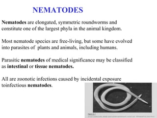
NEMATODES
Nematodes are elongated, symmetric roundworms and
constitute one of the largest phyla in the animal kingdom.

Most nematode species are free-living, but some have evolved
into parasites of plants and animals, including humans.

Parasitic nematodes of medical significance may be classified
as intestinal or tissue nematodes.

All are zoonotic infections caused by incidental exposure
toinfectious nematodes.
INTESTINAL NEMATODES


• More than a billion people worldwide are
  infected with one or more species of
  intestinal nematodes.
• These parasites are most common in regions
  with poor fecal sanitation, particularly in
  developing countries in the tropics and
  subtropics
• Although nematode infections are not
  usually fatal, they contribute to malnutrition
  and diminished work capacity.
• Humans may on occasion be infected with
  nematode parasites that ordinarily infect
  animals; these are zoonotic parasites.
Trichinellosis / Trichinosis
          Трихинелоза

• Develops after the ingestion of meat
  containing larves of Trichinella spiralis -
  pork or meat from a carnivore.

• While most infections are mild and
   asymptomatic, but heavy infections can
   cause:
- severe enteritis,
- periorbital edema,
- myositis,
- death
Five species of Trichinella are now recognized as causes of
    infection in humans. Two species are distributed worldwide:
-    Trichinella spiralis, which is found in a great variety of
    carnivorous and omnivorous animals,
-   Trichinella pseudospiralis, which is found in mammals and
    birds.
-   Trichinella nativa is present in Arctic regions and infects
    bears;
-   Trichinella nelsoni is found in equatorial Africa, where it is
    common among felid predators and scavengers such as
    hyenas and bush pigs;
-   Trichinella britovi is found in temperate areas of Europe
    and western Asia among carnivores but not among domestic
    swine.
Life Cycle

-   After the consumption of meat
    by the host, encysted larvae are
    liberated by digestive acid and
    pepsin.
-   The larvae invade the small –
    bowel mucosa and mature
    rapidly into adult worms.
-   After about 1 week, female
    worms release newborn larvae
    that migrate via the circulation
    to striated muscle.
-   The larvae of all species except
    T. pseudospiralis then encyst in
    the muscle cell
• Human trichinosis is most often caused by the
  ingestion of infected pork products and thus can
  occur in almost any location where the meat of
  domestic or wild swine is eaten.

• Human trichinosis also may be acquired from the
   meat of other animals, including:
- dogs (in parts of Asia and Africa),
- horses (in Italy and France), and
- bears and walruses (in northern regions).
Pathogenesis And Clinical Features
•   Clinical symptoms of trichinosis arise from the
    successive phases of parasite enteric invasion, larval
    migration, and muscle encystment.
•   Most light infections (those with fewer than 10
    larvae per gram of muscle) are asymptomatic,
•   whereas heavy infections (which can involve more
    than 50 larvae per gram of muscle) can be life-
    threatening.
•   Invasion by large numbers of parasites provokes
    diarrhea during the first week after infection.
•    Abdominal pain, constipation, nausea, or vomiting
    also may be prominent.
•   The prolonged and fulminant diarrhea noted
    probably reflects a response to repeated infection.
The migrating Trichinella larvae provoke a
  marked
local and systemic hypersensitivity reaction:
- fever
- hypereosinophilia,
- Periorbital and facial edema
- hemorrhages in the subconjunctivae, retina,
- nail beds ("splinter" hemorrhages).
Hypersensitivity reaction:


- Periorbital and facial
  edema
• dysphagia sometimes develops

• Myocarditis with tachyarrhythmias or heart failure,

• less commonly, encephalitis or pneumonitis may
  develop and accounts for most deaths of patients
  with trichinosis.

• A maculopapular rash, headache, cough, dyspnea
2 to 3 weeks after infection there are symptoms:
•   myositis with myalgias,
•   muscle edema,
•   weakness develop, usually
•   with the inflammatory reactions to migrating larvae.

•   The most commonly involved muscle groups include:
-   the extraocular muscles;
-   the biceps; and
-   the muscles of the jaw, neck, lower back, and diaphragm

    Peaking about 3 weeks after infection, symptoms subside
    only gradually during a prolonged convalescence.
Laboratory Findings And Diagnosis

• Blood eosinophilia develops in more than 90 % of
  patients with symptomatic trichinosis and may peak at a
  level of greater than 50 %
• Serum levels of IgE and
• muscle enzymes, including
- creatine phosphokinase,
- lactate dehydrogenase, and
- aspartate aminotransferase, are elevated in most
  symptomatic patients.
Epidemiological Diagnosis
-   Patients should be questioned about their consumption of
    pork or wild-animal meat and about illness in other
    individuals who ate the same meat.
-   A presumptive clinical diagnosis can be based on fevers,
    eosinophilia, periorbital edema, and myalgias after a
    suspect meal.
-   Arise in the titer of parasite-specific antibody, which
    usually does not occur until after the third week of
    infection, confirms the diagnosis.
-   Alternatively, a definitive diagnosis requires surgical
    biopsy of at least 1 g of involved muscle; the yields are
    highest near tendon insertions. The fresh muscle tissue
    should be compressed between glass slides and examined
    microscopically
TREATMENT

    Current anthelmintic drugs are ineffective against
    Trichinella larvae in muscle.
    Glucocorticoids like prednisone (1mg/kg daily for 5 days)
    are beneficial for severe myositis and myocarditis.
    Mebendazole, like thiabendazole, appears to be active
    against enteric stages of the parasite, but its efficacy against
    encysted larvae has not been conclusively demonstrated.
•   Mebendazole (Vermox) tb. 0,1 gr., 20 mg/kg
    /day, 3x1, 10-14 days; children - 5 mg/ kg/day
•   Albendazole (Zentel) tb. 0,2 gr, 10 mg/kg /day, 7-10 days
•   Pyrantel (Combartin) tb. 0,25 gr 10 mg/kg/twice a day, 5
    days
•   antipyretics, and analgesics.
Prevention

  Larvae may be killed by:

- cooking pork until it is no longer pink or
- by freezing it at -15°C for 3 weeks.
- However, Arctic T. nativa larvae in walrus or bear meat
  are relatively resistant and may remain viable despite
  freezing.
Ascariasis
/Ascaridosis/

Ascaris lumbricoides is the
largest intestinal nematode
parasite of humans, reaching
up to 40 cm in length.
1 billion people are infected
worldwide.
Most infected individuals
have low worm burdens and
are asymptomatic. Clinical
disease arises from
pulmonary and intestinal
complications.
Life Cycle
• Adult worms live in the lumen of
  the small intestine.
• Mature female Ascaris worms are
  producing up to 240,000 eggs a
  day, which pass with the feces.
• Ascarid eggs, which are
  remarkably resistant to
  environmental stresses, become
  infective after several weeks of
  maturation in the soil and can
  remain infective for years.
• After infective eggs are
  swallowed, larvae hatched in the
  intestine invade the
  mucosa, migrate via the circulation
  to the lungs, break into the
  alveoli, ascend the bronchial
  tree, and return via swallowing to
  the small intestine, where they
  develop into adult worms.
Between 2 and 3 months elapse between initial
  infection and egg production.
• The adult worms live for approximately 1 to 2 years.

Epidemiology

• Ascaris is widely distributed in tropical and
  subtropical regions as well as in other humid areas.
• Transmission typically occurs via fecally
  contaminated soil
Clinical Features

    During the lung phase of larval migration, about 9 to 12
    days after egg ingestion, patients may develop an:
-   irritating nonproductive cough and
-   burning substernal discomfort that is aggravated by
    coughing or deep inspiration.
-   Dyspnea and blood-tinged sputum are less common.
-   Fever is usually reported, with temperatures sometimes
    exceeding 38.5°C.
-   Chest x-rays may reveal evidence of eosinophilic
    pneumonitis (Loeffler's syndrome), with round or oval
    infiltrates a few millimeters to several centimeters in size.
    These infiltrates may be transient and intermittent.
• Adult worms in the small intestine usually cause no
  symptoms.
• In heavy infections, particularly in children, a large bolus of
  entangled worms can cause pain and small-bowel
  obstruction,
• complicated by perforation
• A large worm can enter in the biliary tree, causing biliary
  colic, cholecystitis, holangitis, pancreatitis, and
  intrahepatic abscesses.
• Migration of an adult worm up the esophagus can provoke
  coughing and oral expulsion of the worm.
• intestinal and biliary ascariasis can rival acute appendicitis
  and gallstones as causes of surgical acute abdomen.
Laboratory Findings

• Microscopic detection of
  characteristic Ascaris
  eggs (65 by 45 um) in
  fecal samples.
• Larvae can be found in
  sputum
• A plain abdominal film
• Worms can be detected
  by ultrasound and
  cholangiopancreatograpy
TREATMENT


- Mebendazole 2 x 0,2 gr. 3 days
- Albendazole 0,4 gr.
   These benzimidazoles are contraindicated in
   pregnancy
• Pyrantel pamoate – 10 mg/kg and
• piperazine citrate are safe in pregnancy
TRICHURIASIS

Most invasions with the
whipworm Trichuris trichiura
are asymptomatic, but
heavy infections may cause
gastrointestinal symptoms.

Like the other soil-transmitted
helminths, whipworm is
distributed globally in the
tropics and subtropics and is
most common among poor
children.
Life Cycle

The adult worms reside in the
colon and cecum, into the
superficial mucosa.
Thousands of eggs laid daily by
adult female worms pass via the
feces and mature in the soil.
After ingestion, infective eggs
hatch in the duodenum, releasing
larvae that mature before
migrating to the large bowel.
The entire cycle takes about 3
months, and adult worms may live
for several years.
Clinical Features

Most infected individuals have no symptoms
Heavy infections may result in abdominal
pain, anorexia, and bloody or mucoid diarrhea resembling
inflammatory bowel disease.

Rectal prolapse can result from massive infections in
children, who often suffer from malnourishment and other
diarrheal illnesses.
Diagnosis
    The characteristic - 50- by 20-um lemon-shaped
    whipworm eggs are readily detected on stool
    examination.

    Treatment
•   Adult worms, which are 3 to 5 cm long, occasionally
    can be seen on proctoscopy.
-   Mebendazole
-   Albendazole
-   Pyrantel pamoate
ENTEROBIOSIS
  PINWORM

  Enterobius vermicularis is
  more common in temperate
  countries than in the tropics.
• Enterobius adult worms are about 1
  cm long and dwell in the bowel
  lumen.
• The gravid female worm migrates
  nocturnally out into the perianal
  region and releases up to 10,000
  immature eggs.
Life Cycle
   The eggs become infective
   within hours and are transmitted
   via hand-to-mouth passage.
This life cycle takes about 1 month,
   and adult worms survive for
   about 2 months.
Self- infection results from
   perianal scratching and transport
   of infective eggs on the hands or
   under the nails to the mouth.
Owing to the ease of person-to-
   person spread, pinworm
   infections are common among
   family members
   andinstitutionalized populations.
Clinical Features
•   Most pinworm infections are asymptomatic.
•   Perianal pruritus is the cardinal symptom. The itching is
    often worse at night owing to the nocturnal migration of the
    female worms, and it may lead to excoriation and bacterial
    superinfection.
•   Heavy infections have been claimed to cause abdominal
    pain and weight loss.
•   On rare occasions, pinworms invade the female genital
    tract, causing vulvovaginitis and pelvic or peritoneal
    granulomas.
•   Eosinophilia or elevated levels of serum IgE are rare.
Diagnosis

Since pinworm eggs are not usually
released in the bowel, the diagnosis
cannot be made by looking for eggs
in the feces.

Instead, eggs deposited in the
perianal region are detected by the
application of clear cellulose tape to
the perianal region in the morning.

After the tape is transferred to a
microscope slide, will reveal the
characteristic pinworm eggs, which
are oval, measure 55 by 25 um, and
are flattened along one side.
TREATMENT

Mebendazole 2 x 0,2 gr. 3 days
Albendazole 0,4 gr.
  These benzimidazoles are contraindicated in pregnancy
Pyrantel pamoate – 10 mg/kg and
piperazine citrate are safe in pregnancy
 All affected individuals should be given a dose of
  mebendazole or pyrantel pamoate, with treatment repeated
  after 10 to 14 days.
• Treatment of household members is also advocated to
  eliminate asymptomatic reservoirs of potential reinfection.

Nematodoses10

  • 1.
    NEMATODES Nematodes are elongated,symmetric roundworms and constitute one of the largest phyla in the animal kingdom. Most nematode species are free-living, but some have evolved into parasites of plants and animals, including humans. Parasitic nematodes of medical significance may be classified as intestinal or tissue nematodes. All are zoonotic infections caused by incidental exposure toinfectious nematodes.
  • 2.
    INTESTINAL NEMATODES • Morethan a billion people worldwide are infected with one or more species of intestinal nematodes. • These parasites are most common in regions with poor fecal sanitation, particularly in developing countries in the tropics and subtropics • Although nematode infections are not usually fatal, they contribute to malnutrition and diminished work capacity. • Humans may on occasion be infected with nematode parasites that ordinarily infect animals; these are zoonotic parasites.
  • 3.
    Trichinellosis / Trichinosis Трихинелоза • Develops after the ingestion of meat containing larves of Trichinella spiralis - pork or meat from a carnivore. • While most infections are mild and asymptomatic, but heavy infections can cause: - severe enteritis, - periorbital edema, - myositis, - death
  • 4.
    Five species ofTrichinella are now recognized as causes of infection in humans. Two species are distributed worldwide: - Trichinella spiralis, which is found in a great variety of carnivorous and omnivorous animals, - Trichinella pseudospiralis, which is found in mammals and birds. - Trichinella nativa is present in Arctic regions and infects bears; - Trichinella nelsoni is found in equatorial Africa, where it is common among felid predators and scavengers such as hyenas and bush pigs; - Trichinella britovi is found in temperate areas of Europe and western Asia among carnivores but not among domestic swine.
  • 5.
    Life Cycle - After the consumption of meat by the host, encysted larvae are liberated by digestive acid and pepsin. - The larvae invade the small – bowel mucosa and mature rapidly into adult worms. - After about 1 week, female worms release newborn larvae that migrate via the circulation to striated muscle. - The larvae of all species except T. pseudospiralis then encyst in the muscle cell
  • 6.
    • Human trichinosisis most often caused by the ingestion of infected pork products and thus can occur in almost any location where the meat of domestic or wild swine is eaten. • Human trichinosis also may be acquired from the meat of other animals, including: - dogs (in parts of Asia and Africa), - horses (in Italy and France), and - bears and walruses (in northern regions).
  • 7.
    Pathogenesis And ClinicalFeatures • Clinical symptoms of trichinosis arise from the successive phases of parasite enteric invasion, larval migration, and muscle encystment. • Most light infections (those with fewer than 10 larvae per gram of muscle) are asymptomatic, • whereas heavy infections (which can involve more than 50 larvae per gram of muscle) can be life- threatening. • Invasion by large numbers of parasites provokes diarrhea during the first week after infection. • Abdominal pain, constipation, nausea, or vomiting also may be prominent. • The prolonged and fulminant diarrhea noted probably reflects a response to repeated infection.
  • 8.
    The migrating Trichinellalarvae provoke a marked local and systemic hypersensitivity reaction: - fever - hypereosinophilia, - Periorbital and facial edema - hemorrhages in the subconjunctivae, retina, - nail beds ("splinter" hemorrhages).
  • 9.
  • 10.
    • dysphagia sometimesdevelops • Myocarditis with tachyarrhythmias or heart failure, • less commonly, encephalitis or pneumonitis may develop and accounts for most deaths of patients with trichinosis. • A maculopapular rash, headache, cough, dyspnea
  • 11.
    2 to 3weeks after infection there are symptoms: • myositis with myalgias, • muscle edema, • weakness develop, usually • with the inflammatory reactions to migrating larvae. • The most commonly involved muscle groups include: - the extraocular muscles; - the biceps; and - the muscles of the jaw, neck, lower back, and diaphragm Peaking about 3 weeks after infection, symptoms subside only gradually during a prolonged convalescence.
  • 12.
    Laboratory Findings AndDiagnosis • Blood eosinophilia develops in more than 90 % of patients with symptomatic trichinosis and may peak at a level of greater than 50 % • Serum levels of IgE and • muscle enzymes, including - creatine phosphokinase, - lactate dehydrogenase, and - aspartate aminotransferase, are elevated in most symptomatic patients.
  • 13.
    Epidemiological Diagnosis - Patients should be questioned about their consumption of pork or wild-animal meat and about illness in other individuals who ate the same meat. - A presumptive clinical diagnosis can be based on fevers, eosinophilia, periorbital edema, and myalgias after a suspect meal. - Arise in the titer of parasite-specific antibody, which usually does not occur until after the third week of infection, confirms the diagnosis. - Alternatively, a definitive diagnosis requires surgical biopsy of at least 1 g of involved muscle; the yields are highest near tendon insertions. The fresh muscle tissue should be compressed between glass slides and examined microscopically
  • 14.
    TREATMENT Current anthelmintic drugs are ineffective against Trichinella larvae in muscle. Glucocorticoids like prednisone (1mg/kg daily for 5 days) are beneficial for severe myositis and myocarditis. Mebendazole, like thiabendazole, appears to be active against enteric stages of the parasite, but its efficacy against encysted larvae has not been conclusively demonstrated. • Mebendazole (Vermox) tb. 0,1 gr., 20 mg/kg /day, 3x1, 10-14 days; children - 5 mg/ kg/day • Albendazole (Zentel) tb. 0,2 gr, 10 mg/kg /day, 7-10 days • Pyrantel (Combartin) tb. 0,25 gr 10 mg/kg/twice a day, 5 days • antipyretics, and analgesics.
  • 15.
    Prevention Larvaemay be killed by: - cooking pork until it is no longer pink or - by freezing it at -15°C for 3 weeks. - However, Arctic T. nativa larvae in walrus or bear meat are relatively resistant and may remain viable despite freezing.
  • 16.
    Ascariasis /Ascaridosis/ Ascaris lumbricoides isthe largest intestinal nematode parasite of humans, reaching up to 40 cm in length. 1 billion people are infected worldwide. Most infected individuals have low worm burdens and are asymptomatic. Clinical disease arises from pulmonary and intestinal complications.
  • 17.
    Life Cycle • Adultworms live in the lumen of the small intestine. • Mature female Ascaris worms are producing up to 240,000 eggs a day, which pass with the feces. • Ascarid eggs, which are remarkably resistant to environmental stresses, become infective after several weeks of maturation in the soil and can remain infective for years. • After infective eggs are swallowed, larvae hatched in the intestine invade the mucosa, migrate via the circulation to the lungs, break into the alveoli, ascend the bronchial tree, and return via swallowing to the small intestine, where they develop into adult worms.
  • 18.
    Between 2 and3 months elapse between initial infection and egg production. • The adult worms live for approximately 1 to 2 years. Epidemiology • Ascaris is widely distributed in tropical and subtropical regions as well as in other humid areas. • Transmission typically occurs via fecally contaminated soil
  • 19.
    Clinical Features During the lung phase of larval migration, about 9 to 12 days after egg ingestion, patients may develop an: - irritating nonproductive cough and - burning substernal discomfort that is aggravated by coughing or deep inspiration. - Dyspnea and blood-tinged sputum are less common. - Fever is usually reported, with temperatures sometimes exceeding 38.5°C. - Chest x-rays may reveal evidence of eosinophilic pneumonitis (Loeffler's syndrome), with round or oval infiltrates a few millimeters to several centimeters in size. These infiltrates may be transient and intermittent.
  • 20.
    • Adult wormsin the small intestine usually cause no symptoms. • In heavy infections, particularly in children, a large bolus of entangled worms can cause pain and small-bowel obstruction, • complicated by perforation • A large worm can enter in the biliary tree, causing biliary colic, cholecystitis, holangitis, pancreatitis, and intrahepatic abscesses. • Migration of an adult worm up the esophagus can provoke coughing and oral expulsion of the worm. • intestinal and biliary ascariasis can rival acute appendicitis and gallstones as causes of surgical acute abdomen.
  • 21.
    Laboratory Findings • Microscopicdetection of characteristic Ascaris eggs (65 by 45 um) in fecal samples. • Larvae can be found in sputum • A plain abdominal film • Worms can be detected by ultrasound and cholangiopancreatograpy
  • 22.
    TREATMENT - Mebendazole 2x 0,2 gr. 3 days - Albendazole 0,4 gr. These benzimidazoles are contraindicated in pregnancy • Pyrantel pamoate – 10 mg/kg and • piperazine citrate are safe in pregnancy
  • 23.
    TRICHURIASIS Most invasions withthe whipworm Trichuris trichiura are asymptomatic, but heavy infections may cause gastrointestinal symptoms. Like the other soil-transmitted helminths, whipworm is distributed globally in the tropics and subtropics and is most common among poor children.
  • 24.
    Life Cycle The adultworms reside in the colon and cecum, into the superficial mucosa. Thousands of eggs laid daily by adult female worms pass via the feces and mature in the soil. After ingestion, infective eggs hatch in the duodenum, releasing larvae that mature before migrating to the large bowel. The entire cycle takes about 3 months, and adult worms may live for several years.
  • 25.
    Clinical Features Most infectedindividuals have no symptoms Heavy infections may result in abdominal pain, anorexia, and bloody or mucoid diarrhea resembling inflammatory bowel disease. Rectal prolapse can result from massive infections in children, who often suffer from malnourishment and other diarrheal illnesses.
  • 26.
    Diagnosis The characteristic - 50- by 20-um lemon-shaped whipworm eggs are readily detected on stool examination. Treatment • Adult worms, which are 3 to 5 cm long, occasionally can be seen on proctoscopy. - Mebendazole - Albendazole - Pyrantel pamoate
  • 27.
    ENTEROBIOSIS PINWORM Enterobius vermicularis is more common in temperate countries than in the tropics. • Enterobius adult worms are about 1 cm long and dwell in the bowel lumen. • The gravid female worm migrates nocturnally out into the perianal region and releases up to 10,000 immature eggs.
  • 28.
    Life Cycle The eggs become infective within hours and are transmitted via hand-to-mouth passage. This life cycle takes about 1 month, and adult worms survive for about 2 months. Self- infection results from perianal scratching and transport of infective eggs on the hands or under the nails to the mouth. Owing to the ease of person-to- person spread, pinworm infections are common among family members andinstitutionalized populations.
  • 29.
    Clinical Features • Most pinworm infections are asymptomatic. • Perianal pruritus is the cardinal symptom. The itching is often worse at night owing to the nocturnal migration of the female worms, and it may lead to excoriation and bacterial superinfection. • Heavy infections have been claimed to cause abdominal pain and weight loss. • On rare occasions, pinworms invade the female genital tract, causing vulvovaginitis and pelvic or peritoneal granulomas. • Eosinophilia or elevated levels of serum IgE are rare.
  • 30.
    Diagnosis Since pinworm eggsare not usually released in the bowel, the diagnosis cannot be made by looking for eggs in the feces. Instead, eggs deposited in the perianal region are detected by the application of clear cellulose tape to the perianal region in the morning. After the tape is transferred to a microscope slide, will reveal the characteristic pinworm eggs, which are oval, measure 55 by 25 um, and are flattened along one side.
  • 31.
    TREATMENT Mebendazole 2 x0,2 gr. 3 days Albendazole 0,4 gr. These benzimidazoles are contraindicated in pregnancy Pyrantel pamoate – 10 mg/kg and piperazine citrate are safe in pregnancy All affected individuals should be given a dose of mebendazole or pyrantel pamoate, with treatment repeated after 10 to 14 days. • Treatment of household members is also advocated to eliminate asymptomatic reservoirs of potential reinfection.