SlideShare a Scribd company logo
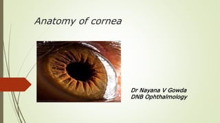
Anatomy of cornea
Dr Nayana V Gowda
DNB Ophthalmology
Contents
 Brief introduction
 Topography
 Embryology
 Function
 Histology
 Transparency
 Blood and nerve supply
 Limbus
INTRODUCTION
Defintion: Transparent, avascular, anterior coat of the eye that
covers the iris, pupil and anterior chamber being continuous with the
sclera at the periphery.
Forms anterior one-sixth of fibrous coat of the eyeball
Refractive power: Anterior surface of cornea has a refractory
power of +48D and it’s posterior surface of
about -5D. Hence the net refractory power of cornea is
+43D which is 3/4th of the total refractory power of the eye (+60D).
• Refractive index is 1.37
DIMENSIONS
• Anterior surface of cornea is horizontally
elliptical 11.75 mm. by 11 mm.
• Posterior surface of cornea is circular with
diameter of 11.5 mm.
DIMENSIONS ctd…….
• Cornea is 0.52 mm. thick at the center which
gradually increases to 0.67mm at the
periphery.
• The radius of curvature of the central part of
cornea is 7.8 mm anteriorly and 6.5 mm.
posteriorly.
TOPOGRAPHY
• Steeper in infants.
• Greater flattening nasally than temporally.
• Near the limbus, the curvature increases before entering the trough
like contour of limbal zone.
• Flattens on convergence.
• Knowledge of topography important for CL fitting
EMBRYOLOGY
EPITHELIUM: Surface ectoderm
BOWMAN’S LAYER: Laid down by anterior stromal keratocytes
STROMA: Mesenchymal cells
ENDOTHELIUM AND DESCEMET’S MEMBRANE: neural crest cells
EMBROLOGY OF CORNEA
• Mesenchymal mass of neural crest origin is now considered to give rise to cornea , iris
and anterior chamber angle.
• Three waves of tissue come forward between surface ectoderm and developing lens,
from the developing undifferentiated mesenchymal mass of NCC origin to contribute to
structures of anterior segment origin
EMBRYOLOGY: EPITHELIUM
• 40 days POG: Superficial squamous layer & a
basal cuboidal layer.
• 2-3rd month POG: Superficial cells increase in
size & are covered by microvilli & microplicae.
• 5-6 months POG: corneal epithelium attains Adult morphology
EMBRYOLOGY: BOWMAN’S ZONE
• Not formed until 4th month POG.
• Always acellular.
• Superficial keratocytes synthesize & lay down the
ground substance & collagen.
• At this stage the hemidesmosomes & anchoring
filaments in the basal lamina are partially developed.
•By 26 wks they have an adult appearance
EMBRYOLOGY: STROMA
• 7 weeks POG: Anterior extension of mesenchymal cells
migrates btw epithelium and endothelium & contributes to
growth of stroma.
• Initially acellular but ingrowing cells differentiate to
keratocytes that secrete type 1 collagen & stromal matrix.
• Fibrils are organized as lamellae and successive lamellae are
formed.
• Posterior layers of stroma are confluent with the condensed
tissue of the future sclera.
EMBRYOLOGY: DM & ENDOTHELIUM
Formed from mesenchymal cells derived from neural crest , which are
situated at margins of rim of optic cup. These cells migrate into
developing eye beneath the basal lamina of corneal epithelium and
form primordial corneal endothelium
EMBRYOLOGY: DM & ENDOTHELIUM ctd…..
• At about 40 days of POG – corneal endothelium consist of 2 layered
flattened cells
• By 3rd month endothelium in the centre of cornea becomes a single
layer of flattened cells that rest on the interrupted basal lamina the
future DM
• Apices of endothelial cells are joined by zona occludens in the middle
of 4th month
• 6th months of gestation DM demarcated clearly
FUNCTIONS
Refraction - 70% of total refractory power of eye is due to
smooth surface tear film & regular anterior
Curvature (Power = +43.1D , RI = 1.376)
Contains IOP - Function of collagen fibres in stroma
Transparency - Mainly due to Corneal endothelium which
continuously pumps out excess water
Protection - Acts as mechanical barrier to external
noxious stimuli.
EPITHELIUM
 Epithelium type-Stratified squamous
 non keratinized
 Thickness-50-90 microns,
 5-6 layers of nucleated cells.
 Continuous with conjunctiva at limbus but devoid of goblet cells.
 Consists of 5-6 layers of nucleated cells resting on a basal lamina, namely –
basal cells , wing cells , surface cells
BASAL LAYER
 Deepest layer
 Cells are arranged in palisade manner
 Germinativelayer of epithelium
 Cells are columnar with rounded heads & flat bases
 Nucleus is oval & oriented parallel to long axis of the cell
WING CELLS
 Second layer of epithelium (1-2 layers) Polyhedral cells
 Convex anteriorly forming cap over basal cells & send processes
between them
 Nucleus is oval & oriented parallel to corneal surface
SURFACE CELLS
 Most superficial layer (2-3 layers)
 Cells are polyhedral & become wider & flatter towards the surface
 Flattened nuclei project backwards leaving the surface perfectly smooth
 Most superficial cells are mostly hexagonal & exhibit surface microvilli /
microplicae
BASAL LAMINA
Secreted by basal cells.
•Made up of collagen (*Type7 & glycoprotein
•Structurally attached with the underlying Bowman’s layer by short anchoring
filaments forming anchoring plaques
•Cohesion between basal lamina & Bowman’s zone maybe loosened by lipid
solvents, edema or inflammation but it remains attached to basal cells.
•Thicker in periphery and in diabetes & certain corneal disorders
Thickened with old age.
EPITHELIAL TURNOVER
 The germinative region lies at the limbus, the stem cells, and cells migrate at a very
slower rate (123 μm/week) to the centre of cornea.
 The XYZ hypothesis:ThoftR. and Friend J. (1983) proposed on the basis of experimental
evidence ,that both limbal basal and corneal basal cells are the source for corneal
epithelial cells,and there is a balance among division, migration & shedding.
 The corneal epithelium is maintained by a balance among sloughing (Z) of cells from
the corneal surface, cell division (X) in the basal layer and renewal of basal cells by
centripetal migration (Y) of new basal cells originating from the limbalstem cells.
BOWMANS MEMBRANE
 Modified region of anterior stroma
 Acellular homogeneous zone
 8 –14 μm thick
 Ant. surface is smooth & parallel with corneal surface
 It delineates the anterior junction between cornea and limbus
 Perimeter delineates anterior junction btw the cornea & the limbus &
is clinically marked by summits of marginal arcades of
limbalcapillaries.
 Ultrastructurallyit is a felted meshwork of fine collagen fibrils of
uniform size in a ground substance
 Posteriorly it becomes blended & interweaving with fibrils of ant.
Stroma
 Compact arrangement of collagen gives it great strength and relatively
resistant to trauma both mechanical and infective
 Perforated in many places by corneal nerves.
 Convex ridges may generate over surface if its tension is relaxed during
indentation, hypotonyor manipulation causes ant.cornealmosaic,
polygonal or chicken-wire pattern over surface
 No regeneration and replaced by coarse scar tissue
STROMA
 About 500 μm thick (about 90% of corneal thickness)
 Two components : 1) Lamellae 2) Cells
 stroma consists of regularly arranged lamellae of collagen bundles, lie in
proteoglycan ground substance.
 In the anterior 1/3rd there is more interweave & some lamellae pass
forward to be inserted into Bowman’s layer
 In the deep stroma lamellae form strap like ribbons which run
approxright angle to consecutive layers. Approx300 lamellae.
 Interfibrillar separation is equal to the fibril diameter. This separation
decreases with age.
 Precise ordering is responsible for transparency of the stroma.
 CELLS-
1)Keratocytesmake up 2.5 –5% of the stromal volume and are responsible
for synthesis of collagen & proteoglycan during development &
maintaining it thereafter.
2) Other cells found in stroma occasionally: lymphocytes,macrophages and
very rarely polymorphonuclearlymphocytes.
DUA’S LAYER
 Named after Dr.Harminder Dua, who
discovered it
 Acellular layer between stroma & DM , 6-15 μ
thick
 Consists of about 5-8 lamellae of type-1
collagen bundles
 Bundle spacing is similar to stromal tissue but
devoid of keratocytes
 Very strong layer
 Can withstand upto200 kPa pressure
DESCEMET’S MEMBRANE
 It is the basal lamina of corneal endothelium
 First appears at 2nd month of gestation and synthesis continue
throughout adult life
 Thickness –at birth :-3 –4 μm at childhood :-about 5 μm
 at adult :-10 –12 μm
 Acts as a strong resistant sheet
 It thickens with age and in some corneal degenerative conditions
 Major protein of DM is Type IV collagen
ENDOTHELIUM
 It is a single layer of hexagonal, cuboidal cells attached posterior aspect of
DM
 It is neuroectodermalin origin
 Corneal endothelial cells production is relatively fixed
 Endothelial cells density –about 6000cells/mm² at birth
 26%lost in 1styear, Further 26% lost over next 11years
 cell loss slows stabilizes around middle age about 2500 cells/mm²
 If cells density falls upto500 cells/mm² corneal oedema develops and
transparency reduced
CORNEAL TRANSPARENCY
The cornea transmits nearly 100% of the light that enters it. Transparency achieved by –
 1. Arrangement of stromal lamellae
Two theories –
i)Maurice(1957): The transparency of the stroma is due to the lattice arrangement of
collagen fibrils. He explained, because of their small diameter and regularity of separation,
back scattered light would be almost completely suppressed by destructive interference
ii) Goldman et al. (1968): He suggest, a perfect crystalline lattice periodicity is not always
necessary for sufficient destructive interference. He explained, if fibril separation and
diameter is less than a third of the wavelength of incident light, then almost perfect
transparency will ensue. This is the situation which obtains in normal cornea.
 2.Corneal epithelium & tear film
•Epithelial non-keratinization
•Regular & uniform arrangement of corneal epithelium
•Junctions between cells & its compactness and also tear film
maintain a homogenicityof its refractive index
 3.Relative deturgescencestate of normal cornea
 4.Corneal avascularity
 5.Non myelenatednerve fibres
BLOOD SUPPLY
• Avascular
• Loops of anterior ciliary artery invade the
subconjunctival tissue at corneal periphery by 1mm.
• Many pathologic processes show corneal
vascularisation.
• Superficial vessels are bright red, arborescent and
may raise the epithelium
• Deep vessels are greyish red, run parallel and do not raise the
epithelium.
NERVE SUPPLY OF CORNEA
LIMBUS
Anatomically , the limbus refers to circumcorneal transition zone of
conjunctivo-corneal and corneoscleral
Junction
 1.5 mm wide in horizontal plane and 2mm in the vertical plane.
 Limbus is the weakest region in the corneoscleralenvelope.
CONJUNCTIVOCORNEAL JUNCTION
 At this point bulbar conjunctiva is firmly adherent to the underlying
structures
 Epithelium becomes several layers thick here and arranged
irregularly at limbus
 Limbal epithelial basal cells are arranged in a peculiar pattern of
palisade of vogt, containing stem cells
SCLEROCORNEAL JUNCTION
At this point transparent corneal lamellae becomes continuous with
the oblique , circular and opaque fibres of sclera
SURGICAL LIMBUS
Its 2mm wide circumcorneal transition zone between the clear cornea
on one side and the opaque sclera on the other side
BORDERS OF SURGICAL LIMBUS
 Anterior limbal border- marked by insertion of conjunctiva and tenon’s capsule into
cornea , forms anterior border of surgical limbus
 Mid limbal line-junction of blue and white zone overlies termination of Descemet’s
membrane
 Posterior limbal border-lies 1mm posterior to mid limbal line. It overlies the scleral spur
and can only be seen with the use of sclerotic scatter illumination
ZONES OF SURGICAL LIMBUS
 Blue limbal zone – translucent zone seen posterior to anterior limbal border
dissecting limbus free of conjunctiva and tenon’s capsule
 Extent - superior quadrant – 1mm
Inferior quadrant– 0.8mm
nasal and temporal – 0.4mm
WHITE LIMBAL ZONE
 Its 1mm wide whitish area lies between mid-limbal line and the posterior limbal
border
 Overlies trabecular meshwork
 Its width is constant in all quadrant 1mm , so surgical limbus is greatest in superior
quadrant where width of blue zone is 1mm
Cornea anatomy simplified

More Related Content

What's hot

Conjunctiva
ConjunctivaConjunctiva
Conjunctiva
JESLIN JOSE
 
Anatomy of optic nerve
Anatomy of optic nerveAnatomy of optic nerve
Anatomy of optic nerve
Sudheer Kumar
 
Tear film
Tear filmTear film
Tear film
Meenakshi Jha
 
Vitreous
VitreousVitreous
Anatomy and embryology of anterior chamber angle ppt new
Anatomy and embryology of anterior chamber angle ppt newAnatomy and embryology of anterior chamber angle ppt new
Anatomy and embryology of anterior chamber angle ppt new
anupama manoharan
 
Keratoconus
KeratoconusKeratoconus
Keratoconus
SSSIHMS-PG
 
Cornea Anatomy and Physiology
Cornea Anatomy and PhysiologyCornea Anatomy and Physiology
Cornea Anatomy and Physiology
Shandilyan Mbbs
 
Physiology of the cornea
Physiology of the corneaPhysiology of the cornea
Physiology of the cornea
Desta Genete
 
anatomy & physiology of lens
anatomy & physiology of lensanatomy & physiology of lens
anatomy & physiology of lens
rakesh jaiswal
 
Crystalline Lens
Crystalline LensCrystalline Lens
Crystalline Lens
Mero Eye
 
HUMAN Lens ANATOMY
HUMAN Lens ANATOMYHUMAN Lens ANATOMY
HUMAN Lens ANATOMY
SSSIHMS-PG
 
Lens
LensLens
Anatomy of Cornea
Anatomy of CorneaAnatomy of Cornea
Anatomy of Cornea
SristiThakur
 
Lens anatomy sivateja
Lens anatomy sivatejaLens anatomy sivateja
Lens anatomy sivateja
Sivateja Challa
 
Anatomy of the eyelids
Anatomy of the eyelidsAnatomy of the eyelids
Anatomy of the eyelids
SSSIHMS-PG
 
Tear film dynamics
Tear film dynamicsTear film dynamics
Tear film dynamics
Ashish Badgujar
 
Aqueous humor outflow
Aqueous humor outflowAqueous humor outflow
Aqueous humor outflow
Jagdish Dukre
 
The orbit of eye
The orbit of eyeThe orbit of eye
The orbit of eye
Idris Siddiqui
 
Corneal metabolism
Corneal metabolismCorneal metabolism
Eyelid anatomy and physiology
Eyelid anatomy and physiologyEyelid anatomy and physiology
Eyelid anatomy and physiology
Najara Thapa
 

What's hot (20)

Conjunctiva
ConjunctivaConjunctiva
Conjunctiva
 
Anatomy of optic nerve
Anatomy of optic nerveAnatomy of optic nerve
Anatomy of optic nerve
 
Tear film
Tear filmTear film
Tear film
 
Vitreous
VitreousVitreous
Vitreous
 
Anatomy and embryology of anterior chamber angle ppt new
Anatomy and embryology of anterior chamber angle ppt newAnatomy and embryology of anterior chamber angle ppt new
Anatomy and embryology of anterior chamber angle ppt new
 
Keratoconus
KeratoconusKeratoconus
Keratoconus
 
Cornea Anatomy and Physiology
Cornea Anatomy and PhysiologyCornea Anatomy and Physiology
Cornea Anatomy and Physiology
 
Physiology of the cornea
Physiology of the corneaPhysiology of the cornea
Physiology of the cornea
 
anatomy & physiology of lens
anatomy & physiology of lensanatomy & physiology of lens
anatomy & physiology of lens
 
Crystalline Lens
Crystalline LensCrystalline Lens
Crystalline Lens
 
HUMAN Lens ANATOMY
HUMAN Lens ANATOMYHUMAN Lens ANATOMY
HUMAN Lens ANATOMY
 
Lens
LensLens
Lens
 
Anatomy of Cornea
Anatomy of CorneaAnatomy of Cornea
Anatomy of Cornea
 
Lens anatomy sivateja
Lens anatomy sivatejaLens anatomy sivateja
Lens anatomy sivateja
 
Anatomy of the eyelids
Anatomy of the eyelidsAnatomy of the eyelids
Anatomy of the eyelids
 
Tear film dynamics
Tear film dynamicsTear film dynamics
Tear film dynamics
 
Aqueous humor outflow
Aqueous humor outflowAqueous humor outflow
Aqueous humor outflow
 
The orbit of eye
The orbit of eyeThe orbit of eye
The orbit of eye
 
Corneal metabolism
Corneal metabolismCorneal metabolism
Corneal metabolism
 
Eyelid anatomy and physiology
Eyelid anatomy and physiologyEyelid anatomy and physiology
Eyelid anatomy and physiology
 

Similar to Cornea anatomy simplified

Cornea
Cornea Cornea
anatomy and physiology of cornea-.pdf gfhh
anatomy and physiology of cornea-.pdf gfhhanatomy and physiology of cornea-.pdf gfhh
anatomy and physiology of cornea-.pdf gfhh
epicsoundever
 
Anatomy and physiology of cornea
Anatomy and physiology of corneaAnatomy and physiology of cornea
Anatomy and physiology of cornea
SSSIHMS-PG
 
cornea, conjunctiva and sclera
cornea, conjunctiva and scleracornea, conjunctiva and sclera
cornea, conjunctiva and sclera
Fawaz Alzweimel
 
Cornea
CorneaCornea
Cornea Applied anatomy.pptx
Cornea Applied anatomy.pptxCornea Applied anatomy.pptx
Cornea Applied anatomy.pptx
Dr. Pradeep Bastola
 
CORNEA.pptx
CORNEA.pptxCORNEA.pptx
CORNEA.pptx
BARNABASMUGABI
 
Anatomy of cornea
Anatomy of corneaAnatomy of cornea
Anatomy of cornea
Kanwal Perveen
 
CORNEA anat and physio.pptx
CORNEA anat and physio.pptxCORNEA anat and physio.pptx
CORNEA anat and physio.pptx
ManoAarthi1
 
Anatomy of cornea & corneal tranparency dr.ysr
Anatomy of cornea &  corneal tranparency dr.ysrAnatomy of cornea &  corneal tranparency dr.ysr
Anatomy of cornea & corneal tranparency dr.ysr
DrYajuvendra Rathore
 
CORNEA.pptx
CORNEA.pptxCORNEA.pptx
CORNEA.pptx
NISHA S.S
 
Anatomy of crystalline lens by Dr. Aayush Tandon
Anatomy of crystalline lens by Dr. Aayush Tandon Anatomy of crystalline lens by Dr. Aayush Tandon
Anatomy of crystalline lens by Dr. Aayush Tandon
Aayush Tandon
 
Lect.cornea
Lect.corneaLect.cornea
Lect.cornea
Yumna Tariq
 
anatomyofthecornea-161101224539-converted.pptx
anatomyofthecornea-161101224539-converted.pptxanatomyofthecornea-161101224539-converted.pptx
anatomyofthecornea-161101224539-converted.pptx
Arun Kumar A
 
Anatomy and embryology of crystalline lens DrBP
Anatomy and embryology of crystalline lens DrBPAnatomy and embryology of crystalline lens DrBP
Anatomy and embryology of crystalline lens DrBP
drbhushan17
 
Anatomy and physiology of cornea
Anatomy and physiology of corneaAnatomy and physiology of cornea
Anatomy and physiology of cornea
Lhacha
 
Corneal anatomy physiology and wound healing
Corneal anatomy physiology and wound healingCorneal anatomy physiology and wound healing
Corneal anatomy physiology and wound healing
Socrates Narvaez
 
Anatomy of cornea of dr. sohel mahmud
Anatomy of cornea of dr. sohel mahmudAnatomy of cornea of dr. sohel mahmud
Anatomy of cornea of dr. sohel mahmud
Sohel Sumon
 
Anatomi fisiologi kornea
Anatomi fisiologi korneaAnatomi fisiologi kornea
Anatomi fisiologi kornea
hasya arianda
 
Minimalinvasiveglaucomasurgerylllll.pptx
Minimalinvasiveglaucomasurgerylllll.pptxMinimalinvasiveglaucomasurgerylllll.pptx
Minimalinvasiveglaucomasurgerylllll.pptx
AmitMirre
 

Similar to Cornea anatomy simplified (20)

Cornea
Cornea Cornea
Cornea
 
anatomy and physiology of cornea-.pdf gfhh
anatomy and physiology of cornea-.pdf gfhhanatomy and physiology of cornea-.pdf gfhh
anatomy and physiology of cornea-.pdf gfhh
 
Anatomy and physiology of cornea
Anatomy and physiology of corneaAnatomy and physiology of cornea
Anatomy and physiology of cornea
 
cornea, conjunctiva and sclera
cornea, conjunctiva and scleracornea, conjunctiva and sclera
cornea, conjunctiva and sclera
 
Cornea
CorneaCornea
Cornea
 
Cornea Applied anatomy.pptx
Cornea Applied anatomy.pptxCornea Applied anatomy.pptx
Cornea Applied anatomy.pptx
 
CORNEA.pptx
CORNEA.pptxCORNEA.pptx
CORNEA.pptx
 
Anatomy of cornea
Anatomy of corneaAnatomy of cornea
Anatomy of cornea
 
CORNEA anat and physio.pptx
CORNEA anat and physio.pptxCORNEA anat and physio.pptx
CORNEA anat and physio.pptx
 
Anatomy of cornea & corneal tranparency dr.ysr
Anatomy of cornea &  corneal tranparency dr.ysrAnatomy of cornea &  corneal tranparency dr.ysr
Anatomy of cornea & corneal tranparency dr.ysr
 
CORNEA.pptx
CORNEA.pptxCORNEA.pptx
CORNEA.pptx
 
Anatomy of crystalline lens by Dr. Aayush Tandon
Anatomy of crystalline lens by Dr. Aayush Tandon Anatomy of crystalline lens by Dr. Aayush Tandon
Anatomy of crystalline lens by Dr. Aayush Tandon
 
Lect.cornea
Lect.corneaLect.cornea
Lect.cornea
 
anatomyofthecornea-161101224539-converted.pptx
anatomyofthecornea-161101224539-converted.pptxanatomyofthecornea-161101224539-converted.pptx
anatomyofthecornea-161101224539-converted.pptx
 
Anatomy and embryology of crystalline lens DrBP
Anatomy and embryology of crystalline lens DrBPAnatomy and embryology of crystalline lens DrBP
Anatomy and embryology of crystalline lens DrBP
 
Anatomy and physiology of cornea
Anatomy and physiology of corneaAnatomy and physiology of cornea
Anatomy and physiology of cornea
 
Corneal anatomy physiology and wound healing
Corneal anatomy physiology and wound healingCorneal anatomy physiology and wound healing
Corneal anatomy physiology and wound healing
 
Anatomy of cornea of dr. sohel mahmud
Anatomy of cornea of dr. sohel mahmudAnatomy of cornea of dr. sohel mahmud
Anatomy of cornea of dr. sohel mahmud
 
Anatomi fisiologi kornea
Anatomi fisiologi korneaAnatomi fisiologi kornea
Anatomi fisiologi kornea
 
Minimalinvasiveglaucomasurgerylllll.pptx
Minimalinvasiveglaucomasurgerylllll.pptxMinimalinvasiveglaucomasurgerylllll.pptx
Minimalinvasiveglaucomasurgerylllll.pptx
 

Recently uploaded

Ketone bodies and metabolism-biochemistry
Ketone bodies and metabolism-biochemistryKetone bodies and metabolism-biochemistry
Ketone bodies and metabolism-biochemistry
Dhayanithi C
 
THERAPEUTIC ANTISENSE MOLECULES .pptx
THERAPEUTIC ANTISENSE MOLECULES    .pptxTHERAPEUTIC ANTISENSE MOLECULES    .pptx
THERAPEUTIC ANTISENSE MOLECULES .pptx
70KRISHPATEL
 
OCT Training Course for clinical practice Part 1
OCT Training Course for clinical practice Part 1OCT Training Course for clinical practice Part 1
OCT Training Course for clinical practice Part 1
KafrELShiekh University
 
CHEMOTHERAPY_RDP_CHAPTER 6_Anti Malarial Drugs.pdf
CHEMOTHERAPY_RDP_CHAPTER 6_Anti Malarial Drugs.pdfCHEMOTHERAPY_RDP_CHAPTER 6_Anti Malarial Drugs.pdf
CHEMOTHERAPY_RDP_CHAPTER 6_Anti Malarial Drugs.pdf
rishi2789
 
A Classical Text Review on Basavarajeeyam
A Classical Text Review on BasavarajeeyamA Classical Text Review on Basavarajeeyam
A Classical Text Review on Basavarajeeyam
Dr. Jyothirmai Paindla
 
CHEMOTHERAPY_RDP_CHAPTER 2 _LEPROSY.pdf1
CHEMOTHERAPY_RDP_CHAPTER 2 _LEPROSY.pdf1CHEMOTHERAPY_RDP_CHAPTER 2 _LEPROSY.pdf1
CHEMOTHERAPY_RDP_CHAPTER 2 _LEPROSY.pdf1
rishi2789
 
Clinic ^%[+27633867063*Abortion Pills For Sale In Tembisa Central
Clinic ^%[+27633867063*Abortion Pills For Sale In Tembisa CentralClinic ^%[+27633867063*Abortion Pills For Sale In Tembisa Central
Clinic ^%[+27633867063*Abortion Pills For Sale In Tembisa Central
19various
 
Complementary feeding in infant IAP PROTOCOLS
Complementary feeding in infant IAP PROTOCOLSComplementary feeding in infant IAP PROTOCOLS
Complementary feeding in infant IAP PROTOCOLS
chiranthgowda16
 
Top 10 Best Ayurvedic Kidney Stone Syrups in India
Top 10 Best Ayurvedic Kidney Stone Syrups in IndiaTop 10 Best Ayurvedic Kidney Stone Syrups in India
Top 10 Best Ayurvedic Kidney Stone Syrups in India
Swastik Ayurveda
 
REGULATION FOR COMBINATION PRODUCTS AND MEDICAL DEVICES.pptx
REGULATION FOR COMBINATION PRODUCTS AND MEDICAL DEVICES.pptxREGULATION FOR COMBINATION PRODUCTS AND MEDICAL DEVICES.pptx
REGULATION FOR COMBINATION PRODUCTS AND MEDICAL DEVICES.pptx
LaniyaNasrink
 
Hemodialysis: Chapter 4, Dialysate Circuit - Dr.Gawad
Hemodialysis: Chapter 4, Dialysate Circuit - Dr.GawadHemodialysis: Chapter 4, Dialysate Circuit - Dr.Gawad
Hemodialysis: Chapter 4, Dialysate Circuit - Dr.Gawad
NephroTube - Dr.Gawad
 
The Best Ayurvedic Antacid Tablets in India
The Best Ayurvedic Antacid Tablets in IndiaThe Best Ayurvedic Antacid Tablets in India
The Best Ayurvedic Antacid Tablets in India
Swastik Ayurveda
 
Journal Article Review on Rasamanikya
Journal Article Review on RasamanikyaJournal Article Review on Rasamanikya
Journal Article Review on Rasamanikya
Dr. Jyothirmai Paindla
 
Part II - Body Grief: Losing parts of ourselves and our identity before, duri...
Part II - Body Grief: Losing parts of ourselves and our identity before, duri...Part II - Body Grief: Losing parts of ourselves and our identity before, duri...
Part II - Body Grief: Losing parts of ourselves and our identity before, duri...
bkling
 
The Electrocardiogram - Physiologic Principles
The Electrocardiogram - Physiologic PrinciplesThe Electrocardiogram - Physiologic Principles
The Electrocardiogram - Physiologic Principles
MedicoseAcademics
 
Tests for analysis of different pharmaceutical.pptx
Tests for analysis of different pharmaceutical.pptxTests for analysis of different pharmaceutical.pptx
Tests for analysis of different pharmaceutical.pptx
taiba qazi
 
Chapter 11 Nutrition and Chronic Diseases.pptx
Chapter 11 Nutrition and Chronic Diseases.pptxChapter 11 Nutrition and Chronic Diseases.pptx
Chapter 11 Nutrition and Chronic Diseases.pptx
Earlene McNair
 
Adhd Medication Shortage Uk - trinexpharmacy.com
Adhd Medication Shortage Uk - trinexpharmacy.comAdhd Medication Shortage Uk - trinexpharmacy.com
Adhd Medication Shortage Uk - trinexpharmacy.com
reignlana06
 
Artificial Intelligence Symposium (THAIS)
Artificial Intelligence Symposium (THAIS)Artificial Intelligence Symposium (THAIS)
Artificial Intelligence Symposium (THAIS)
Josep Vidal-Alaball
 
8 Surprising Reasons To Meditate 40 Minutes A Day That Can Change Your Life.pptx
8 Surprising Reasons To Meditate 40 Minutes A Day That Can Change Your Life.pptx8 Surprising Reasons To Meditate 40 Minutes A Day That Can Change Your Life.pptx
8 Surprising Reasons To Meditate 40 Minutes A Day That Can Change Your Life.pptx
Holistified Wellness
 

Recently uploaded (20)

Ketone bodies and metabolism-biochemistry
Ketone bodies and metabolism-biochemistryKetone bodies and metabolism-biochemistry
Ketone bodies and metabolism-biochemistry
 
THERAPEUTIC ANTISENSE MOLECULES .pptx
THERAPEUTIC ANTISENSE MOLECULES    .pptxTHERAPEUTIC ANTISENSE MOLECULES    .pptx
THERAPEUTIC ANTISENSE MOLECULES .pptx
 
OCT Training Course for clinical practice Part 1
OCT Training Course for clinical practice Part 1OCT Training Course for clinical practice Part 1
OCT Training Course for clinical practice Part 1
 
CHEMOTHERAPY_RDP_CHAPTER 6_Anti Malarial Drugs.pdf
CHEMOTHERAPY_RDP_CHAPTER 6_Anti Malarial Drugs.pdfCHEMOTHERAPY_RDP_CHAPTER 6_Anti Malarial Drugs.pdf
CHEMOTHERAPY_RDP_CHAPTER 6_Anti Malarial Drugs.pdf
 
A Classical Text Review on Basavarajeeyam
A Classical Text Review on BasavarajeeyamA Classical Text Review on Basavarajeeyam
A Classical Text Review on Basavarajeeyam
 
CHEMOTHERAPY_RDP_CHAPTER 2 _LEPROSY.pdf1
CHEMOTHERAPY_RDP_CHAPTER 2 _LEPROSY.pdf1CHEMOTHERAPY_RDP_CHAPTER 2 _LEPROSY.pdf1
CHEMOTHERAPY_RDP_CHAPTER 2 _LEPROSY.pdf1
 
Clinic ^%[+27633867063*Abortion Pills For Sale In Tembisa Central
Clinic ^%[+27633867063*Abortion Pills For Sale In Tembisa CentralClinic ^%[+27633867063*Abortion Pills For Sale In Tembisa Central
Clinic ^%[+27633867063*Abortion Pills For Sale In Tembisa Central
 
Complementary feeding in infant IAP PROTOCOLS
Complementary feeding in infant IAP PROTOCOLSComplementary feeding in infant IAP PROTOCOLS
Complementary feeding in infant IAP PROTOCOLS
 
Top 10 Best Ayurvedic Kidney Stone Syrups in India
Top 10 Best Ayurvedic Kidney Stone Syrups in IndiaTop 10 Best Ayurvedic Kidney Stone Syrups in India
Top 10 Best Ayurvedic Kidney Stone Syrups in India
 
REGULATION FOR COMBINATION PRODUCTS AND MEDICAL DEVICES.pptx
REGULATION FOR COMBINATION PRODUCTS AND MEDICAL DEVICES.pptxREGULATION FOR COMBINATION PRODUCTS AND MEDICAL DEVICES.pptx
REGULATION FOR COMBINATION PRODUCTS AND MEDICAL DEVICES.pptx
 
Hemodialysis: Chapter 4, Dialysate Circuit - Dr.Gawad
Hemodialysis: Chapter 4, Dialysate Circuit - Dr.GawadHemodialysis: Chapter 4, Dialysate Circuit - Dr.Gawad
Hemodialysis: Chapter 4, Dialysate Circuit - Dr.Gawad
 
The Best Ayurvedic Antacid Tablets in India
The Best Ayurvedic Antacid Tablets in IndiaThe Best Ayurvedic Antacid Tablets in India
The Best Ayurvedic Antacid Tablets in India
 
Journal Article Review on Rasamanikya
Journal Article Review on RasamanikyaJournal Article Review on Rasamanikya
Journal Article Review on Rasamanikya
 
Part II - Body Grief: Losing parts of ourselves and our identity before, duri...
Part II - Body Grief: Losing parts of ourselves and our identity before, duri...Part II - Body Grief: Losing parts of ourselves and our identity before, duri...
Part II - Body Grief: Losing parts of ourselves and our identity before, duri...
 
The Electrocardiogram - Physiologic Principles
The Electrocardiogram - Physiologic PrinciplesThe Electrocardiogram - Physiologic Principles
The Electrocardiogram - Physiologic Principles
 
Tests for analysis of different pharmaceutical.pptx
Tests for analysis of different pharmaceutical.pptxTests for analysis of different pharmaceutical.pptx
Tests for analysis of different pharmaceutical.pptx
 
Chapter 11 Nutrition and Chronic Diseases.pptx
Chapter 11 Nutrition and Chronic Diseases.pptxChapter 11 Nutrition and Chronic Diseases.pptx
Chapter 11 Nutrition and Chronic Diseases.pptx
 
Adhd Medication Shortage Uk - trinexpharmacy.com
Adhd Medication Shortage Uk - trinexpharmacy.comAdhd Medication Shortage Uk - trinexpharmacy.com
Adhd Medication Shortage Uk - trinexpharmacy.com
 
Artificial Intelligence Symposium (THAIS)
Artificial Intelligence Symposium (THAIS)Artificial Intelligence Symposium (THAIS)
Artificial Intelligence Symposium (THAIS)
 
8 Surprising Reasons To Meditate 40 Minutes A Day That Can Change Your Life.pptx
8 Surprising Reasons To Meditate 40 Minutes A Day That Can Change Your Life.pptx8 Surprising Reasons To Meditate 40 Minutes A Day That Can Change Your Life.pptx
8 Surprising Reasons To Meditate 40 Minutes A Day That Can Change Your Life.pptx
 

Cornea anatomy simplified

  • 1. Anatomy of cornea Dr Nayana V Gowda DNB Ophthalmology
  • 2. Contents  Brief introduction  Topography  Embryology  Function  Histology  Transparency  Blood and nerve supply  Limbus
  • 3. INTRODUCTION Defintion: Transparent, avascular, anterior coat of the eye that covers the iris, pupil and anterior chamber being continuous with the sclera at the periphery. Forms anterior one-sixth of fibrous coat of the eyeball Refractive power: Anterior surface of cornea has a refractory power of +48D and it’s posterior surface of about -5D. Hence the net refractory power of cornea is +43D which is 3/4th of the total refractory power of the eye (+60D). • Refractive index is 1.37
  • 4. DIMENSIONS • Anterior surface of cornea is horizontally elliptical 11.75 mm. by 11 mm. • Posterior surface of cornea is circular with diameter of 11.5 mm.
  • 5. DIMENSIONS ctd……. • Cornea is 0.52 mm. thick at the center which gradually increases to 0.67mm at the periphery. • The radius of curvature of the central part of cornea is 7.8 mm anteriorly and 6.5 mm. posteriorly.
  • 6. TOPOGRAPHY • Steeper in infants. • Greater flattening nasally than temporally. • Near the limbus, the curvature increases before entering the trough like contour of limbal zone. • Flattens on convergence. • Knowledge of topography important for CL fitting
  • 7. EMBRYOLOGY EPITHELIUM: Surface ectoderm BOWMAN’S LAYER: Laid down by anterior stromal keratocytes STROMA: Mesenchymal cells ENDOTHELIUM AND DESCEMET’S MEMBRANE: neural crest cells
  • 8. EMBROLOGY OF CORNEA • Mesenchymal mass of neural crest origin is now considered to give rise to cornea , iris and anterior chamber angle. • Three waves of tissue come forward between surface ectoderm and developing lens, from the developing undifferentiated mesenchymal mass of NCC origin to contribute to structures of anterior segment origin
  • 9.
  • 10. EMBRYOLOGY: EPITHELIUM • 40 days POG: Superficial squamous layer & a basal cuboidal layer. • 2-3rd month POG: Superficial cells increase in size & are covered by microvilli & microplicae. • 5-6 months POG: corneal epithelium attains Adult morphology
  • 11.
  • 12. EMBRYOLOGY: BOWMAN’S ZONE • Not formed until 4th month POG. • Always acellular. • Superficial keratocytes synthesize & lay down the ground substance & collagen. • At this stage the hemidesmosomes & anchoring filaments in the basal lamina are partially developed. •By 26 wks they have an adult appearance
  • 13. EMBRYOLOGY: STROMA • 7 weeks POG: Anterior extension of mesenchymal cells migrates btw epithelium and endothelium & contributes to growth of stroma. • Initially acellular but ingrowing cells differentiate to keratocytes that secrete type 1 collagen & stromal matrix. • Fibrils are organized as lamellae and successive lamellae are formed. • Posterior layers of stroma are confluent with the condensed tissue of the future sclera.
  • 14. EMBRYOLOGY: DM & ENDOTHELIUM Formed from mesenchymal cells derived from neural crest , which are situated at margins of rim of optic cup. These cells migrate into developing eye beneath the basal lamina of corneal epithelium and form primordial corneal endothelium
  • 15. EMBRYOLOGY: DM & ENDOTHELIUM ctd….. • At about 40 days of POG – corneal endothelium consist of 2 layered flattened cells • By 3rd month endothelium in the centre of cornea becomes a single layer of flattened cells that rest on the interrupted basal lamina the future DM • Apices of endothelial cells are joined by zona occludens in the middle of 4th month • 6th months of gestation DM demarcated clearly
  • 16. FUNCTIONS Refraction - 70% of total refractory power of eye is due to smooth surface tear film & regular anterior Curvature (Power = +43.1D , RI = 1.376) Contains IOP - Function of collagen fibres in stroma Transparency - Mainly due to Corneal endothelium which continuously pumps out excess water Protection - Acts as mechanical barrier to external noxious stimuli.
  • 17.
  • 18. EPITHELIUM  Epithelium type-Stratified squamous  non keratinized  Thickness-50-90 microns,  5-6 layers of nucleated cells.  Continuous with conjunctiva at limbus but devoid of goblet cells.  Consists of 5-6 layers of nucleated cells resting on a basal lamina, namely – basal cells , wing cells , surface cells
  • 19. BASAL LAYER  Deepest layer  Cells are arranged in palisade manner  Germinativelayer of epithelium  Cells are columnar with rounded heads & flat bases  Nucleus is oval & oriented parallel to long axis of the cell
  • 20. WING CELLS  Second layer of epithelium (1-2 layers) Polyhedral cells  Convex anteriorly forming cap over basal cells & send processes between them  Nucleus is oval & oriented parallel to corneal surface
  • 21. SURFACE CELLS  Most superficial layer (2-3 layers)  Cells are polyhedral & become wider & flatter towards the surface  Flattened nuclei project backwards leaving the surface perfectly smooth  Most superficial cells are mostly hexagonal & exhibit surface microvilli / microplicae
  • 22. BASAL LAMINA Secreted by basal cells. •Made up of collagen (*Type7 & glycoprotein •Structurally attached with the underlying Bowman’s layer by short anchoring filaments forming anchoring plaques •Cohesion between basal lamina & Bowman’s zone maybe loosened by lipid solvents, edema or inflammation but it remains attached to basal cells. •Thicker in periphery and in diabetes & certain corneal disorders Thickened with old age.
  • 23. EPITHELIAL TURNOVER  The germinative region lies at the limbus, the stem cells, and cells migrate at a very slower rate (123 μm/week) to the centre of cornea.  The XYZ hypothesis:ThoftR. and Friend J. (1983) proposed on the basis of experimental evidence ,that both limbal basal and corneal basal cells are the source for corneal epithelial cells,and there is a balance among division, migration & shedding.  The corneal epithelium is maintained by a balance among sloughing (Z) of cells from the corneal surface, cell division (X) in the basal layer and renewal of basal cells by centripetal migration (Y) of new basal cells originating from the limbalstem cells.
  • 24.
  • 25. BOWMANS MEMBRANE  Modified region of anterior stroma  Acellular homogeneous zone  8 –14 μm thick  Ant. surface is smooth & parallel with corneal surface  It delineates the anterior junction between cornea and limbus  Perimeter delineates anterior junction btw the cornea & the limbus & is clinically marked by summits of marginal arcades of limbalcapillaries.
  • 26.  Ultrastructurallyit is a felted meshwork of fine collagen fibrils of uniform size in a ground substance  Posteriorly it becomes blended & interweaving with fibrils of ant. Stroma  Compact arrangement of collagen gives it great strength and relatively resistant to trauma both mechanical and infective  Perforated in many places by corneal nerves.  Convex ridges may generate over surface if its tension is relaxed during indentation, hypotonyor manipulation causes ant.cornealmosaic, polygonal or chicken-wire pattern over surface  No regeneration and replaced by coarse scar tissue
  • 27. STROMA  About 500 μm thick (about 90% of corneal thickness)  Two components : 1) Lamellae 2) Cells  stroma consists of regularly arranged lamellae of collagen bundles, lie in proteoglycan ground substance.  In the anterior 1/3rd there is more interweave & some lamellae pass forward to be inserted into Bowman’s layer  In the deep stroma lamellae form strap like ribbons which run approxright angle to consecutive layers. Approx300 lamellae.
  • 28.  Interfibrillar separation is equal to the fibril diameter. This separation decreases with age.  Precise ordering is responsible for transparency of the stroma.  CELLS- 1)Keratocytesmake up 2.5 –5% of the stromal volume and are responsible for synthesis of collagen & proteoglycan during development & maintaining it thereafter. 2) Other cells found in stroma occasionally: lymphocytes,macrophages and very rarely polymorphonuclearlymphocytes.
  • 29. DUA’S LAYER  Named after Dr.Harminder Dua, who discovered it  Acellular layer between stroma & DM , 6-15 μ thick  Consists of about 5-8 lamellae of type-1 collagen bundles  Bundle spacing is similar to stromal tissue but devoid of keratocytes  Very strong layer  Can withstand upto200 kPa pressure
  • 30. DESCEMET’S MEMBRANE  It is the basal lamina of corneal endothelium  First appears at 2nd month of gestation and synthesis continue throughout adult life  Thickness –at birth :-3 –4 μm at childhood :-about 5 μm  at adult :-10 –12 μm  Acts as a strong resistant sheet  It thickens with age and in some corneal degenerative conditions  Major protein of DM is Type IV collagen
  • 31. ENDOTHELIUM  It is a single layer of hexagonal, cuboidal cells attached posterior aspect of DM  It is neuroectodermalin origin  Corneal endothelial cells production is relatively fixed  Endothelial cells density –about 6000cells/mm² at birth  26%lost in 1styear, Further 26% lost over next 11years  cell loss slows stabilizes around middle age about 2500 cells/mm²  If cells density falls upto500 cells/mm² corneal oedema develops and transparency reduced
  • 32. CORNEAL TRANSPARENCY The cornea transmits nearly 100% of the light that enters it. Transparency achieved by –  1. Arrangement of stromal lamellae Two theories – i)Maurice(1957): The transparency of the stroma is due to the lattice arrangement of collagen fibrils. He explained, because of their small diameter and regularity of separation, back scattered light would be almost completely suppressed by destructive interference ii) Goldman et al. (1968): He suggest, a perfect crystalline lattice periodicity is not always necessary for sufficient destructive interference. He explained, if fibril separation and diameter is less than a third of the wavelength of incident light, then almost perfect transparency will ensue. This is the situation which obtains in normal cornea.
  • 33.  2.Corneal epithelium & tear film •Epithelial non-keratinization •Regular & uniform arrangement of corneal epithelium •Junctions between cells & its compactness and also tear film maintain a homogenicityof its refractive index  3.Relative deturgescencestate of normal cornea  4.Corneal avascularity  5.Non myelenatednerve fibres
  • 34. BLOOD SUPPLY • Avascular • Loops of anterior ciliary artery invade the subconjunctival tissue at corneal periphery by 1mm. • Many pathologic processes show corneal vascularisation. • Superficial vessels are bright red, arborescent and may raise the epithelium • Deep vessels are greyish red, run parallel and do not raise the epithelium.
  • 35.
  • 36.
  • 37. NERVE SUPPLY OF CORNEA
  • 38.
  • 39.
  • 40. LIMBUS Anatomically , the limbus refers to circumcorneal transition zone of conjunctivo-corneal and corneoscleral Junction  1.5 mm wide in horizontal plane and 2mm in the vertical plane.  Limbus is the weakest region in the corneoscleralenvelope.
  • 41.
  • 42. CONJUNCTIVOCORNEAL JUNCTION  At this point bulbar conjunctiva is firmly adherent to the underlying structures  Epithelium becomes several layers thick here and arranged irregularly at limbus  Limbal epithelial basal cells are arranged in a peculiar pattern of palisade of vogt, containing stem cells
  • 43. SCLEROCORNEAL JUNCTION At this point transparent corneal lamellae becomes continuous with the oblique , circular and opaque fibres of sclera
  • 44. SURGICAL LIMBUS Its 2mm wide circumcorneal transition zone between the clear cornea on one side and the opaque sclera on the other side
  • 45. BORDERS OF SURGICAL LIMBUS  Anterior limbal border- marked by insertion of conjunctiva and tenon’s capsule into cornea , forms anterior border of surgical limbus  Mid limbal line-junction of blue and white zone overlies termination of Descemet’s membrane  Posterior limbal border-lies 1mm posterior to mid limbal line. It overlies the scleral spur and can only be seen with the use of sclerotic scatter illumination
  • 46. ZONES OF SURGICAL LIMBUS  Blue limbal zone – translucent zone seen posterior to anterior limbal border dissecting limbus free of conjunctiva and tenon’s capsule  Extent - superior quadrant – 1mm Inferior quadrant– 0.8mm nasal and temporal – 0.4mm
  • 47. WHITE LIMBAL ZONE  Its 1mm wide whitish area lies between mid-limbal line and the posterior limbal border  Overlies trabecular meshwork  Its width is constant in all quadrant 1mm , so surgical limbus is greatest in superior quadrant where width of blue zone is 1mm