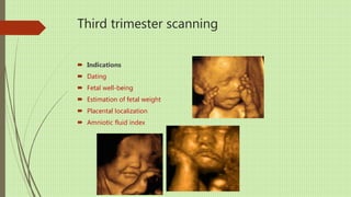
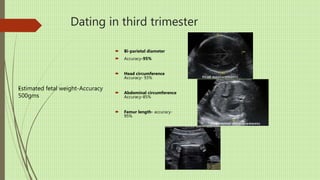
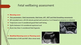
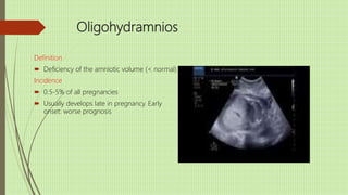
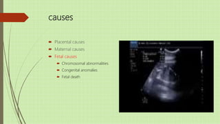
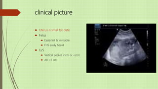
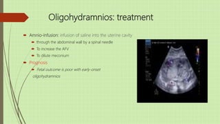
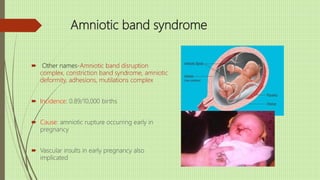
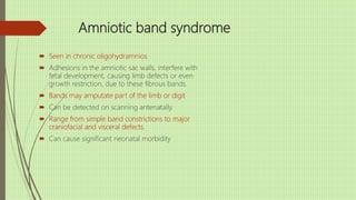
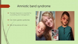
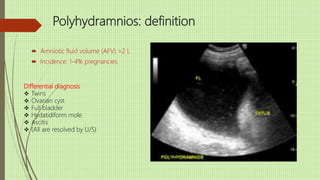
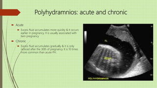
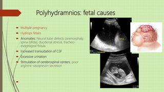
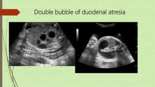
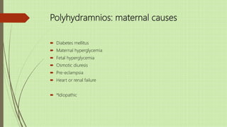
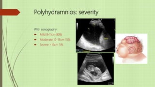
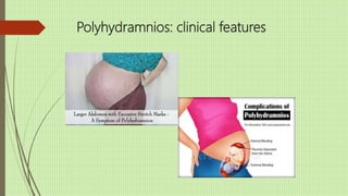
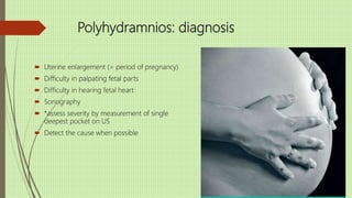
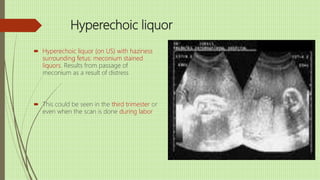
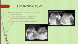
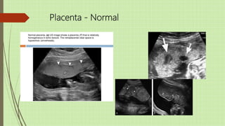
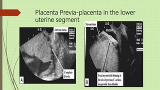
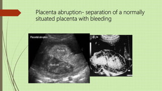
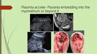
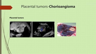
Third trimester
scanning,
Placenta and
Amniotic fluid
abnormalities
Mini Sood
Contents
 Third trimester scans
 Placenta-Normal and abnormal
 Liqour amnii normal and abnormal
Third trimester scanning
 Indications
 Dating
 Fetal well-being
 Estimation of fetal weight
 Placental localization
 Amniotic fluid index
Dating in third trimester
 Bi-parietal diameter
 Accuracy-95%
 Head circumference
Accuracy- 93%
 Abdominal circumference
Accuracy-85%
 Femur length- accuracy-
95%
Estimated fetal weight-Accuracy
500gms
-
Fetal wellbeing assessment
 Manning score
 Five parameters- Fetal movements, fetal tone, AFI , NST and fetal breathing movements.
 All recorded over a 20-30 minute period and scored as 2 or 0 based on presence or absence.
 Total score over 8 considered good fetal well-being,
 Score between 6-8 considered equivocal and
 Score less than 4 considered fetal hypoxia.
 Modified Manning score or Planning score
Combination of AFI and NST (Non stress test)
Physiology of amniotic fluid: functions
 Derived almost entirely from the fetus
 Protects fetus from trauma
 Cushions the umbilical cord from compression
 Antibacterial properties
 Reservoir of fluid, nutrients for the fetus
 Provides growth factors for development of systems (esp. lungs, musculoskeletal,
gastrointestinal)
Physiology of amniotic fluid: source
 Produced from fetal urination, nasal and oral secretions, placental and
amniotic membrane surfaces, intramembranous and trans membranous flow
and fetal lung secretions
 The entire amniotic fluid recycles in three hours.
 Derangements in volume adversely affect pregnancy, both when it is i or h,
or if it is meconium stained
 The amniotic fluid is 98-99% water and 1-2% solids like proteins,
carbohydrates lipids , phospholipids, enzymes, hormones, pigments and
chemicals like urea, uric acid, creatinine and electrolytes
 There many be fetal cells, amnion cells, hair, blood cells and meconium as
well
Physiology of amniotic fluid: content
 Daily amniotic volume flows in the near term fetus are:
 Fetal urine 800-1200ml/day
 Fetal lung liquid secretions-170ml/day
 Fetal swallowing-500-1000ml/day
 Intramembranous flow 200-400ml/day
 Oral-nasal secretions -25ml/day
 Trans-membranous flow- 10ml/day
Physiology of amniotic fluid: normal levels
 Amniotic volume increases to 1L or more by 36 wks
 In post-term there may by only 100-200 ml
 Normal volume varies with the duration of pregnancy
 Average 12 weeks: 50 ml
24 weeks: 500 ml
36 weeks: 1000 ml, and decreases thereafter
 Term: normally (singleton pregnancy): 500-1500 ml
Oligohydramnios
Definition
 Deficiency of the amniotic volume (< normal)
Incidence
 0.5-5% of all pregnancies
 Usually develops late in pregnancy. Early
onset: worse prognosis
causes
 Placental causes
 Abruption
 IUGR
 Post-term
 PROM
 Twin-twin transfusion
 Utero-placental insufficiency
 Maternal causes
 Fetal causes
causes
 Placental causes
 Maternal causes
 Hypertension, diabetes
 Infections, fevers
 Drugs
 Prostaglandin synthetase inhibitors,
 Angiotensin converting enzyme inhibitors
 Idiopathic
 Fetal causes
causes
 Placental causes
 Maternal causes
 Fetal causes
 Chromosomal abnormalities
 Congenital anomalies
 Fetal death
clinical picture
 Uterus is small for date
 Fetus
 Easily felt & immobile
 FHS easily heard
 U/S
 Vertical pocket <1cm or <2cm
 AFI <5 cm
Oligohydramnios: complications
 During pregnancy
 Fetal hypoxia (cord compression)
 Persistent position of the fetus
 Limb deformities:(pressure or amniotic bands)
 Talipes (clubfoot)
 Ankylosis of joins
 Pulmonary hypoplasia
 During labor
Increased variable deceleration
Increased cesarean section rate
Oligohydramnios: treatment
 Amnio-infusion: infusion of saline into the uterine cavity
 through the abdominal wall by a spinal needle
 To increase the AFV
 To dilute meconium
 Prognosis
 Fetal outcome is poor with early-onset
oligohydramnios
Amniotic band syndrome
 Other names-Amniotic band disruption
complex, constriction band syndrome, amniotic
deformity, adhesions, mutilations complex
 Incidence: 0.89/10,000 births
 Cause: amniotic rupture occurring early in
pregnancy
 Vascular insults in early pregnancy also
implicated
Amniotic band syndrome
 Seen in chronic oligohydramnios
 Adhesions in the amniotic sac walls, interfere with
fetal development, causing limb defects or even
growth restriction, due to these fibrous bands.
 Bands may amputate part of the limb or digit
 Can be detected on scanning antenatally.
 Range from simple band constrictions to major
craniofacial and visceral defects.
 Can cause significant neonatal morbidity
Amniotic band syndrome
 Prenatal diagnosis is essential for
counselling and management
 Can mimic genetic syndromes
 Risk of recurrence of is low
Polyhydramnios: definition
 Amniotic fluid volume (AFV) >2 L
 Incidence: 1-4% pregnancies.
Differential diagnosis
 Twins
 Ovarian cyst
 Full bladder
 Hydatidiform mole
 Ascitis
 (All are resolved by U/S)
Polyhydramnios: acute and chronic
 Acute
 Excess fluid accumulates more quickly & it occurs
earlier in pregnancy. It is usually associated with
twin pregnancy
 Chronic
 Excess fluid accumulates gradually & it is only
noticed after the 30th of pregnancy. It is 10 times
more common than acute PH.
Polyhydramnios: fetal causes
 Multiple pregnancy
 Hydrops fetalis
 Anomalies: Neural tube defects (anencephaly,
spina bifida), duodenal atresia, tracheo-
esophageal fistula
 Increased transudation of CSF
 Excessive urination
 Stimulation of cerebrospinal centers, poor
arginine vasopressin secretion

Double bubble of duodenal atresia
Polyhydramnios: maternal causes
 Diabetes mellitus
 Maternal hyperglycemia
 Fetal hyperglycemia
 Osmotic diuresis
 Pre-eclampsia
 Heart or renal failure
 *Idiopathic
Polyhydramnios: severity
With sonography:
 Mild 8-11cm 80%
 Moderate 12-15cm 15%
 Severe >16cm 5%
Polyhydramnios: clinical features
 Dyspnea
 Edema
 Oliguria
 Dyspepsia
Polyhydramnios: diagnosis
 Uterine enlargement (> period of pregnancy)
 Difficulty in palpating fetal parts
 Difficulty in hearing fetal heart
 Sonography
 *assess severity by measurement of single
deepest pocket on US
 Detect the cause when possible
Polyhydramnios: treatment
 Amniocentesis - 1500-2000 ml/day
 Indomethacin
 Decreases lung liquid production
 Decreases fetal urine production
 Increases fluid movement across fetal membranes
 Mild polyhydramnios rarely requires treatment
 Moderate: can usually manage until labor
 Severe (dyspnea, abdominal pain, ):  higher
perinatal mortality rate  hospitalization
Hyperechoic liquor
 Hyperechoic liquor (on US) with haziness
surrounding fetus: meconium stained
liquors. Results from passage of
meconium as a result of distress
 This could be seen in the third trimester or
even when the scan is done during labor
Hyperechoic liquor
 When hazy liquor is detected, try to find the
underlying cause
 Amnioscopy (if the cervix is dilated), can
confirm the diagnosis of meconium.
 Delivery on an urgent basis to rescue the
fetus.
Placenta - Normal
Placenta Previa-placenta in the lower
uterine segment
Placenta abruption- separation of a normally
situated placenta with bleeding
Placenta accrete- Placenta embedding into the
myometrium or beyond it
Placental tumors-Chorioangioma
Placental tumors
Abnormalities of placenta
 Circumvallate Placenta
 Succenturiate lobe
Conclusions
 Liquor abnormality is a common US feature.
 Amniotic fluid abnormalities can increase perinatal mortality and morbidity.
 Oligohydramnios can be due to infections, preterm membrane rupture, or fetal anomaly. It
leads to fetal lung hypoplasia, amniotic band syndrome, preterm labor, fetal demise
 Polyhydramnios can result from maternal diabetes or fetal anomaly. It causes maternal and fetal
complications.
 Meconium stained liquor is a consequence of acute or chronic fetal distress and results in
meconium aspiration syndrome with respiratory distress in the newborn and can be fatal.
 The ultrasonic evaluation of fetal wellbeing includes the assessment of the amniotic fluid index.
It accurately detects fetal distress, and diagnoses oligohydramnios as well as polyhydramnios
Conclusions
 Placenta Previa minor and major are detected by ultrasound with an over 99% accuracy.
 Associated placenta accrete can be detected and when there is a doubt an MRI can be done
to confirm accrete.
 Placental abruption is recognized by the presence of the retro-placental clot along with the
clinical picture.
 Rare placenta structural defects and tumors too can be accurately diagnosed.
amniotic fluid normal and abnormal

amniotic fluid normal and abnormal

  • 1.
  • 2.
    Contents  Third trimesterscans  Placenta-Normal and abnormal  Liqour amnii normal and abnormal
  • 3.
    Third trimester scanning Indications  Dating  Fetal well-being  Estimation of fetal weight  Placental localization  Amniotic fluid index
  • 4.
    Dating in thirdtrimester  Bi-parietal diameter  Accuracy-95%  Head circumference Accuracy- 93%  Abdominal circumference Accuracy-85%  Femur length- accuracy- 95% Estimated fetal weight-Accuracy 500gms -
  • 5.
    Fetal wellbeing assessment Manning score  Five parameters- Fetal movements, fetal tone, AFI , NST and fetal breathing movements.  All recorded over a 20-30 minute period and scored as 2 or 0 based on presence or absence.  Total score over 8 considered good fetal well-being,  Score between 6-8 considered equivocal and  Score less than 4 considered fetal hypoxia.  Modified Manning score or Planning score Combination of AFI and NST (Non stress test)
  • 6.
    Physiology of amnioticfluid: functions  Derived almost entirely from the fetus  Protects fetus from trauma  Cushions the umbilical cord from compression  Antibacterial properties  Reservoir of fluid, nutrients for the fetus  Provides growth factors for development of systems (esp. lungs, musculoskeletal, gastrointestinal)
  • 7.
    Physiology of amnioticfluid: source  Produced from fetal urination, nasal and oral secretions, placental and amniotic membrane surfaces, intramembranous and trans membranous flow and fetal lung secretions  The entire amniotic fluid recycles in three hours.  Derangements in volume adversely affect pregnancy, both when it is i or h, or if it is meconium stained  The amniotic fluid is 98-99% water and 1-2% solids like proteins, carbohydrates lipids , phospholipids, enzymes, hormones, pigments and chemicals like urea, uric acid, creatinine and electrolytes  There many be fetal cells, amnion cells, hair, blood cells and meconium as well
  • 8.
    Physiology of amnioticfluid: content  Daily amniotic volume flows in the near term fetus are:  Fetal urine 800-1200ml/day  Fetal lung liquid secretions-170ml/day  Fetal swallowing-500-1000ml/day  Intramembranous flow 200-400ml/day  Oral-nasal secretions -25ml/day  Trans-membranous flow- 10ml/day
  • 9.
    Physiology of amnioticfluid: normal levels  Amniotic volume increases to 1L or more by 36 wks  In post-term there may by only 100-200 ml  Normal volume varies with the duration of pregnancy  Average 12 weeks: 50 ml 24 weeks: 500 ml 36 weeks: 1000 ml, and decreases thereafter  Term: normally (singleton pregnancy): 500-1500 ml
  • 10.
    Oligohydramnios Definition  Deficiency ofthe amniotic volume (< normal) Incidence  0.5-5% of all pregnancies  Usually develops late in pregnancy. Early onset: worse prognosis
  • 11.
    causes  Placental causes Abruption  IUGR  Post-term  PROM  Twin-twin transfusion  Utero-placental insufficiency  Maternal causes  Fetal causes
  • 12.
    causes  Placental causes Maternal causes  Hypertension, diabetes  Infections, fevers  Drugs  Prostaglandin synthetase inhibitors,  Angiotensin converting enzyme inhibitors  Idiopathic  Fetal causes
  • 13.
    causes  Placental causes Maternal causes  Fetal causes  Chromosomal abnormalities  Congenital anomalies  Fetal death
  • 14.
    clinical picture  Uterusis small for date  Fetus  Easily felt & immobile  FHS easily heard  U/S  Vertical pocket <1cm or <2cm  AFI <5 cm
  • 15.
    Oligohydramnios: complications  Duringpregnancy  Fetal hypoxia (cord compression)  Persistent position of the fetus  Limb deformities:(pressure or amniotic bands)  Talipes (clubfoot)  Ankylosis of joins  Pulmonary hypoplasia  During labor Increased variable deceleration Increased cesarean section rate
  • 16.
    Oligohydramnios: treatment  Amnio-infusion:infusion of saline into the uterine cavity  through the abdominal wall by a spinal needle  To increase the AFV  To dilute meconium  Prognosis  Fetal outcome is poor with early-onset oligohydramnios
  • 17.
    Amniotic band syndrome Other names-Amniotic band disruption complex, constriction band syndrome, amniotic deformity, adhesions, mutilations complex  Incidence: 0.89/10,000 births  Cause: amniotic rupture occurring early in pregnancy  Vascular insults in early pregnancy also implicated
  • 18.
    Amniotic band syndrome Seen in chronic oligohydramnios  Adhesions in the amniotic sac walls, interfere with fetal development, causing limb defects or even growth restriction, due to these fibrous bands.  Bands may amputate part of the limb or digit  Can be detected on scanning antenatally.  Range from simple band constrictions to major craniofacial and visceral defects.  Can cause significant neonatal morbidity
  • 19.
    Amniotic band syndrome Prenatal diagnosis is essential for counselling and management  Can mimic genetic syndromes  Risk of recurrence of is low
  • 20.
    Polyhydramnios: definition  Amnioticfluid volume (AFV) >2 L  Incidence: 1-4% pregnancies. Differential diagnosis  Twins  Ovarian cyst  Full bladder  Hydatidiform mole  Ascitis  (All are resolved by U/S)
  • 21.
    Polyhydramnios: acute andchronic  Acute  Excess fluid accumulates more quickly & it occurs earlier in pregnancy. It is usually associated with twin pregnancy  Chronic  Excess fluid accumulates gradually & it is only noticed after the 30th of pregnancy. It is 10 times more common than acute PH.
  • 22.
    Polyhydramnios: fetal causes Multiple pregnancy  Hydrops fetalis  Anomalies: Neural tube defects (anencephaly, spina bifida), duodenal atresia, tracheo- esophageal fistula  Increased transudation of CSF  Excessive urination  Stimulation of cerebrospinal centers, poor arginine vasopressin secretion 
  • 23.
    Double bubble ofduodenal atresia
  • 24.
    Polyhydramnios: maternal causes Diabetes mellitus  Maternal hyperglycemia  Fetal hyperglycemia  Osmotic diuresis  Pre-eclampsia  Heart or renal failure  *Idiopathic
  • 25.
    Polyhydramnios: severity With sonography: Mild 8-11cm 80%  Moderate 12-15cm 15%  Severe >16cm 5%
  • 26.
    Polyhydramnios: clinical features Dyspnea  Edema  Oliguria  Dyspepsia
  • 27.
    Polyhydramnios: diagnosis  Uterineenlargement (> period of pregnancy)  Difficulty in palpating fetal parts  Difficulty in hearing fetal heart  Sonography  *assess severity by measurement of single deepest pocket on US  Detect the cause when possible
  • 28.
    Polyhydramnios: treatment  Amniocentesis- 1500-2000 ml/day  Indomethacin  Decreases lung liquid production  Decreases fetal urine production  Increases fluid movement across fetal membranes  Mild polyhydramnios rarely requires treatment  Moderate: can usually manage until labor  Severe (dyspnea, abdominal pain, ):  higher perinatal mortality rate  hospitalization
  • 29.
    Hyperechoic liquor  Hyperechoicliquor (on US) with haziness surrounding fetus: meconium stained liquors. Results from passage of meconium as a result of distress  This could be seen in the third trimester or even when the scan is done during labor
  • 30.
    Hyperechoic liquor  Whenhazy liquor is detected, try to find the underlying cause  Amnioscopy (if the cervix is dilated), can confirm the diagnosis of meconium.  Delivery on an urgent basis to rescue the fetus.
  • 31.
  • 32.
    Placenta Previa-placenta inthe lower uterine segment
  • 33.
    Placenta abruption- separationof a normally situated placenta with bleeding
  • 34.
    Placenta accrete- Placentaembedding into the myometrium or beyond it
  • 35.
  • 36.
    Abnormalities of placenta Circumvallate Placenta  Succenturiate lobe
  • 37.
    Conclusions  Liquor abnormalityis a common US feature.  Amniotic fluid abnormalities can increase perinatal mortality and morbidity.  Oligohydramnios can be due to infections, preterm membrane rupture, or fetal anomaly. It leads to fetal lung hypoplasia, amniotic band syndrome, preterm labor, fetal demise  Polyhydramnios can result from maternal diabetes or fetal anomaly. It causes maternal and fetal complications.  Meconium stained liquor is a consequence of acute or chronic fetal distress and results in meconium aspiration syndrome with respiratory distress in the newborn and can be fatal.  The ultrasonic evaluation of fetal wellbeing includes the assessment of the amniotic fluid index. It accurately detects fetal distress, and diagnoses oligohydramnios as well as polyhydramnios
  • 38.
    Conclusions  Placenta Previaminor and major are detected by ultrasound with an over 99% accuracy.  Associated placenta accrete can be detected and when there is a doubt an MRI can be done to confirm accrete.  Placental abruption is recognized by the presence of the retro-placental clot along with the clinical picture.  Rare placenta structural defects and tumors too can be accurately diagnosed.