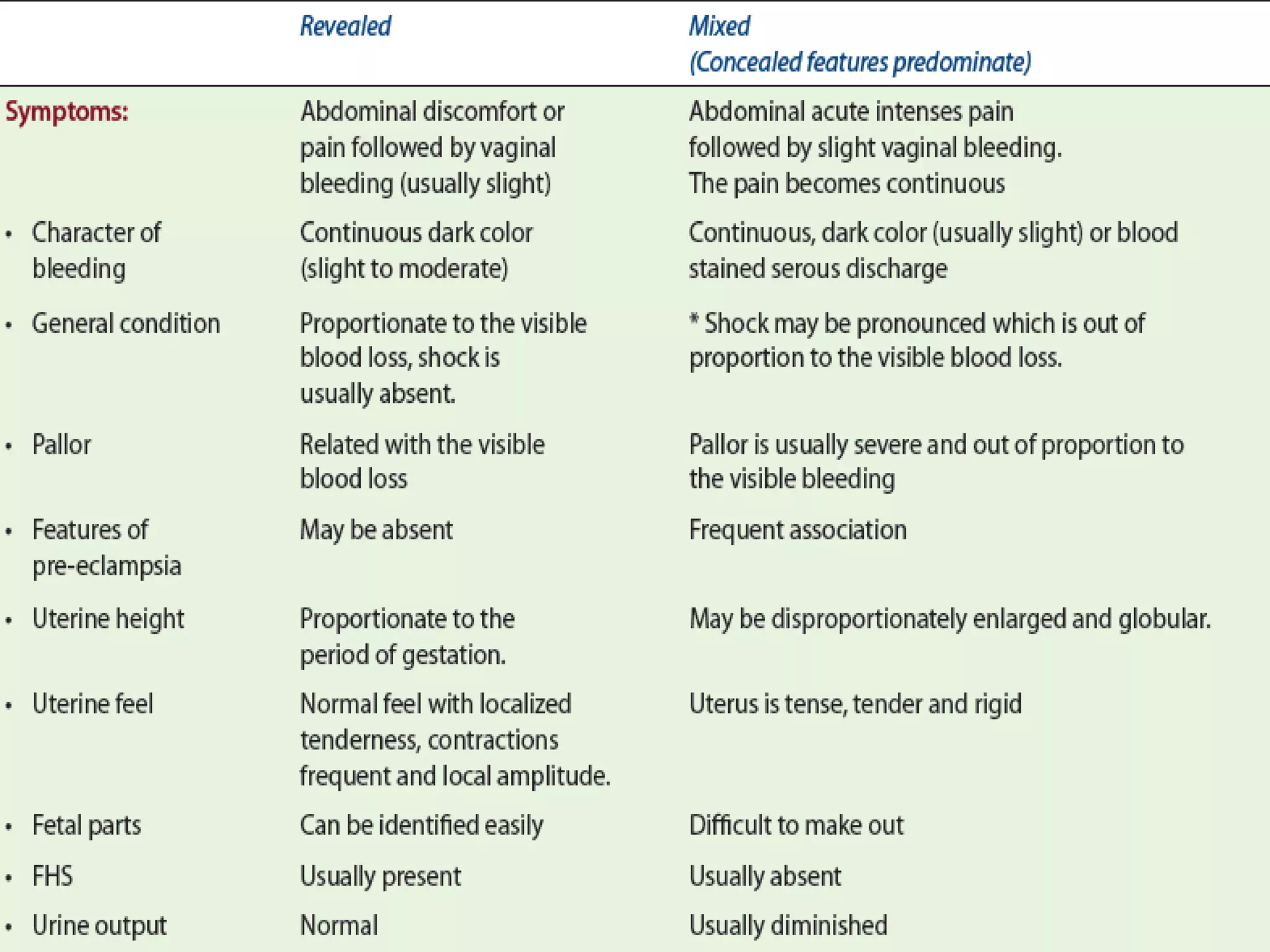
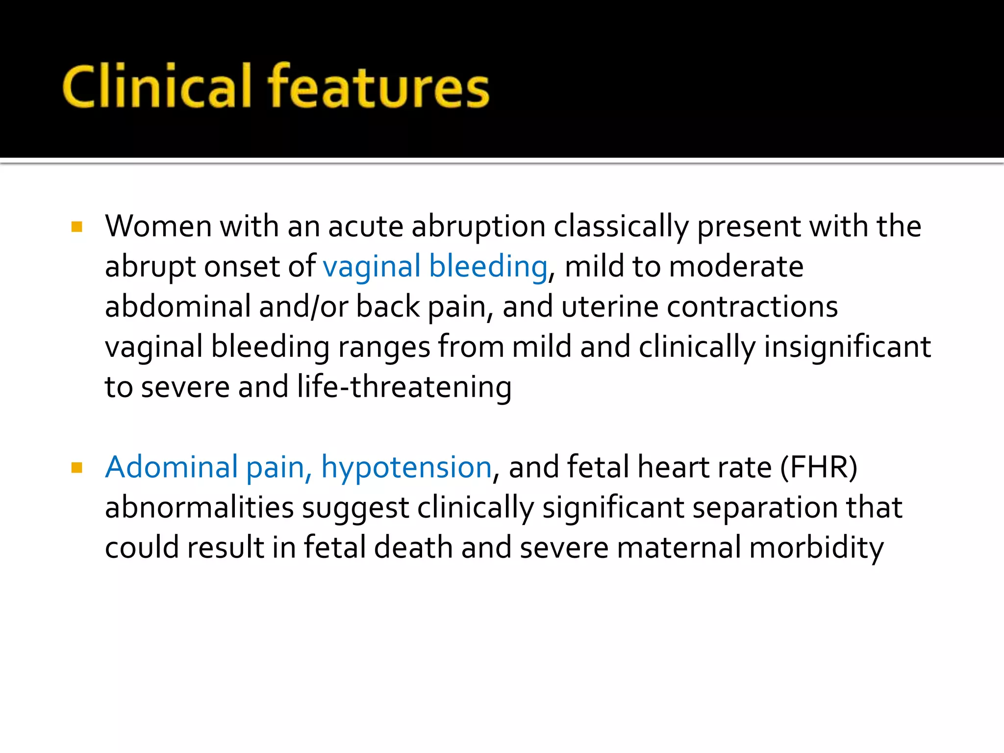
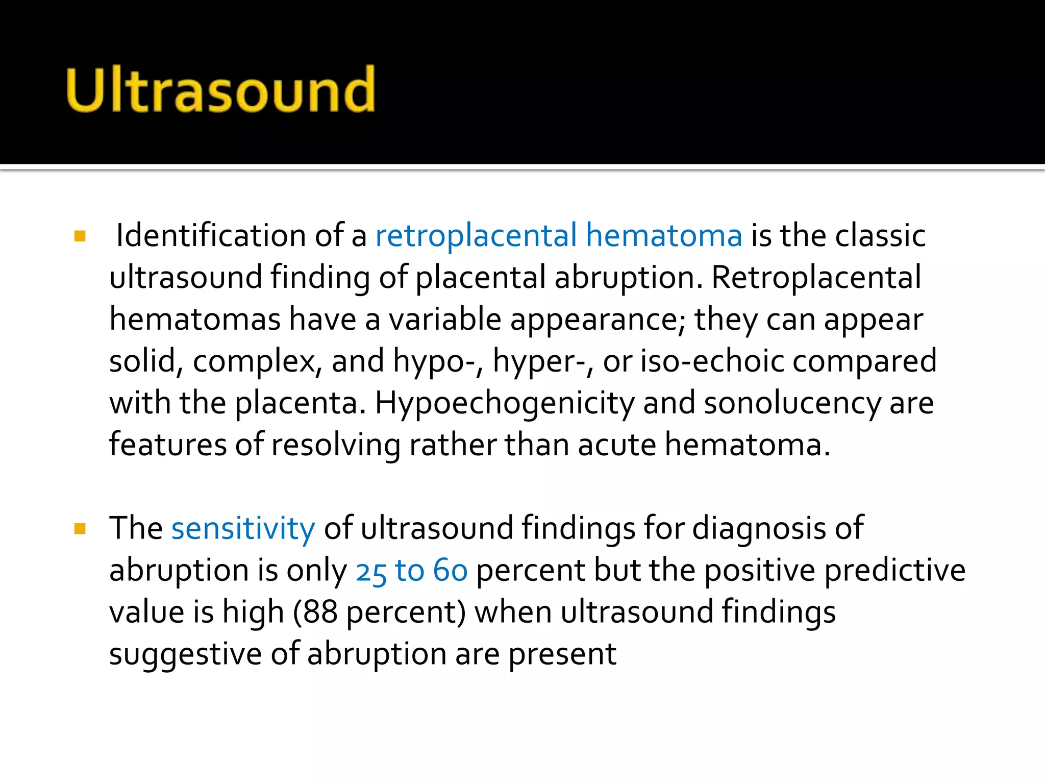
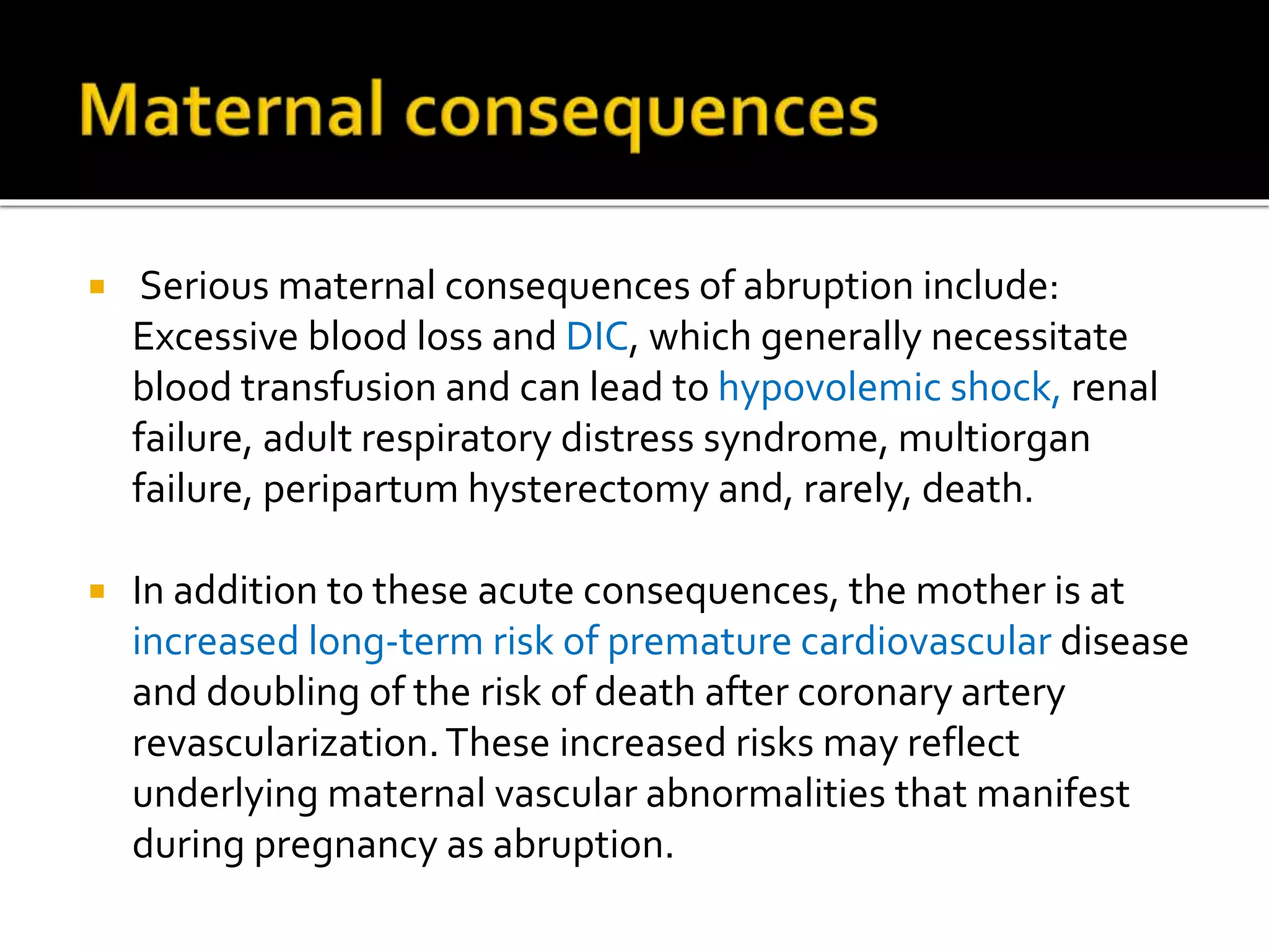
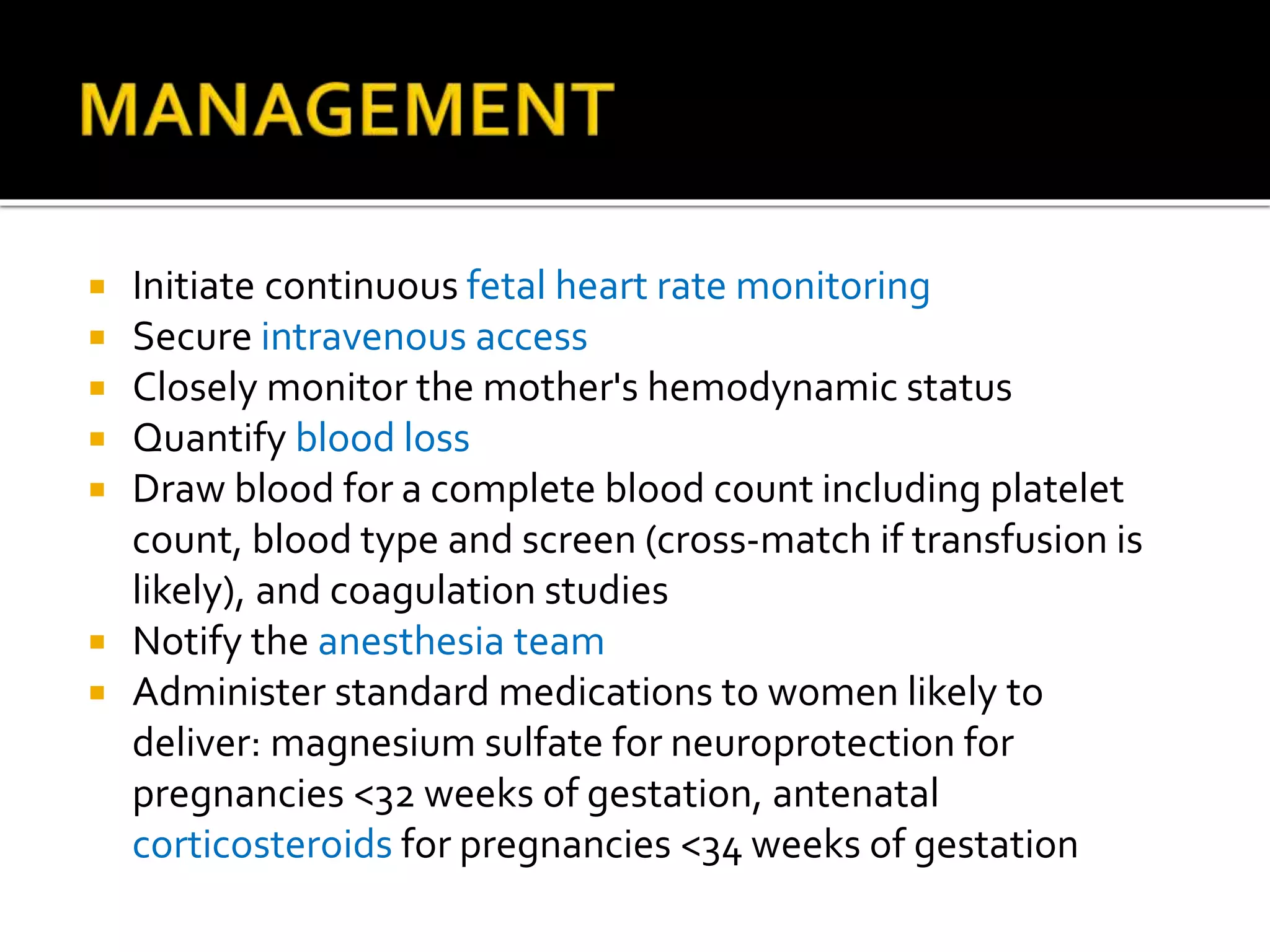
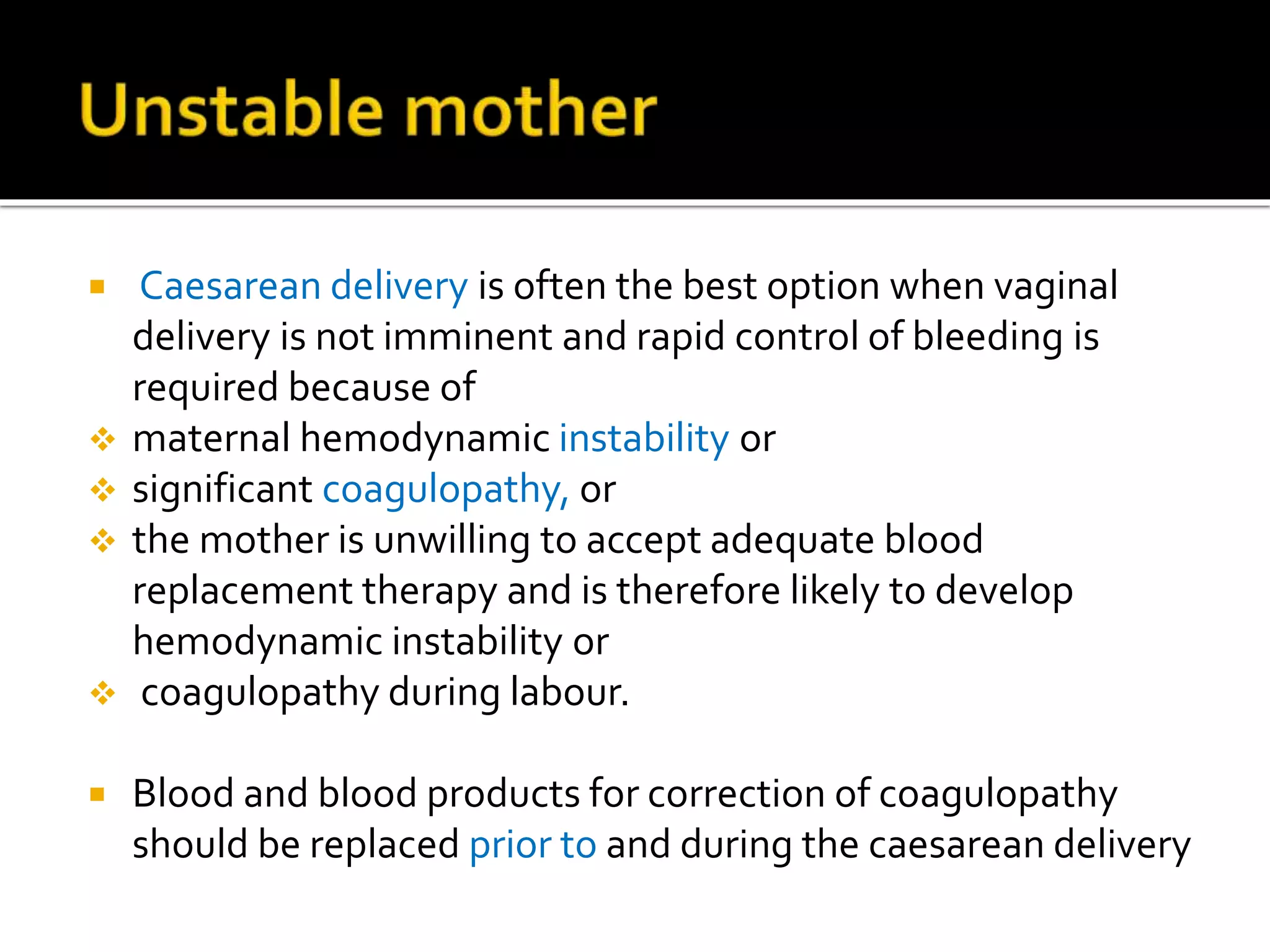
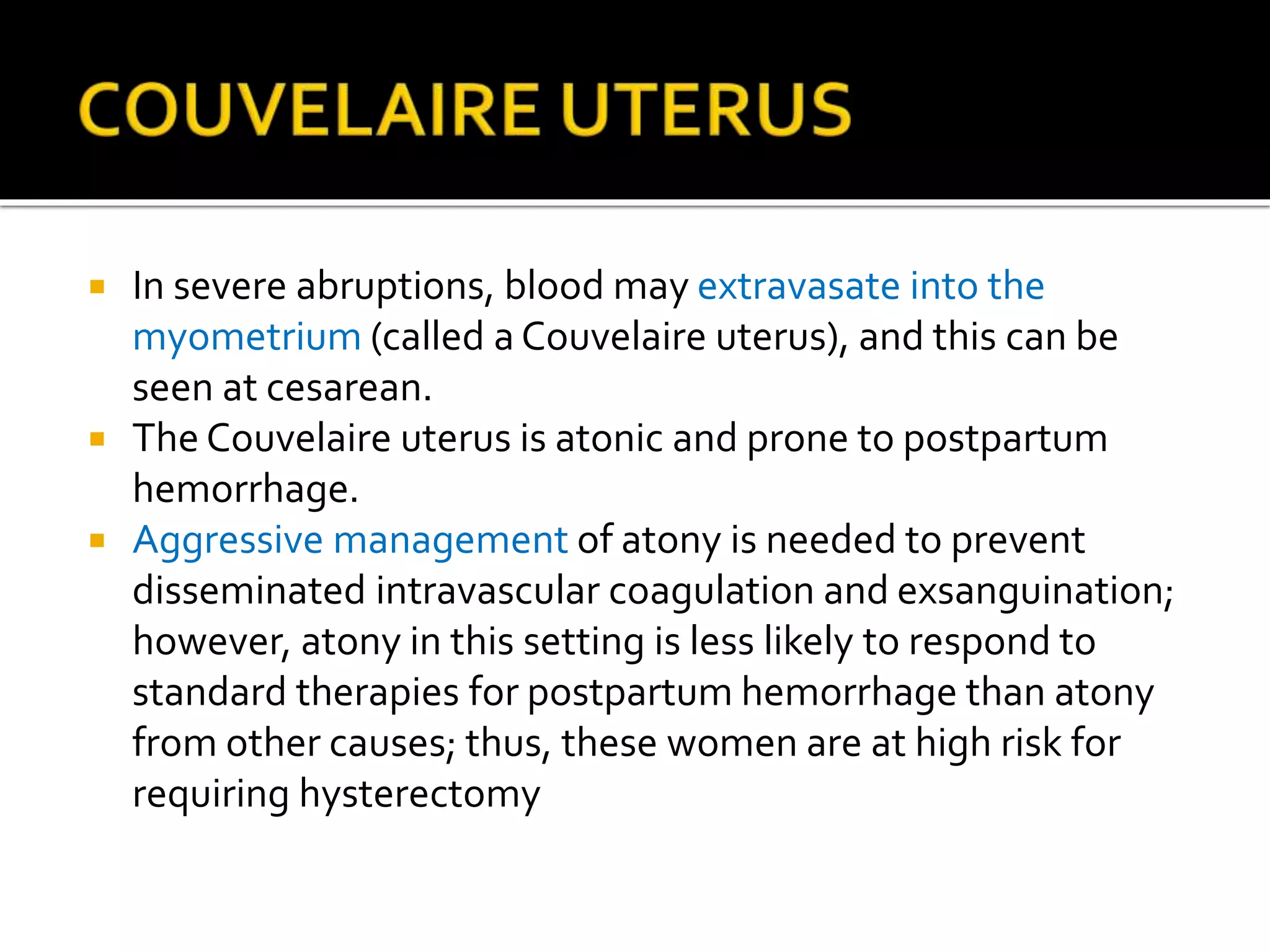
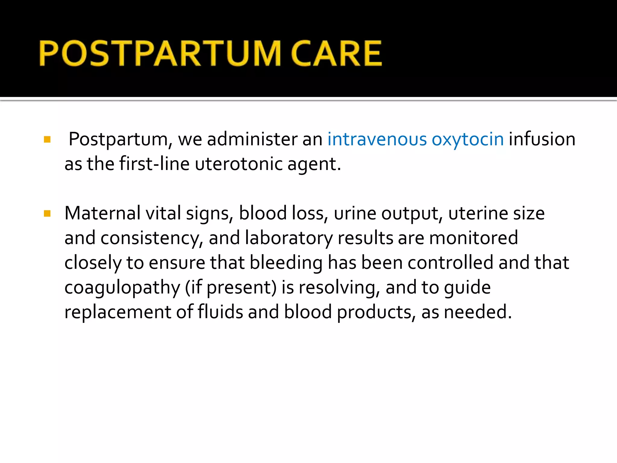
Placental abruption occurs when the placenta detaches from the uterus prior to delivery of the baby. It can lead to significant maternal and neonatal morbidity and mortality. Risk factors include hypertension, smoking, and prior abruption. Symptoms may include vaginal bleeding, abdominal pain, and changes in fetal heart rate. Ultrasound can show a retroplacental hematoma. Management involves monitoring, administering medications to help the baby's lungs/brain if preterm, and delivery depending on gestational age and stability of the mother and baby. Women with a history of abruption have a higher risk of recurrence.
Prelabour Rupture of Membrane (PROM) by Sunil Kumar Dahasunil kumar daha
Please find the power point on Prelabour Rupture of Membrane (PROM). I tried to present it on understandable way and all the contents are reviewed by experts and from very reliable references. Thank you
A serious pregnancy complication in which the placenta detaches from the womb (uterus).
Placental abruption occurs when the placenta detaches from the inner wall of the womb before delivery. The condition can deprive the baby of oxygen and nutrients.
Symptoms include vaginal bleeding, stomach pain and back pain in the last 12 weeks of pregnancy.
Depending on the degree of placental separation and how close the baby is to full-term, treatment may include bed rest or a Caesarean (C-section).
Please find the power point on Management of Preterm labor. I tried to present it on understandable way and all the contents are reviewed by experts and from very reliable references. Thank you
Retained placenta can be defined as lack of placental expulsion within 30 minutes of delivery of an infant. it is more common in preterm. Retained Placenta can lead to massive PPH and increase maternal morbidity and mortality.
Case Report on Invasive Mole. Gestational Trophoblastic Neoplasia (GTN) encom...Niranjan Chavan
Gestational Trophoblastic Neoplasia (GTN) encompasses a suite of rare but significant gynecological malignancies arising from aberrant placental trophoblast cells. As medical professionals and researchers, our comprehension of GTN's complexities is crucial for accurate diagnosis and effective treatment. This introduction serves to illuminate the key features, diagnostic procedures, and treatment protocols associated with GTN, helping to navigate the intricate landscape of this disease.
Peripartum cardiomyopathy (PPCM) is a rare form of heart failure that occurs during the last month of pregnancy or within the first five months postpartum. It presents significant challenges in diagnosis and treatment due to its overlap with symptoms of normal pregnancy and postpartum changes. This condition varies in incidence across different racial groups and geographical locations, with a notable occurrence in the United States and southern India.
DR. NNC LAPAROSCOPY IN PREGNANCY IAGE VARANASI, 17TH MARCH 2024.pptxNiranjan Chavan
Our journey will navigate the evolution of laparoscopy in the context of pregnancy, detailing key milestones, breakthroughs, and advancements in technology and techniques. The presentation highlights how laparoscopy has revolutionized the diagnosis and treatment of conditions such as ectopic pregnancy, ovarian cysts and other gynecological disorders during pregnancy.
Optimising Delivery Of 1kg Fetus - Special Considerations.pptxNiranjan Chavan
After an uncomplicated vaginal birth in a health facility, healthy mothers and newborns should receive care in the facility for at least 24 hours after birth.
VACCINE IN WOMEN TOWARDS SDG 2030 DR.N N CHAVAN 10012024 AICOG HYDERABAD.pptxNiranjan Chavan
In our presentation today, we will unravel the transformative power of vaccines in women, aligning with the Sustainable Development Goals (SDGs) for 2030. By exploring the pivotal role of vaccinations, we aim to elucidate how they contribute to women's health, empowerment, and overall well-being. Through this lens, we envision a future where widespread vaccine access propels us closer to achieving the SDGs and ensures a healthier, more equitable world for women globally.
RRRR IN OBSTETRIC HEMORRHAGE 09012024 AICOG 2024 HEYDERABAD.pptxNiranjan Chavan
This presentation focuses on a critical aspect of maternal care: "Reducing Maternal Mortality through Rapid Response in Obstetric Haemorrhage" (RRRR). As we navigate through this presentation, let us collectively work towards advancing our understanding and application of RRRR in obstetric care to safeguard the well-being of mothers during childbirth.
Anemia is a condition in which the number of red blood cells and/OR their oxy...Niranjan Chavan
Anemia is a condition in which the number of red blood cells and/OR their
oxygen-carrying capacity is insufficient to meet the body’s physiological needs.
HELLP syndrome is a pregnancy complication. It is a type of preeclampsia. It ...Niranjan Chavan
HELLP syndrome is a pregnancy complication. It is a type of preeclampsia. It usually occurs during the third trimester of pregnancy. But it also can develop in the first week after childbirth
Guidelines & Identification of Early Sepsis DR. NN CHAVAN 02122023.pptxNiranjan Chavan
Here is a highly informative session on guidelines and identification of early sepsis as it is critical for timely intervention and improved patient outcomes.
PAST, PRESENT AND FUTURE IN OBGYN INFECTIONS 01102023.pptxNiranjan Chavan
Today, we face new infectious threats; but also benefit from advanced diagnostics and treatments. Looking ahead, it’s crucial to continue
adapting to emerging pathogens, implement stringent preventive measures, and
leverage cutting-edge technologies to ensure the safety and well-being of our patients in the ever-evolving landscape of obstetrics and gynecology.
Vaccination during pregnancy is crucial to protect both the mother and the developing baby. It helps prevent serious complications and ensures a healthier start in life. #VaccinateForTwo 🤰💉
Explore a comprehensive presentation on Invasive Cervical Carcinoma, shedding light on its causes, symptoms, diagnosis, treatment options, and preventive measures.
- Video recording of this lecture in English language: https://youtu.be/kqbnxVAZs-0
- Video recording of this lecture in Arabic language: https://youtu.be/SINlygW1Mpc
- Link to download the book free: https://nephrotube.blogspot.com/p/nephrotube-nephrology-books.html
- Link to NephroTube website: www.NephroTube.com
- Link to NephroTube social media accounts: https://nephrotube.blogspot.com/p/join-nephrotube-on-social-media.html
Title: Sense of Taste
Presenter: Dr. Faiza, Assistant Professor of Physiology
Qualifications:
MBBS (Best Graduate, AIMC Lahore)
FCPS Physiology
ICMT, CHPE, DHPE (STMU)
MPH (GC University, Faisalabad)
MBA (Virtual University of Pakistan)
Learning Objectives:
Describe the structure and function of taste buds.
Describe the relationship between the taste threshold and taste index of common substances.
Explain the chemical basis and signal transduction of taste perception for each type of primary taste sensation.
Recognize different abnormalities of taste perception and their causes.
Key Topics:
Significance of Taste Sensation:
Differentiation between pleasant and harmful food
Influence on behavior
Selection of food based on metabolic needs
Receptors of Taste:
Taste buds on the tongue
Influence of sense of smell, texture of food, and pain stimulation (e.g., by pepper)
Primary and Secondary Taste Sensations:
Primary taste sensations: Sweet, Sour, Salty, Bitter, Umami
Chemical basis and signal transduction mechanisms for each taste
Taste Threshold and Index:
Taste threshold values for Sweet (sucrose), Salty (NaCl), Sour (HCl), and Bitter (Quinine)
Taste index relationship: Inversely proportional to taste threshold
Taste Blindness:
Inability to taste certain substances, particularly thiourea compounds
Example: Phenylthiocarbamide
Structure and Function of Taste Buds:
Composition: Epithelial cells, Sustentacular/Supporting cells, Taste cells, Basal cells
Features: Taste pores, Taste hairs/microvilli, and Taste nerve fibers
Location of Taste Buds:
Found in papillae of the tongue (Fungiform, Circumvallate, Foliate)
Also present on the palate, tonsillar pillars, epiglottis, and proximal esophagus
Mechanism of Taste Stimulation:
Interaction of taste substances with receptors on microvilli
Signal transduction pathways for Umami, Sweet, Bitter, Sour, and Salty tastes
Taste Sensitivity and Adaptation:
Decrease in sensitivity with age
Rapid adaptation of taste sensation
Role of Saliva in Taste:
Dissolution of tastants to reach receptors
Washing away the stimulus
Taste Preferences and Aversions:
Mechanisms behind taste preference and aversion
Influence of receptors and neural pathways
Impact of Sensory Nerve Damage:
Degeneration of taste buds if the sensory nerve fiber is cut
Abnormalities of Taste Detection:
Conditions: Ageusia, Hypogeusia, Dysgeusia (parageusia)
Causes: Nerve damage, neurological disorders, infections, poor oral hygiene, adverse drug effects, deficiencies, aging, tobacco use, altered neurotransmitter levels
Neurotransmitters and Taste Threshold:
Effects of serotonin (5-HT) and norepinephrine (NE) on taste sensitivity
Supertasters:
25% of the population with heightened sensitivity to taste, especially bitterness
Increased number of fungiform papillae
Recomendações da OMS sobre cuidados maternos e neonatais para uma experiência pós-natal positiva.
Em consonância com os ODS – Objetivos do Desenvolvimento Sustentável e a Estratégia Global para a Saúde das Mulheres, Crianças e Adolescentes, e aplicando uma abordagem baseada nos direitos humanos, os esforços de cuidados pós-natais devem expandir-se para além da cobertura e da simples sobrevivência, de modo a incluir cuidados de qualidade.
Estas diretrizes visam melhorar a qualidade dos cuidados pós-natais essenciais e de rotina prestados às mulheres e aos recém-nascidos, com o objetivo final de melhorar a saúde e o bem-estar materno e neonatal.
Uma “experiência pós-natal positiva” é um resultado importante para todas as mulheres que dão à luz e para os seus recém-nascidos, estabelecendo as bases para a melhoria da saúde e do bem-estar a curto e longo prazo. Uma experiência pós-natal positiva é definida como aquela em que as mulheres, pessoas que gestam, os recém-nascidos, os casais, os pais, os cuidadores e as famílias recebem informação consistente, garantia e apoio de profissionais de saúde motivados; e onde um sistema de saúde flexível e com recursos reconheça as necessidades das mulheres e dos bebês e respeite o seu contexto cultural.
Estas diretrizes consolidadas apresentam algumas recomendações novas e já bem fundamentadas sobre cuidados pós-natais de rotina para mulheres e neonatos que recebem cuidados no pós-parto em unidades de saúde ou na comunidade, independentemente dos recursos disponíveis.
É fornecido um conjunto abrangente de recomendações para cuidados durante o período puerperal, com ênfase nos cuidados essenciais que todas as mulheres e recém-nascidos devem receber, e com a devida atenção à qualidade dos cuidados; isto é, a entrega e a experiência do cuidado recebido. Estas diretrizes atualizam e ampliam as recomendações da OMS de 2014 sobre cuidados pós-natais da mãe e do recém-nascido e complementam as atuais diretrizes da OMS sobre a gestão de complicações pós-natais.
O estabelecimento da amamentação e o manejo das principais intercorrências é contemplada.
Recomendamos muito.
Vamos discutir essas recomendações no nosso curso de pós-graduação em Aleitamento no Instituto Ciclos.
Esta publicação só está disponível em inglês até o momento.
Prof. Marcus Renato de Carvalho
www.agostodourado.com
Basavarajeeyam is a Sreshta Sangraha grantha (Compiled book ), written by Neelkanta kotturu Basavaraja Virachita. It contains 25 Prakaranas, First 24 Chapters related to Rogas& 25th to Rasadravyas.
micro teaching on communication m.sc nursing.pdfAnurag Sharma
Microteaching is a unique model of practice teaching. It is a viable instrument for the. desired change in the teaching behavior or the behavior potential which, in specified types of real. classroom situations, tends to facilitate the achievement of specified types of objectives.
Knee anatomy and clinical tests 2024.pdfvimalpl1234
This includes all relevant anatomy and clinical tests compiled from standard textbooks, Campbell,netter etc..It is comprehensive and best suited for orthopaedicians and orthopaedic residents.
3. Placental abruption (also referred to as abruptio
placentae) refers to partial or complete placental
detachment prior to delivery of the fetus.
The diagnosis is typically reserved for
pregnancies over 20 weeks of gestation.
Abruption is a significant cause of both maternal
morbidity and neonatal morbidity and mortality,
particularly when it occurs preterm
4. Placental abruption complicates
approximately 1 in 100 to 120 pregnancies,
with two-thirds classified as severe based
on associated maternal, fetal, and
neonatal morbidity
https://www.uptodate.com/contents/placental-abruption-pathophysiology-
clinical-features-diagnosis-and-consequences/abstract/1
5. The immediate cause of the premature placental
separation is rupture of maternal vessels in the
decidua basalis.
Rarely, the bleeding originates from the fetal-
placental vessels.
The accumulating blood splits the decidua, separating
off a thin layer of decidua with its placental
attachment.
Complete or nearly complete placental separations
are caused by high pressure arterial hemorrhage in
the central area of the placenta that extensively
dissect through the placental-decidual interface
6. Thrombin plays a key role in the clinical consequences of
placental abruption, and may be important in its
pathogenesis, as well.
It is formed via two pathways:
-Decidual bleeding leads to release of tissue factor
(thromboplastin) from decidual cells, which generates
thrombin
-Decidual hypoxia induces production of vascular
endothelial growth factor (VEGF), which acts directly on
decidual endothelial cells to induce aberrant expression of
tissue factor, which then generates thrombin
7. Uterine hypertonus and contractions
Enhanced expression of matrix metalloproteinases, up-
regulation of genes involved in apoptosis , and induced
expression of inflammatory cytokines (predominantly
interleukin-8), leading to tissue necrosis and degradation of
extracellular matrix
Triggering of coagulation
Functional progesterone withdrawal by reduced expression
of progesterone receptors in decidual cells
8. The prevalence is more with
(a) high birth order pregnancies with gravida 5 and above —
three times more common than in first birth
(b) advancing age of the mother
(c) poor socio-economic condition
(d) Malnutrition
(e) smoking (vaso-spasm).
Hypertension in pregnancy is the most important predisposing
factor. Pre-eclampsia, gestational hypertension and essential
hypertension, all are associated with placental abruption.
The association of pre-eclampsia in abruptio placenta varies
from10-50 percent.
9. The mechanism of the placental separation in pre-
eclampsia is : Spasm of the vessels in the utero placental
bed (decidual spiral artery) → anoxic endothelial damage →
rupture of vessels or extravasation of blood in the decidua
basalis (retroplacental hematoma).
Trauma:Traumatic separation of the placenta usually
leads to its marginal separation with escape of blood
outside. The trauma may be due to: (i) Attempted external
cephalic version specially under anesthesia using great force
(ii) Road traffic accidents or blow on the abdomen (iii)
Needle puncture at amniocentesis
10. Sudden uterine decompression: Sudden decompression of
the uterus leads to diminished surface area of the uterus
adjacent to the placental attachment and results in
separation of the placenta.This may occur following—(a)
delivery of the first baby of twins (b) sudden escape of liquor
amnii in hydramnios and (c) premature rupture of
membranes.
Short cord, either relative or absolute, can bring about
placental separation during labor by mechanical pull.
11. Placental anomaly: Circumvallate placenta .
Sick placenta: Poor placentation, evidenced by abnormal
uterine artery Doppler waveforms is associated with
placental abruption.
Folic acid deficiency even without evidence of overt
megaloblastic erythropoiesis — this has been observed to be
associated.
Uterine factor: Placenta implanted over a septum (Septate
Uterus) or a submucous fibroid.
12. Torsion of the uterus leads to increased venous pressure and
rupture of the veins with separation of the placenta.
Cocaine abuse is associated with increased risk of transient
hypertension, vasospasm and placental abruption.
Thrombophilias inherited or acquired have been associated
with increased risk of placental infarcts or
abruption.
Prior abruption: Risk of recurrence for a woman with
previous abruption varies between 5 to 17%.
14. Blood may accumulate behind the placenta when it is
totally separated from the uterine wall except at the margin
— concealed type.
Blood may dissect downwards in between the membranes
and the uterine wall and ultimately escapes out through
the cervix or may be kept concealed by the pressure of
the fetal head on the lower uterine segment
— revealed type.
Blood may gain access to the amniotic cavity after rupturing
the membranes
Blood may percolate through the layers of myometrium
upto the serous coat – couvelaire uterus
15. Grade—0: Clinical features may be absent.The diagnosis is
made after inspection of placenta following delivery.
Grade—1 (40%): (i)Vaginal bleeding is slight (ii) Uterus:
irritable, tenderness may be minimal or absent (iii) Maternal
BP and fibrinogen levels unaffected (iv) FHS is good.
16. Grade—2 (45%): (i)Vaginal bleeding mild to moderate (ii)
Uterine tenderness is always present (iii) Maternal pulse ↑,
BP is maintained (iv) Fibrinogen level may be decreased (v)
Shock is absent (vi) Fetal distress or even fetal death occurs.
Grade—3 (15%): (i) Bleeding is moderate to severe or may be
concealed (ii) Uterine tenderness is marked (iii) Shock is
pronounced (iv) Fetal death is the rule (v) Associated
coagulation defect or anuria may complicate.
17.
18.
19. Women with an acute abruption classically present with the
abrupt onset of vaginal bleeding, mild to moderate
abdominal and/or back pain, and uterine contractions
vaginal bleeding ranges from mild and clinically insignificant
to severe and life-threatening
Adominal pain, hypotension, and fetal heart rate (FHR)
abnormalities suggest clinically significant separation that
could result in fetal death and severe maternal morbidity
20. Identification of a retroplacental hematoma is the classic
ultrasound finding of placental abruption. Retroplacental
hematomas have a variable appearance; they can appear
solid, complex, and hypo-, hyper-, or iso-echoic compared
with the placenta. Hypoechogenicity and sonolucency are
features of resolving rather than acute hematoma.
The sensitivity of ultrasound findings for diagnosis of
abruption is only 25 to 60 percent but the positive predictive
value is high (88 percent) when ultrasound findings
suggestive of abruption are present
21.
22. Serious maternal consequences of abruption include:
Excessive blood loss and DIC, which generally necessitate
blood transfusion and can lead to hypovolemic shock, renal
failure, adult respiratory distress syndrome, multiorgan
failure, peripartum hysterectomy and, rarely, death.
In addition to these acute consequences, the mother is at
increased long-term risk of premature cardiovascular disease
and doubling of the risk of death after coronary artery
revascularization.These increased risks may reflect
underlying maternal vascular abnormalities that manifest
during pregnancy as abruption.
23. Serious fetal and neonatal consequences of abruption
include:
Increased perinatal morbidity and mortality related to
hypoxemia, asphyxia, low birth weight, and/or preterm
delivery.
Fetal growth restriction (with chronic abruption)
Fetal asphyxia, preterm birth, and growth restriction can be
associated with short- and long-term sequelae, and
abruption appears to compound the risk
24. Initiate continuous fetal heart rate monitoring
Secure intravenous access
Closely monitor the mother's hemodynamic status
Quantify blood loss
Draw blood for a complete blood count including platelet
count, blood type and screen (cross-match if transfusion is
likely), and coagulation studies
Notify the anesthesia team
Administer standard medications to women likely to
deliver: magnesium sulfate for neuroprotection for
pregnancies <32 weeks of gestation, antenatal
corticosteroids for pregnancies <34 weeks of gestation
25. The most important factors impacting the decision to deliver
a patient with placental abruption versus expectant
management are:
Gestational age
Fetal and maternal status, which reflect the severity of the
abruption.
26. The optimal route of delivery in these cases minimizes the
risk of maternal morbidity or mortality, since fetal well-being
is no longer a factor.
Blood and blood product replacement is often necessary and
expeditious delivery is desirable because the frequency of
coagulopathy and continuous heavy bleeding is much higher
in abruptions in which fetal death has occurred.
Placental separation is often greater than 50 percent.
27. Caesarean delivery is often the best option when vaginal
delivery is not imminent and rapid control of bleeding is
required because of
maternal hemodynamic instability or
significant coagulopathy, or
the mother is unwilling to accept adequate blood
replacement therapy and is therefore likely to develop
hemodynamic instability or
coagulopathy during labour.
Blood and blood products for correction of coagulopathy
should be replaced prior to and during the caesarean delivery
28. When the fetus and mother are both stable, the decision to
deliver depends primarily on gestational age, with
consideration of ongoing maternal symptoms
Less than 34 weeks of gestation —When the fetus and
mother are both stable and there is no evidence of ongoing
major blood loss or coagulopathy, conservative
management with the aim of delivering a more mature fetus
is the main goal before 34 weeks of gestation .
29. We take the following approach:
Administer corticosteroids – Corticosteroids to promote
fetal lung maturation and reduce complications of
prematurity are administered to pregnancies at 23 to 34
weeks of gestation, given the increased risk of need for
preterm delivery.
Tocolysis – For women in preterm labor, we administer a 48-
hour course of nifedipine to enable administration of a full
course of corticosteroids.
30.
31. In severe abruptions, blood may extravasate into the
myometrium (called a Couvelaire uterus), and this can be
seen at cesarean.
The Couvelaire uterus is atonic and prone to postpartum
hemorrhage.
Aggressive management of atony is needed to prevent
disseminated intravascular coagulation and exsanguination;
however, atony in this setting is less likely to respond to
standard therapies for postpartum hemorrhage than atony
from other causes; thus, these women are at high risk for
requiring hysterectomy
32. Naked eye features:
The uterus is of dark port wine colour which may be patchy
or diffuse.
It tends to occur initially on the cornua before spreading to
other areas, more specially over the placental site.
Subperitoneal petechial haemorrhages are found
under the uterine peritoneum and may extend into the broad
ligament.
There may be free blood in the peritoneal cavity or
broad ligament hematoma.
33. Microscopic appearance:
The uterine muscles over the affected area are necrosed and
there is infiltration of blood and fluid in between the muscle
bundles.
The serosa may split on occasions, to allow the blood to
enter the peritoneal cavity.
The blood vessels show acute degenerative changes with
thrombosis.
Uterus as observed during cesarean section is not an
indication per se for hysterectomy.
34. Postpartum, we administer an intravenous oxytocin infusion
as the first-line uterotonic agent.
Maternal vital signs, blood loss, urine output, uterine size
and consistency, and laboratory results are monitored
closely to ensure that bleeding has been controlled and that
coagulopathy (if present) is resolving, and to guide
replacement of fluids and blood products, as needed.
35. Women with placental abruption are at several fold higher
risk of abruption in a subsequent pregnancy.
3 to 15 percent of women have a recurrence, compared with
a baseline incidence of 0.4 to 1.3 percent in the general
population.
In one longitudinal population-based study, the risk of
placental abruption in a subsequent pregnancy was about 6
percent in women with an abruption in their first pregnancy
versus 0.06 percent in women without an abruption.