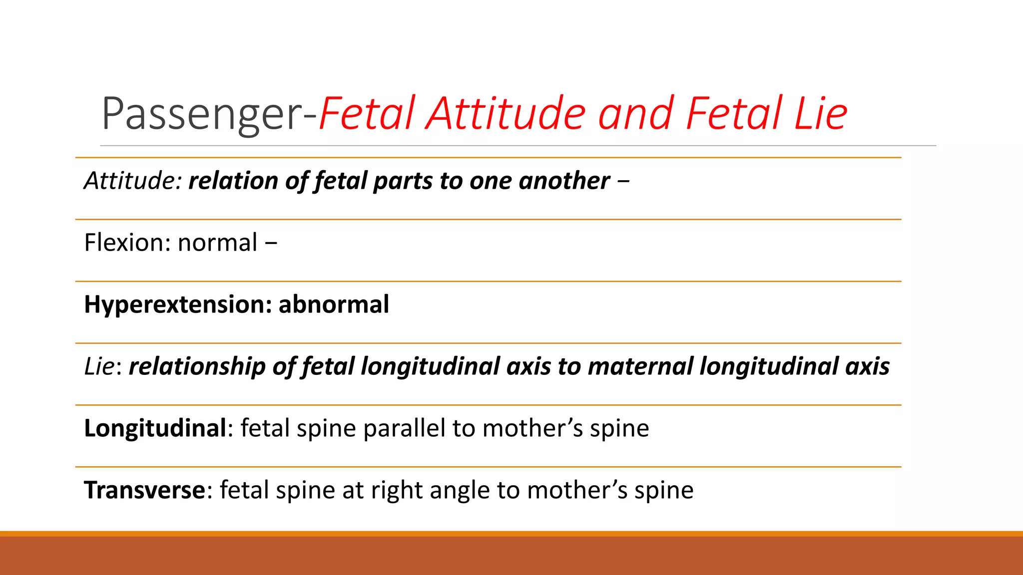
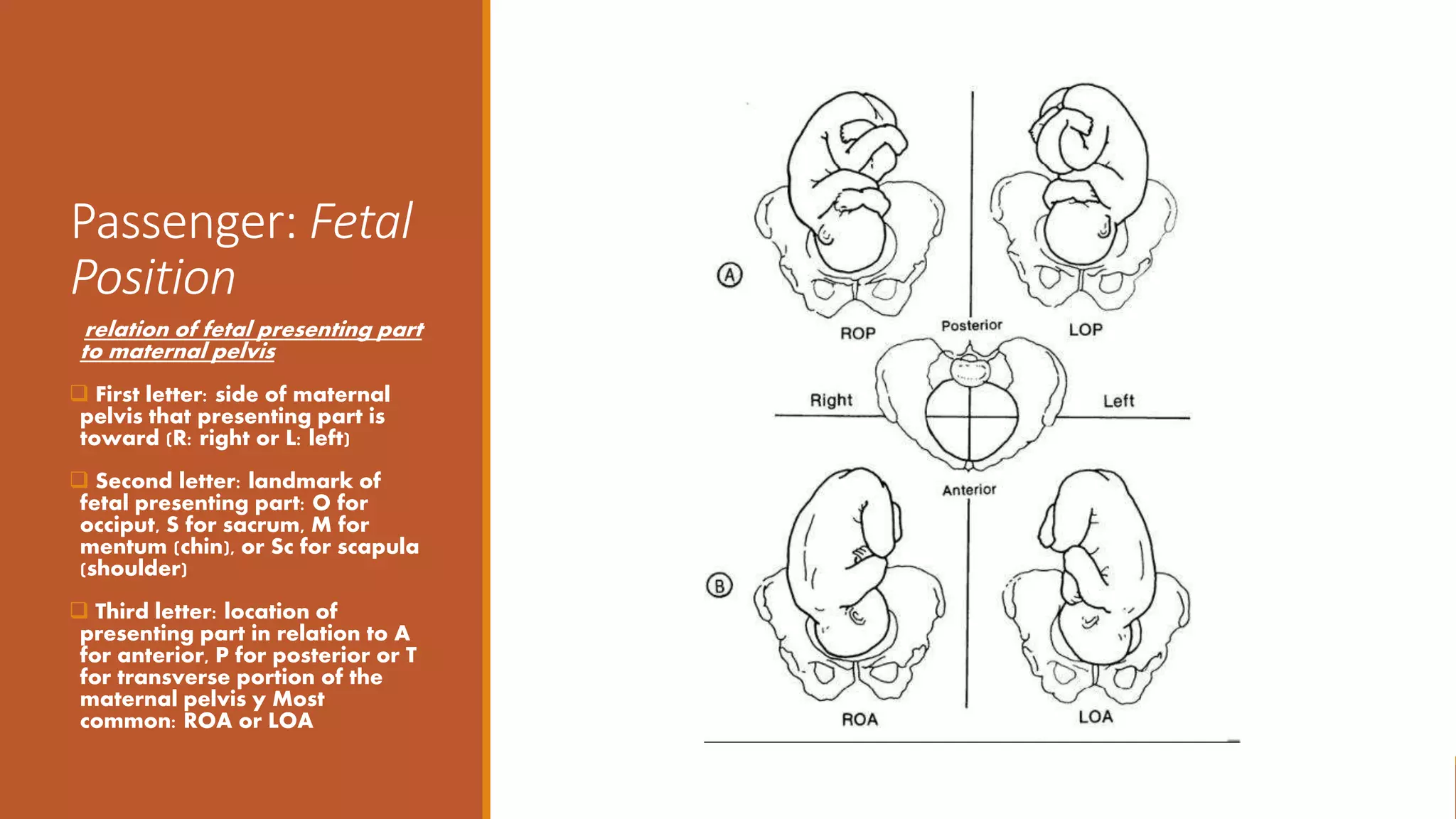
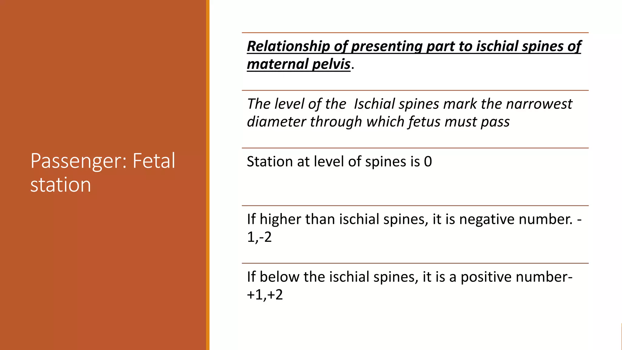
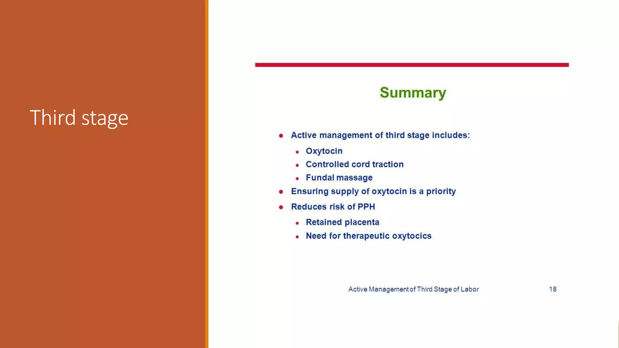
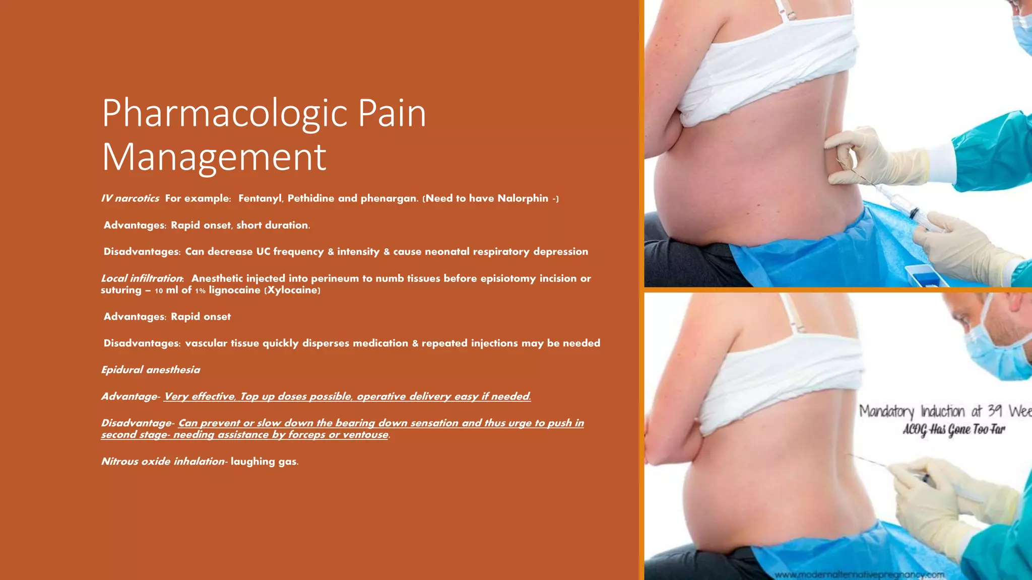
NORMAL LABOR AND
BIRTH.
Objectives
Stages of labor
The five Ps of labor
Positioning of labor
The cardinal movements of labor
Management
The Five “Ps” of
Labor
Passageway:
Pelvis -maternal bony and tissues
Passenger: the fetus
Powers: primary and secondary forces of labor
Position: maternal position y
Psyche: psychological component of mother
Maternal pelvis
3-Diameters (inlet)
Anteroposterior- 12cms
Transverse -13cms
Oblique-11cms
Mid-pelvis
Anteroposterior-11-12cms
Transverse -11-12cms
Oblique-12cms
Outlet
Anteroposterior 13cms
Transverse-11-12cms
Diagonal Conjugate- 12-13.5cms
Passageway:
Pelvis
Pelvis: Fusion of ilium, ischium,
pubis & sacral bones
– False pelvis: above pelvic
inlet
– True pelvis: inlet, mid-pelvis
and outlet
4 main pelvic types:
 Gynecoid: round, most
common type, vaginal birth
(female type)
 Android: heart-shaped,
usually have C/S(male type)
 Anthropoid: oval, vaginal
birth (often forceps)(Ape like)
 Platypelloid: flat, least
common, vaginal birth.
Fetal Skull
Anatomy
 Bones: frontal, occipital, 2
parietal, and 2 temporal
 Molding: overlapping of fetal
skull bones during labor;
resolved by 3 days after birth
 Sutures: membranous tissue
between bones of skull
 Fontanels: membrane-filled
spaces where sutures intersect: ·
Anterior: large; diamond
shaped (junction of 4 sutures);
closes by 18 months after birth ·
 Posterior: small; triangular
(junction of 3 sutures); closes 6 to
8 weeks after birth
Passenger-Fetal Attitude and Fetal Lie
Attitude: relation of fetal parts to one another −
Flexion: normal −
Hyperextension: abnormal
Lie: relationship of fetal longitudinal axis to maternal longitudinal axis
Longitudinal: fetal spine parallel to mother’s spine
Transverse: fetal spine at right angle to mother’s spine
Passenger-Fetal
Presentation
The Fetal part entering pelvis first (felt on
vaginal exam) –
Cephalic: occiput presents in vertex (brow,
face or military are other cephalic
presentations)
Breech: sacrum presents, complete, frank or
footling breech
Shoulder: scapula presents
Passenger: Fetal
Position
relation of fetal presenting part
to maternal pelvis
 First letter: side of maternal
pelvis that presenting part is
toward (R: right or L: left)
 Second letter: landmark of
fetal presenting part: O for
occiput, S for sacrum, M for
mentum (chin), or Sc for scapula
(shoulder)
 Third letter: location of
presenting part in relation to A
for anterior, P for posterior or T
for transverse portion of the
maternal pelvis y Most
common: ROA or LOA
Passenger: Fetal
station
Relationship of presenting part to ischial spines of
maternal pelvis.
The level of the Ischial spines mark the narrowest
diameter through which fetus must pass
Station at level of spines is 0
If higher than ischial spines, it is negative number. -
1,-2
If below the ischial spines, it is a positive number-
+1,+2
Passenger:
Engagement –
Fetal head
Engagement is when largest diameter of
presenting part (BPD) crosses the pelvic inlet and
can be detected by vaginal exam
Floating: presenting part directed towards pelvis,
but can easily be moved out of the inlet. Both
fetal poles- occiput and sinciput felt easily.
Ballotable: presenting part dips into inlet, yet is
displaced with upward pressure from
obstetricians fingers
Engaged: presenting part fixed in pelvic inlet;
cannot be dislodged
Power:
Forces of Labor Primary &
secondary powers combine
to expel fetus and placenta
from uterus
Primary (involuntary)
forces: contractions of
uterine muscle fibers
Secondary (voluntary)
forces: use of abdominal
muscles during second
stage of labor to facilitate
descent & delivery of fetus
Primary Forces of Labor
Effacement: Shortening and thinning of cervix
during first stage of labor. Measured from 0% to
100%.
Dilation: Opening or widening of cervix as labor
progresses.
Measured from 0 to 10 cm.
Is complete when cervix not palpable on vaginal
examination
Position of Laboring Woman
Affects circulation, fatigue & comfort
1. Upright position (walking, sitting. Standing, squatting) beneficial –
Promotes descent of fetus
 Improves blood flow
 Relieves backache
 Straightens axis of birth canal
 Increases pelvic outlet
2. Hands & knees(All fours) may relieve back pain, help fetus to rotate
3. Lateral position may help fetus rotate favorably and slow birth
4. Supine position- worst as it compromises blood flow to uterus and fetus –
should be avoided as far as possible.
Psyche
Preparation for childbirth – reading, antenatal exercise
Sociocultural heritage
Previous childbirth experience
Support from significant others- family, husband, friend, student,
staff.
Emotional status – fearful, anxious, confident, relaxed mentally
prepared.
Environmental influence
Role of health personal and treating team, family , friends, and
reading materials.
Cardinal Movements of
Labor
1. Engagement - presenting part fixed into pelvic
inlet
2. Descent –of fetal head into pelvis
3. Flexion – of fetal head on neck as it reaches
levator ani muscle.
4. Internal rotation -by 180degrees of fetal head to
accommodate widest diameter of maternal pelvis
5. Extension - of fetal head as it comes under pubic
symphysis
6. Restitution - as head turns 45° to untwist neck
7. External rotation -viewed as head turns 45° to
align shoulders with widest diameter of pelvis
8. Expulsion- as anterior shoulder slips under pubis
Signs &
Symptoms of
Labor
Preceding labor: – Lightening – Bloody show – Stronger
Braxton Hicks – Cervical ripening – Diarrhea – Energy
burst
Onset of labor: – Regular contractions with progressive
frequency and intensity –
Progressive effacement and dilation of cervix –
Progressive descent of presenting part
Marked on the Partogram
Stages of labor
First stage- onset of labor till full
cervical dilatation
Second stage- delivery of baby
Third stage – placenta delivery
Stages of Labor
First stage: 6-8 hours
multi, 8-10 hours primi.
Begins with onset of true
labor and ends with
complete dilation -
Second stage: 30 minutes
multi, 30 minutes-1 hour
primi
Begins with complete
dilation and ends with
birth of infant
Third stage: 15 minutes
multi, 30 minutes primi
Begins with expulsion of
infant and ends with
expulsion of placenta
Fourth stage:
Begins with expulsion of
placenta, lasting 4 hours -
to monitor vital signs,
vaginal bleeding and urine
passage by the mother.
First Stage of
Labor
Latent Phase
Cervical dilation: 0 - 3 cm
Maternal pain usually under
control
Active Phase
Cervical dilation: 4 - 7 cm
Contractions closer, longer &
stronger.
Mom working to maintain
control
Transition
Cervical dilation: 8 - 10 cm
Increased bloody show; urge
to push Mom may be irritable,
discouraged and tired or
dehydrated
Second Stage of Labor
10 cm to birth of baby
0 - 30 min in multipara; up to 2 hours in nullipara
Bearing down pains- urge to pass stool.
Urge to push increases , as presenting part descends,
with
increased rectal & perineal pressure.
sensation of burning & stretching of vagina & perineum
Mom may be eager or reluctant to push ’
Crowning of fetal head:
bulging and thinning of perineum and opening of vagina
as widest part of head (biparietal diameter) presses
downward onto the perineum and becomes visible prior
to delivery.
Birthing and after
Check baby
Time of birth
Cry
Apgar score at 1 and 5 minutes
Sex
Weight
Anomaly- structural
Head and abd circumference
Ponderal index
Skin color
Reflexes
Passage of urine
Third Stage of
Labor
From birth of infant to birth of
placenta
Usually ≤ 30 minutes •
Uterine surface volume
decreases, shearing placenta
from wall
Signs of placental separation :
Separation causes gush of
blood vaginally ,
 Umbilical cord appears to
lengthen.
 Mild uterine contractions;
feeling of fullness in vagina as
placenta is expelled
 Supra-pubic bulge
Placental separation
Third stage
Episiotomy-
Perineal
incisions
Surgical incision into perineum to enlarge vaginal opening during
2nd stage
Midline:
incision made from vagina toward rectum.
Advantage: muscle fibers split lengthwise with faster/less painful
healing. Disadvantage: 30 % extend into 3rd/4th degree
Medio-lateral:
incision from vagina obliquely toward one buttock.
Advantage: larger episiotomy possible; rectal structures avoided.
Disadvantage: cut across muscle fibers causes more pain during
healing.
Lacerations
Lacerations to perineum or surrounding tissue may occur during childbirth; 3rd and
4th degree lacerations most commonly occur after midline episiotomy performed
1st degree :
involves only epidermal layers; if no bleeding may not need repair
2nd degree:
epidermal and muscle/fascia involvement requires suturing
3rd degree:
extends into rectal sphincter
4th degree:
through rectal mucosa
Non-
pharmacologic
Pain
Management
Position change to decrease weight of fetus
Hydrotherapy: warm tub or shower, hot towels
Breathing techniques to prevent breath-holding, facilitate
O2/CO2 exchange
Hypnotherapy
Relaxation techniques: imagery, verbal instruction,
massage, soft music, therapeutic touch
Childbirth prep: Dick-Read, Bradley, Lamaze
ANTENATAL EXERCISE CLASSES FOR COUPLES
Pharmacologic Pain
Management
IV narcotics For example: Fentanyl, Pethidine and phenargan. (Need to have Nalorphin -)
Advantages: Rapid onset, short duration.
Disadvantages: Can decrease UC frequency & intensity & cause neonatal respiratory depression
Local infiltration: Anesthetic injected into perineum to numb tissues before episiotomy incision or
suturing – 10 ml of 1% lignocaine (Xylocaine)
Advantages: Rapid onset
Disadvantages: vascular tissue quickly disperses medication & repeated injections may be needed
Epidural anesthesia
Advantage- Very effective, Top up doses possible, operative delivery easy if needed.
Disadvantage- Can prevent or slow down the bearing down sensation and thus urge to push in
second stage- needing assistance by forceps or ventouse.
Nitrous oxide inhalation- laughing gas.
Some aspects of patient education, for timely reaching delivery
unit.
Summary of approach to labor patient
Greet and give your name to patient, ask her name, age and also permission to examine verbally
Greet and give
Take history and clinically determine if in true or false labor from history
Take
Do general, complete physical examination, abdominal and vaginal examination to determine stage of labor clinically.
Do
Start the white apron, gowning, gloving, use of googles , opening the delivery pack and checking delivery and episiotomy
instruments.
Start
Keep talking to laboring patient explaining what you plan to do next and to avoid pushing if she has crowned with fetal head
visible at the introitus.
Keep
Summary
Conduct delivery of baby completely till cord
clamping,
assessing condition of baby and placing on
mother’s belly if all good.
Inj. Oxytocin at delivery of anterior shoulder/
after delivery of placenta
Conduction of delivery of placenta by
controlled cord traction, after explain signs of
placental separation – examining of placenta
and membranes.
Examining uterus and vagina – for bleeding,
clots, and contractility of the uterus.
Check perineum for tears, suture episiotomy if
given.
Complete by doing a Per- vaginal and per
rectal examination before transferring patient
to the post deliver area.
Help mother pass urine ,check vitals, bleeding,
fever in the fourth stage.
Encourage breastfeeding,
Discuss briefly contraception before
transferring patient to post natal ward.
Suggested
reading and
homework
Ten teachers
Williams Obstetrics
Look up
Maternal pelvis and fetal head-diameters
Partogram
Care in I st, 2nd 3rd and 4th stages of labor.
Episiotomy types and repairs and complications
Thank You

Labor aspects for students

  • 1.
  • 2.
    Objectives Stages of labor Thefive Ps of labor Positioning of labor The cardinal movements of labor Management
  • 3.
    The Five “Ps”of Labor Passageway: Pelvis -maternal bony and tissues Passenger: the fetus Powers: primary and secondary forces of labor Position: maternal position y Psyche: psychological component of mother
  • 4.
    Maternal pelvis 3-Diameters (inlet) Anteroposterior-12cms Transverse -13cms Oblique-11cms Mid-pelvis Anteroposterior-11-12cms Transverse -11-12cms Oblique-12cms Outlet Anteroposterior 13cms Transverse-11-12cms Diagonal Conjugate- 12-13.5cms
  • 5.
    Passageway: Pelvis Pelvis: Fusion ofilium, ischium, pubis & sacral bones – False pelvis: above pelvic inlet – True pelvis: inlet, mid-pelvis and outlet 4 main pelvic types:  Gynecoid: round, most common type, vaginal birth (female type)  Android: heart-shaped, usually have C/S(male type)  Anthropoid: oval, vaginal birth (often forceps)(Ape like)  Platypelloid: flat, least common, vaginal birth.
  • 6.
    Fetal Skull Anatomy  Bones:frontal, occipital, 2 parietal, and 2 temporal  Molding: overlapping of fetal skull bones during labor; resolved by 3 days after birth  Sutures: membranous tissue between bones of skull  Fontanels: membrane-filled spaces where sutures intersect: · Anterior: large; diamond shaped (junction of 4 sutures); closes by 18 months after birth ·  Posterior: small; triangular (junction of 3 sutures); closes 6 to 8 weeks after birth
  • 7.
    Passenger-Fetal Attitude andFetal Lie Attitude: relation of fetal parts to one another − Flexion: normal − Hyperextension: abnormal Lie: relationship of fetal longitudinal axis to maternal longitudinal axis Longitudinal: fetal spine parallel to mother’s spine Transverse: fetal spine at right angle to mother’s spine
  • 8.
    Passenger-Fetal Presentation The Fetal partentering pelvis first (felt on vaginal exam) – Cephalic: occiput presents in vertex (brow, face or military are other cephalic presentations) Breech: sacrum presents, complete, frank or footling breech Shoulder: scapula presents
  • 9.
    Passenger: Fetal Position relation offetal presenting part to maternal pelvis  First letter: side of maternal pelvis that presenting part is toward (R: right or L: left)  Second letter: landmark of fetal presenting part: O for occiput, S for sacrum, M for mentum (chin), or Sc for scapula (shoulder)  Third letter: location of presenting part in relation to A for anterior, P for posterior or T for transverse portion of the maternal pelvis y Most common: ROA or LOA
  • 10.
    Passenger: Fetal station Relationship ofpresenting part to ischial spines of maternal pelvis. The level of the Ischial spines mark the narrowest diameter through which fetus must pass Station at level of spines is 0 If higher than ischial spines, it is negative number. - 1,-2 If below the ischial spines, it is a positive number- +1,+2
  • 11.
    Passenger: Engagement – Fetal head Engagementis when largest diameter of presenting part (BPD) crosses the pelvic inlet and can be detected by vaginal exam Floating: presenting part directed towards pelvis, but can easily be moved out of the inlet. Both fetal poles- occiput and sinciput felt easily. Ballotable: presenting part dips into inlet, yet is displaced with upward pressure from obstetricians fingers Engaged: presenting part fixed in pelvic inlet; cannot be dislodged
  • 12.
    Power: Forces of LaborPrimary & secondary powers combine to expel fetus and placenta from uterus Primary (involuntary) forces: contractions of uterine muscle fibers Secondary (voluntary) forces: use of abdominal muscles during second stage of labor to facilitate descent & delivery of fetus
  • 13.
    Primary Forces ofLabor Effacement: Shortening and thinning of cervix during first stage of labor. Measured from 0% to 100%. Dilation: Opening or widening of cervix as labor progresses. Measured from 0 to 10 cm. Is complete when cervix not palpable on vaginal examination
  • 14.
    Position of LaboringWoman Affects circulation, fatigue & comfort 1. Upright position (walking, sitting. Standing, squatting) beneficial – Promotes descent of fetus  Improves blood flow  Relieves backache  Straightens axis of birth canal  Increases pelvic outlet 2. Hands & knees(All fours) may relieve back pain, help fetus to rotate 3. Lateral position may help fetus rotate favorably and slow birth 4. Supine position- worst as it compromises blood flow to uterus and fetus – should be avoided as far as possible.
  • 15.
    Psyche Preparation for childbirth– reading, antenatal exercise Sociocultural heritage Previous childbirth experience Support from significant others- family, husband, friend, student, staff. Emotional status – fearful, anxious, confident, relaxed mentally prepared. Environmental influence Role of health personal and treating team, family , friends, and reading materials.
  • 16.
    Cardinal Movements of Labor 1.Engagement - presenting part fixed into pelvic inlet 2. Descent –of fetal head into pelvis 3. Flexion – of fetal head on neck as it reaches levator ani muscle. 4. Internal rotation -by 180degrees of fetal head to accommodate widest diameter of maternal pelvis 5. Extension - of fetal head as it comes under pubic symphysis 6. Restitution - as head turns 45° to untwist neck 7. External rotation -viewed as head turns 45° to align shoulders with widest diameter of pelvis 8. Expulsion- as anterior shoulder slips under pubis
  • 17.
    Signs & Symptoms of Labor Precedinglabor: – Lightening – Bloody show – Stronger Braxton Hicks – Cervical ripening – Diarrhea – Energy burst Onset of labor: – Regular contractions with progressive frequency and intensity – Progressive effacement and dilation of cervix – Progressive descent of presenting part Marked on the Partogram
  • 18.
    Stages of labor Firststage- onset of labor till full cervical dilatation Second stage- delivery of baby Third stage – placenta delivery
  • 19.
    Stages of Labor Firststage: 6-8 hours multi, 8-10 hours primi. Begins with onset of true labor and ends with complete dilation - Second stage: 30 minutes multi, 30 minutes-1 hour primi Begins with complete dilation and ends with birth of infant Third stage: 15 minutes multi, 30 minutes primi Begins with expulsion of infant and ends with expulsion of placenta Fourth stage: Begins with expulsion of placenta, lasting 4 hours - to monitor vital signs, vaginal bleeding and urine passage by the mother.
  • 20.
    First Stage of Labor LatentPhase Cervical dilation: 0 - 3 cm Maternal pain usually under control Active Phase Cervical dilation: 4 - 7 cm Contractions closer, longer & stronger. Mom working to maintain control Transition Cervical dilation: 8 - 10 cm Increased bloody show; urge to push Mom may be irritable, discouraged and tired or dehydrated
  • 21.
    Second Stage ofLabor 10 cm to birth of baby 0 - 30 min in multipara; up to 2 hours in nullipara Bearing down pains- urge to pass stool. Urge to push increases , as presenting part descends, with increased rectal & perineal pressure. sensation of burning & stretching of vagina & perineum Mom may be eager or reluctant to push ’ Crowning of fetal head: bulging and thinning of perineum and opening of vagina as widest part of head (biparietal diameter) presses downward onto the perineum and becomes visible prior to delivery.
  • 22.
    Birthing and after Checkbaby Time of birth Cry Apgar score at 1 and 5 minutes Sex Weight Anomaly- structural Head and abd circumference Ponderal index Skin color Reflexes Passage of urine
  • 23.
    Third Stage of Labor Frombirth of infant to birth of placenta Usually ≤ 30 minutes • Uterine surface volume decreases, shearing placenta from wall Signs of placental separation : Separation causes gush of blood vaginally ,  Umbilical cord appears to lengthen.  Mild uterine contractions; feeling of fullness in vagina as placenta is expelled  Supra-pubic bulge
  • 24.
  • 25.
  • 26.
    Episiotomy- Perineal incisions Surgical incision intoperineum to enlarge vaginal opening during 2nd stage Midline: incision made from vagina toward rectum. Advantage: muscle fibers split lengthwise with faster/less painful healing. Disadvantage: 30 % extend into 3rd/4th degree Medio-lateral: incision from vagina obliquely toward one buttock. Advantage: larger episiotomy possible; rectal structures avoided. Disadvantage: cut across muscle fibers causes more pain during healing.
  • 27.
    Lacerations Lacerations to perineumor surrounding tissue may occur during childbirth; 3rd and 4th degree lacerations most commonly occur after midline episiotomy performed 1st degree : involves only epidermal layers; if no bleeding may not need repair 2nd degree: epidermal and muscle/fascia involvement requires suturing 3rd degree: extends into rectal sphincter 4th degree: through rectal mucosa
  • 28.
    Non- pharmacologic Pain Management Position change todecrease weight of fetus Hydrotherapy: warm tub or shower, hot towels Breathing techniques to prevent breath-holding, facilitate O2/CO2 exchange Hypnotherapy Relaxation techniques: imagery, verbal instruction, massage, soft music, therapeutic touch Childbirth prep: Dick-Read, Bradley, Lamaze ANTENATAL EXERCISE CLASSES FOR COUPLES
  • 29.
    Pharmacologic Pain Management IV narcoticsFor example: Fentanyl, Pethidine and phenargan. (Need to have Nalorphin -) Advantages: Rapid onset, short duration. Disadvantages: Can decrease UC frequency & intensity & cause neonatal respiratory depression Local infiltration: Anesthetic injected into perineum to numb tissues before episiotomy incision or suturing – 10 ml of 1% lignocaine (Xylocaine) Advantages: Rapid onset Disadvantages: vascular tissue quickly disperses medication & repeated injections may be needed Epidural anesthesia Advantage- Very effective, Top up doses possible, operative delivery easy if needed. Disadvantage- Can prevent or slow down the bearing down sensation and thus urge to push in second stage- needing assistance by forceps or ventouse. Nitrous oxide inhalation- laughing gas.
  • 30.
    Some aspects ofpatient education, for timely reaching delivery unit.
  • 31.
    Summary of approachto labor patient Greet and give your name to patient, ask her name, age and also permission to examine verbally Greet and give Take history and clinically determine if in true or false labor from history Take Do general, complete physical examination, abdominal and vaginal examination to determine stage of labor clinically. Do Start the white apron, gowning, gloving, use of googles , opening the delivery pack and checking delivery and episiotomy instruments. Start Keep talking to laboring patient explaining what you plan to do next and to avoid pushing if she has crowned with fetal head visible at the introitus. Keep
  • 32.
    Summary Conduct delivery ofbaby completely till cord clamping, assessing condition of baby and placing on mother’s belly if all good. Inj. Oxytocin at delivery of anterior shoulder/ after delivery of placenta Conduction of delivery of placenta by controlled cord traction, after explain signs of placental separation – examining of placenta and membranes. Examining uterus and vagina – for bleeding, clots, and contractility of the uterus. Check perineum for tears, suture episiotomy if given. Complete by doing a Per- vaginal and per rectal examination before transferring patient to the post deliver area. Help mother pass urine ,check vitals, bleeding, fever in the fourth stage. Encourage breastfeeding, Discuss briefly contraception before transferring patient to post natal ward.
  • 33.
    Suggested reading and homework Ten teachers WilliamsObstetrics Look up Maternal pelvis and fetal head-diameters Partogram Care in I st, 2nd 3rd and 4th stages of labor. Episiotomy types and repairs and complications
  • 34.