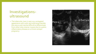
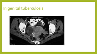
GENITAL
TUBERCULOSIS
Dr. Mini Sood
Epidemiology
• Genital tuberculosis still prevails in
developing countries and is usually a
secondary spread to the genital organs
from a primary in the lungs.
• It can present a long time after the
primary focus has healed.
Incidence
• Almost 1% of all gynecological
admissions are due to genital
tuberculosis.
• It is seen in 2-10% of all patients of
infertility with positive endometrial
aspirations in 4%.
• Infertility is a presenting feature in 90%
of cases and only 10% have conception
after treatment.
Etiology
• Genital tuberculosis is almost always a
secondary infection with the primary
focus in the lungs, lymph nodes, urinary
tract, bones and joints.
• The mode of spread could be via the
blood stream, from the peritoneum, by
direct sexual contact or through the
lymphatics.
• The most frequent site of infection is
the fallopian tube(90%).
Site of infection
• Fallopian tubes - 90%
• Endometrium - 50-60% caseating
lesions, pyometra
• Ovaries – 30%
• Cervix - 5-15%, erosions
• Vagina – 1% Shallow ulcers with
undermined edges
Clinical presentation
• Infertility – 35-60% . Past history
obtained in only 50%, pelvic findings
are negative, and no tubal testing
should be done till a negative
endometrial histology is obtained.
• Menstrual disorders- Menorrhagia in
40% and sec. amenorrhea in 10%.
• Pain – after secondary infection sets in-
pain, nausea, vomiting, fever, tender
fixed lymph nodes
Clinical presentation
• Vaginal discharge-blood stained,
post coital bleeding, leucorrhoea and
painful ulcers
• Abdominal masses- immobile mass
fixed to uterus and appendages
• Fistula formation –Persistent fistula
after a surgery for an abscess.
Clinical presentation
of extra pulmonary
tuberculosis
• Miliary tuberculosis - Tubercle bacilli in
the granulomas of liver, lungs and bone
marrow
• Tuberculous Meningitis Inc. proteins in
CSF, low sugar concentrations and
moderate lymphocytosis. The ELISA
test is -- 80-90% sensitivity.
• Genitourinary Tuberculosis 2-3%Urinary
AFB smears and cultures are positive in
50-70%.
• IVP and cystoscopy reveal calyceal
dilatation, parenchymal calcification,
cavitations, ureteral strictures and
bladder contraction.
Clinical presentation
of extra pulmonary
tuberculosis
• Bone and joint tuberculosis Chronic
mono-articular arthritis, Para spinal
abscess, open joint biopsy or
arthroscopy for tissue diagnosis.
• Genital Tuberculosis 30%Infertility,
menstrual disturbance, abdominal pain
and leucorrhoea. The menstrual blood
is positive for bacilli in 90% of cases.
• Abdominal tuberculosis blind needle
biopsies of the peritoneum, calcified
lymph nodes, ascetic tap smear and
culture
Clinical signs
• General condition , abdomen doughy
• Encysted cyst- immobile and tender
• Pelvic masses-small and fixed or large
and mobile or matted together and fixed
• Recurrent PID – without leukocytosis
• Old healed extra-genital lesion
• Pyometra or postmenopausal bleeding
Diagnostic tests for
tuberculosis
• Clinical Diagnosis
• Radiological Diagnosis – X-ray chest
(apicograms or lateral decubitus films),
Bronchography, CT Scan , Ultrasound
• Laboratory- conventional
demonstration of tubercle bacilli or
smear or animal inoculation studies
• Newer Techniques- serological,
chemical , genetic probes, microphage
typing
• Tuberculin testing
Investigations
• D&C/E.A – Endometrial sampling –histology, PCR, cornual
sample, tubercles, g. pig innoc
• HSG- tubes may have the following changes- Lead pipe
rigidity, non-peristaltic
• Beaded
• Calcifications
• Cornual block
• Jagged tubal outline
• vascular or lymphatic intravasation of the dye
• Hydrosalpinx (bilateral)
• The HSG should be avoided if there is strong suspicion of genital
tuberculosis
•
Investigations
• Diagnostic laparoscopy –Tubercles
• Adhesions, tubes with multiple blocks
or hydrosalpinx, TO masses, abscesses,
ascites.
• Ultrasound
• Mantoux test
TuberculinTest
interpretation: ATS-
CDS
recommendations
•
Induration size
• Positive>5mm HIV Positive women with
Positive Clinical signs( chest x-ray)
• >10mm Health workers, steroids or on
other immunosuppressant drugs,
malnourished patients, Diabetes
Mellitus
• >15mm Low Risk group
Differential
Diagnosis
• Ovarian cysts
• PID
• Ectopic pregnancy
• Carcinoma cervix
• Elephantiasis
Investigations-
ultrasound
• The tubercular mass is seen as a variegated
appearance with cystic and solid areas besides
the uterus and may be difficult to differentiate
from other tubo-ovarian masses due to a pelvic
inflammatory disease or a chronic ectopic
pregnancy
In genital tuberculosis
Magnetic resonance imaging
Magnetic resonance imaging (MRI) findings in genital tuberculosis (TB).
MRI Film showing bilateral tubo-ovarian masses in a confirmed case of
genitalTB
hysteroscopy
• This is seldom done as diagnosis is confirmed by other
methods.
• However, if done for undiagnosed infertility cases, the
findings are of a pale endometrial lining and stenosis
of the tubal ostia.
Laparoscopy
• Laparoscopic findings in genital tuberculosis.
Laparoscopy showing multiple tubal block with
beaded appearance in a proven case genital Koch’s
LAPAROSCOPY
ULTRASOUND
– OVARIAN
CYST
The cystic lesion seen on
the right side of the uterus
which is fluid filled and
anechoic.
Treatment
• Medical treatment- chemotherapeutic
agents
• First line- Isoniazid, rifampicin,
ethambutol
• Second line-
Aminoglycosides,cycloserine, Para-
aminosalicylic acid, ethionamide
• MDRS- Tb- combined resistance to
rifampicin and isoniazid
Treatment
• Surgery
• Indications
• Progression of disease
• Persistent active lesions
• Large inflammatory masses
• Persistence of symptoms
• Persistence of fistula
Contraindications of
surgery
• Active lesions anywhere in the body
• Plastic adhesions of bowels.
Surgery
• Total hysterectomy with bilateral
removal of ovaries and fallopian tubes.
• Follow-up- At least five years
• An annual curettage to check for
reactivation if uterus is still present.
• Prognosis- 90% cure with
chemotherapy
• Decreased fertility restored in 10% of
cases
• 50% tubal pregnancy and 20-30% abort
with 20% having live births.
Controversial issues
in genital
tuberculosis
• Differential diagnosis
• Duration of chemotherapy
• Chemotherapy versus surgery
• Combined chemotherapy
• Resistance
• Role of laparoscopy
THANKYOU

21 08-18 genital tuberculosis

  • 1.
  • 2.
    Epidemiology • Genital tuberculosisstill prevails in developing countries and is usually a secondary spread to the genital organs from a primary in the lungs. • It can present a long time after the primary focus has healed.
  • 3.
    Incidence • Almost 1%of all gynecological admissions are due to genital tuberculosis. • It is seen in 2-10% of all patients of infertility with positive endometrial aspirations in 4%. • Infertility is a presenting feature in 90% of cases and only 10% have conception after treatment.
  • 4.
    Etiology • Genital tuberculosisis almost always a secondary infection with the primary focus in the lungs, lymph nodes, urinary tract, bones and joints. • The mode of spread could be via the blood stream, from the peritoneum, by direct sexual contact or through the lymphatics. • The most frequent site of infection is the fallopian tube(90%).
  • 5.
    Site of infection •Fallopian tubes - 90% • Endometrium - 50-60% caseating lesions, pyometra • Ovaries – 30% • Cervix - 5-15%, erosions • Vagina – 1% Shallow ulcers with undermined edges
  • 6.
    Clinical presentation • Infertility– 35-60% . Past history obtained in only 50%, pelvic findings are negative, and no tubal testing should be done till a negative endometrial histology is obtained. • Menstrual disorders- Menorrhagia in 40% and sec. amenorrhea in 10%. • Pain – after secondary infection sets in- pain, nausea, vomiting, fever, tender fixed lymph nodes
  • 7.
    Clinical presentation • Vaginaldischarge-blood stained, post coital bleeding, leucorrhoea and painful ulcers • Abdominal masses- immobile mass fixed to uterus and appendages • Fistula formation –Persistent fistula after a surgery for an abscess.
  • 8.
    Clinical presentation of extrapulmonary tuberculosis • Miliary tuberculosis - Tubercle bacilli in the granulomas of liver, lungs and bone marrow • Tuberculous Meningitis Inc. proteins in CSF, low sugar concentrations and moderate lymphocytosis. The ELISA test is -- 80-90% sensitivity. • Genitourinary Tuberculosis 2-3%Urinary AFB smears and cultures are positive in 50-70%. • IVP and cystoscopy reveal calyceal dilatation, parenchymal calcification, cavitations, ureteral strictures and bladder contraction.
  • 9.
    Clinical presentation of extrapulmonary tuberculosis • Bone and joint tuberculosis Chronic mono-articular arthritis, Para spinal abscess, open joint biopsy or arthroscopy for tissue diagnosis. • Genital Tuberculosis 30%Infertility, menstrual disturbance, abdominal pain and leucorrhoea. The menstrual blood is positive for bacilli in 90% of cases. • Abdominal tuberculosis blind needle biopsies of the peritoneum, calcified lymph nodes, ascetic tap smear and culture
  • 10.
    Clinical signs • Generalcondition , abdomen doughy • Encysted cyst- immobile and tender • Pelvic masses-small and fixed or large and mobile or matted together and fixed • Recurrent PID – without leukocytosis • Old healed extra-genital lesion • Pyometra or postmenopausal bleeding
  • 11.
    Diagnostic tests for tuberculosis •Clinical Diagnosis • Radiological Diagnosis – X-ray chest (apicograms or lateral decubitus films), Bronchography, CT Scan , Ultrasound • Laboratory- conventional demonstration of tubercle bacilli or smear or animal inoculation studies • Newer Techniques- serological, chemical , genetic probes, microphage typing • Tuberculin testing
  • 12.
    Investigations • D&C/E.A –Endometrial sampling –histology, PCR, cornual sample, tubercles, g. pig innoc • HSG- tubes may have the following changes- Lead pipe rigidity, non-peristaltic • Beaded • Calcifications • Cornual block • Jagged tubal outline • vascular or lymphatic intravasation of the dye • Hydrosalpinx (bilateral) • The HSG should be avoided if there is strong suspicion of genital tuberculosis •
  • 13.
    Investigations • Diagnostic laparoscopy–Tubercles • Adhesions, tubes with multiple blocks or hydrosalpinx, TO masses, abscesses, ascites. • Ultrasound • Mantoux test
  • 14.
    TuberculinTest interpretation: ATS- CDS recommendations • Induration size •Positive>5mm HIV Positive women with Positive Clinical signs( chest x-ray) • >10mm Health workers, steroids or on other immunosuppressant drugs, malnourished patients, Diabetes Mellitus • >15mm Low Risk group
  • 15.
    Differential Diagnosis • Ovarian cysts •PID • Ectopic pregnancy • Carcinoma cervix • Elephantiasis
  • 16.
    Investigations- ultrasound • The tubercularmass is seen as a variegated appearance with cystic and solid areas besides the uterus and may be difficult to differentiate from other tubo-ovarian masses due to a pelvic inflammatory disease or a chronic ectopic pregnancy
  • 17.
    In genital tuberculosis Magneticresonance imaging Magnetic resonance imaging (MRI) findings in genital tuberculosis (TB). MRI Film showing bilateral tubo-ovarian masses in a confirmed case of genitalTB
  • 18.
    hysteroscopy • This isseldom done as diagnosis is confirmed by other methods. • However, if done for undiagnosed infertility cases, the findings are of a pale endometrial lining and stenosis of the tubal ostia.
  • 19.
    Laparoscopy • Laparoscopic findingsin genital tuberculosis. Laparoscopy showing multiple tubal block with beaded appearance in a proven case genital Koch’s
  • 20.
  • 21.
    ULTRASOUND – OVARIAN CYST The cysticlesion seen on the right side of the uterus which is fluid filled and anechoic.
  • 22.
    Treatment • Medical treatment-chemotherapeutic agents • First line- Isoniazid, rifampicin, ethambutol • Second line- Aminoglycosides,cycloserine, Para- aminosalicylic acid, ethionamide • MDRS- Tb- combined resistance to rifampicin and isoniazid
  • 23.
    Treatment • Surgery • Indications •Progression of disease • Persistent active lesions • Large inflammatory masses • Persistence of symptoms • Persistence of fistula
  • 24.
    Contraindications of surgery • Activelesions anywhere in the body • Plastic adhesions of bowels.
  • 25.
    Surgery • Total hysterectomywith bilateral removal of ovaries and fallopian tubes. • Follow-up- At least five years • An annual curettage to check for reactivation if uterus is still present. • Prognosis- 90% cure with chemotherapy • Decreased fertility restored in 10% of cases • 50% tubal pregnancy and 20-30% abort with 20% having live births.
  • 26.
    Controversial issues in genital tuberculosis •Differential diagnosis • Duration of chemotherapy • Chemotherapy versus surgery • Combined chemotherapy • Resistance • Role of laparoscopy
  • 27.