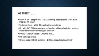
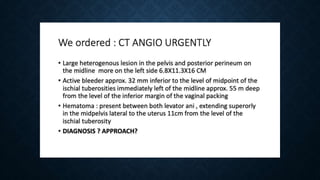
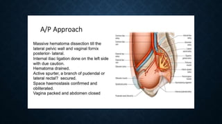
GREETINGS
08-09-2021
HIGHGRAD E3 1
Vulvovaginal Hematoma
Dr Mitra Saxena
MD,DNB,FICOG,FICMH
Chairperson Practical
Obstetrics Committee FOGSI
ANTICIPATE,PREVENT
AND MANAGE ,MANTRA
FOR SAFE OBSTETRICS
20/08/2021
Obstetric Update Bhagalpur 3
20/08/2021
Obstetric Update Bhagalpur 4
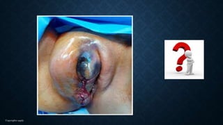
Tackling the bugbear
Vulvovaginal
hematoma
WHAT IS UNIQUE TO VULVOVAGINAL
HEMATOMA
• Insidious in Onset,
• From Innocuous to
• Devastating
• Will happen in VIPs
• COMPLICATE an already
exhausted obstetrician’s life…..
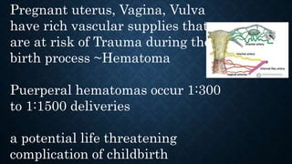
Pregnant uterus, Vagina, Vulva
have rich vascular supplies that
are at risk of Trauma during the
birth process ~Hematoma
Puerperal hematomas occur 1:300
to 1:1500 deliveries
a potential life threatening
complication of childbirth .
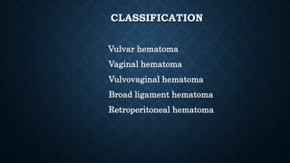
CLASSIFICATION
Vulvar hematoma
Vaginal hematoma
Vulvovaginal hematoma
Broad ligament hematoma
Retroperitoneal hematoma
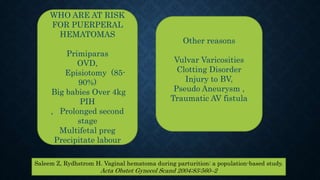
WHO ARE AT RISK
FOR PUERPERAL
HEMATOMAS
Primiparas
OVD,
Episiotomy (85-
90%)
Big babies Over 4kg
PIH
, Prolonged second
stage
Multifetal preg
Precipitate labour
Other reasons
Vulvar Varicosities
Clotting Disorder
Injury to BV,
Pseudo Aneurysm ,
Traumatic AV fistula
Saleem Z, Rydhstrom H. Vaginal hematoma during parturition: a population-based study.
Acta Obstet Gynecol Scand 2004;83:560–2
DETRIMENTAL TO VVH
DELAYED DIAGNOSIS
INCOMPLETE ,INAPPROPRIATE MANAGEMENT
DO IT RIGHT THE FIRST TIME
20/08/2021
Obstetric Update Bhagalpur 12
Copyrights apply
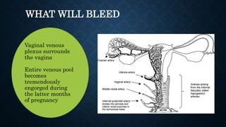
Vaginal venous
plexus surrounds
the vagina
Entire venous pool
becomes
tremendously
engorged during
the latter months
of pregnancy
WHAT WILL BLEED
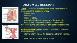
WHAT WILL BLEED?!?
20/08/2021
Obstetric Update Bhagalpur 15
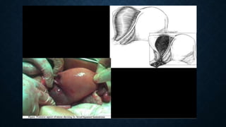
Vulva — Most vulval hematomas result from injuries to
branches of the pudendal artery
(inferior rectal,
perineal,
posterior labial,
and urethral arteries; the artery of the vestibule;
and the deep and dorsal arteries of the clitoris)
that occur during episiotomy or from perineal lacerations
Vaginal/paravaginal hematomas result from injuries to
branches of the
uterine artery, mainly the descending branch ,vaginal.
AND THE RICH VENOUS PLEXUS
Copyrights apply
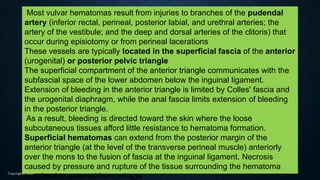
Most vulvar hematomas result from injuries to branches of the pudendal
artery (inferior rectal, perineal, posterior labial, and urethral arteries; the
artery of the vestibule; and the deep and dorsal arteries of the clitoris) that
occur during episiotomy or from perineal lacerations
These vessels are typically located in the superficial fascia of the anterior
(urogenital) or posterior pelvic triangle
The superficial compartment of the anterior triangle communicates with the
subfascial space of the lower abdomen below the inguinal ligament.
Extension of bleeding in the anterior triangle is limited by Colles' fascia and
the urogenital diaphragm, while the anal fascia limits extension of bleeding
in the posterior triangle.
As a result, bleeding is directed toward the skin where the loose
subcutaneous tissues afford little resistance to hematoma formation.
Superficial hematomas can extend from the posterior margin of the
anterior triangle (at the level of the transverse perineal muscle) anteriorly
over the mons to the fusion of fascia at the inguinal ligament. Necrosis
caused by pressure and rupture of the tissue surrounding the hematoma
may lead to external hemorrhage
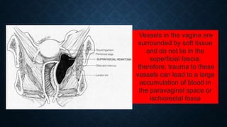
Vessels in the vagina are
surrounded by soft tissue
and do not lie in the
superficial fascia;
therefore, trauma to these
vessels can lead to a large
accumulation of blood in
the paravaginal space or
ischiorectal fossa
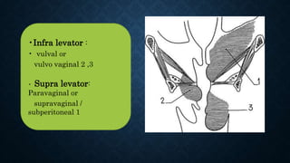
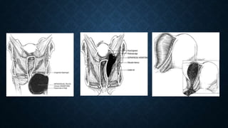
•Infra levator :
• vulval or
vulvo vaginal 2 ,3
• Supra levator:
Paravaginal or
supravaginal /
subperitoneal 1
• Vulvovaginal
haematoma
injury to br of
Pudendal art…
Broad ligament
• haematoma
• Uterine ,Cervical
,vaginal art
20/08/2021
Obstetric Update Bhagalpur 19
Pathogenesis
Two thirds due to failure to
achieve hemostasis particularly at
the upper end of incision / tear deep
ext of episiotomy
Can occur without perineal
laceration / incision due to
stretching and avulsion of vessels
during delivery
Sheikh GN. Perinatal genital hematomas. Obstet Gynecol 1971; 3: 571–5
CLINICAL FEATURES AND DIAGNOSIS
Symptoms develop insidiously in first 24 hrs
Manifest according to the Location
PAIN and MASS effect
Displacement of Vagina, Rectum ,Uterus
HEMODYNAMIC INSTABILITY
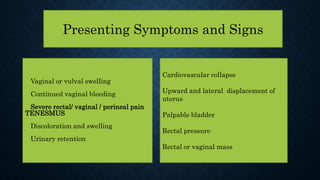
Presenting Symptoms and Signs
Cardiovascular collapse
Upward and lateral displacement of
uterus
Palpable bladder
Rectal pressure
Rectal or vaginal mass
Vaginal or vulval swelling
Continued vaginal bleeding
Severe rectal/ vaginal / perineal pain
TENESMUS
Discoloration and swelling
Urinary retention
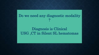
Do we need any diagnostic modality
?
Diagnosis is Clinical
USG ,CT in Silent SL hematomas
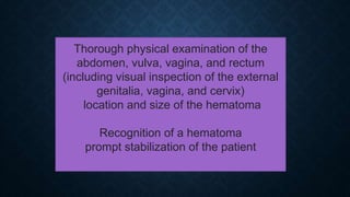
Thorough physical examination of the
abdomen, vulva, vagina, and rectum
(including visual inspection of the external
genitalia, vagina, and cervix)
location and size of the hematoma
Recognition of a hematoma
prompt stabilization of the patient
Copyrights apply
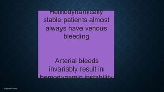
Hemodynamically
stable patients almost
always have venous
bleeding
Arterial bleeds
invariably result in
hemodynamic instability
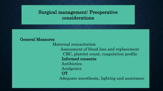
Surgical management: Preoperative
considerations
General Measures
Maternal resuscitation
Assessment of blood loss and replacement
CBC, platelet count, coagulation profile
Informed consents
Antibiotics
Analgesics
OT
Adequate anesthesia, lighting and assistance
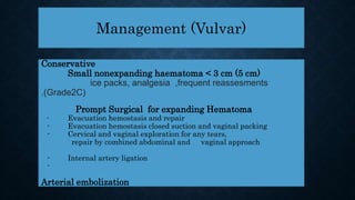
Management (Vulvar)
Conservative
Small nonexpanding haematoma < 3 cm (5 cm)
ice packs, analgesia ,frequent reassesments
.(Grade2C)
Prompt Surgical for expanding Hematoma
- Evacuation hemostasis and repair
- Evacuation hemostasis closed suction and vaginal packing
- Cervical and vaginal exploration for any tears,
repair by combined abdominal and vaginal approach
- Internal artery ligation
-
Arterial embolization
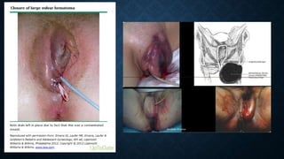
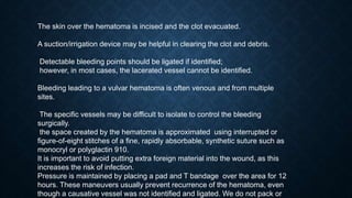
The skin over the hematoma is incised and the clot evacuated.
A suction/irrigation device may be helpful in clearing the clot and debris.
Detectable bleeding points should be ligated if identified;
however, in most cases, the lacerated vessel cannot be identified.
Bleeding leading to a vulvar hematoma is often venous and from multiple
sites.
The specific vessels may be difficult to isolate to control the bleeding
surgically.
the space created by the hematoma is approximated using interrupted or
figure-of-eight stitches of a fine, rapidly absorbable, synthetic suture such as
monocryl or polyglactin 910.
It is important to avoid putting extra foreign material into the wound, as this
increases the risk of infection.
Pressure is maintained by placing a pad and T bandage over the area for 12
hours. These maneuvers usually prevent recurrence of the hematoma, even
though a causative vessel was not identified and ligated. We do not pack or
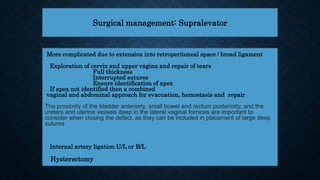
Surgical management: Supralevator
More complicated due to extension into retroperitoneal space / broad ligament
Exploration of cervix and upper vagina and repair of tears
Full thickness
Interrupted sutures
Ensure identification of apex
If apex not identified then a combined
vaginal and abdominal approach for evacuation, hemostasis and repair
The proximity of the bladder anteriorly, small bowel and rectum posteriorly, and the
ureters and uterine vessels deep in the lateral vaginal fornices are important to
consider when closing the defect, as they can be included in placement of large deep
sutures
Internal artery ligation U/L or B/L
Hysterectomy
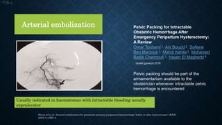
Arterial embolization
Usually indicated in haematomas with intractable bleeding usually
supralevator
Bloom AI et al Arterial embolisation for persistent primary postpartum haemorrhage: before or after hysterectomy? BJOG
2004;111:880–4.
Pelvic Packing for Intractable
Obstetric Hemorrhage After
Emergency Peripartum Hysterectomy:
A Review
Omar Touhami 1, Arij Bouzid 2, Sofiene
Ben Marzouk 3, Mahdi Kehila 4, Mohamed
Badis Channoufi 5, Hayen El Magherbi 6
Oobstet gynecol 2018
Pelvic packing should be part of the
armamentarium available to the
obstetrician whenever intractable pelvic
hemorrhage is encountered
•. 2018
•. 2018
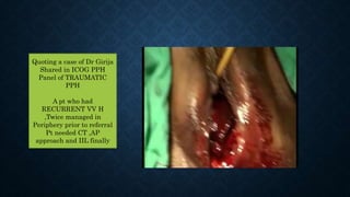
Quoting a case of Dr Girija
Shared in ICOG PPH
Panel of TRAUMATIC
PPH
A pt who had
RECURRENT VV H
,Twice managed in
Periphery prior to referral
Pt needed CT ,AP
approach and IIL finally
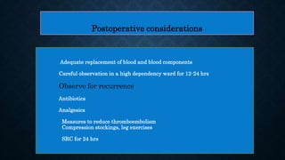
Postoperative considerations
Adequate replacement of blood and blood components
Careful observation in a high dependency ward for 12-24 hrs
Observe for recurrence
Antibiotics
Analgesics
Measures to reduce thromboembolism
Compression stockings, leg exercises
SRC for 24 hrs
Prevention
Ensure complete hemostasis SLOW & STEADY ,EPI
Early detection( Post delivery I hr later PV) Vitals Pain
Correct assessment of blood loss and replacement
Early recourse to surgery DO IT RIGHT FIRST
Antibiotics
Documentation
Complications
Hemorrhage
Ureteric injury
Sepsis
Thromboembolism
Maternal death
Medicolegal litigation
LESSONS LEARNT
The most important factor in correct diagnosis is clinical awareness and
high index of suspicion
Excessive perineal pain is a hallmark symptom: its presence should
prompt examination
Aggressive fluid resuscitation/blood transfusion may be required
Coagulation status should be monitored
Treatment should be carried out in an operating theatre
A urinary catheter should be used to prevent urinary retention and monitor
fluid balance
The threshold for using antibiotics should be low
There is no evidence to support best management, which can be primary
repair or packing, with or without insertion of a drain
Vigilance should be maintained after primary repair / packing, as recurrence
is common
Proper documentation and communication can reduce the chances of
litigation
LESSONS LEARNT
HAVE YOU MANAGED A HEMATOMA OF YOURS OR OF A COLLEAGUE
CAN YOU EVER FORGET THE PATIENTS?
MY TWO PENCE , Delay the suturing but don’t delay the diagnosis
Be a bit slow ..not too fast ..we are the best person to diagnose the hematoma
…So if it’s a big episiotomy more reason not to rush …
DEEP EPISIOTOMY, Close Muscle in two layers, PR,…Post Delivery
Assessment
Don’t ignore vessels or blood filling up..a bleeding vessel doesn’t disappear ..it
will cause havoc if its ignored .Layer by layer HEMOSTASIS.
POST OP PERIOD ..If Pt is C/O unbearable pain Pressure in rectum this could
be a vaginal hematoma
HONEST OPINION POLL
An Rh Negative booked G3P0A2 Full term
preg delivered normally with episiotomy
Was living at a distance, Pt complained of Pain
unrelentlessly
NOD gave injectable Voveran two times and
dismissed the complaints
Perineal care done
Morning round at 8am Pt very uncomfortable
though Happy at having delivered Vaginally
and a healthy baby,
Not ok with pain
PV done Barely could put my finger …..
OT, Anaesthetist, Blood Transfusion Drained
the hematoma,
Ensured Hemostasis …..
A horror
story …..20
years ago
Pt always came back to me but to remind me would mention
..Main Wo HEMATOMA waali pt…
Primi FTP last minute conversion
Platelets 80000
Counselled ,advised referral ,admitted on 27th July
Induced by cerviprime gel ,very fast progress,Took her in OT for
Section Fetal distress ,Bearing down ,Delivered in OT by vacuum
and Episiotomy .
Despite Careful ,meticulous suturing ,Hematoma diagnosed within
2 hours
Investigations sent hemogram ,Coagulation profile ,Consent ,In OT
again ,Under Spinal anesthesia ,with a colleague ,Drained the
hematoma ,and closed dead space ensuring hemostasis .
I was mentally ,Physically ,Medicolegally prepared for NEXT
PROCEDURE IIAL in this case
This Photo by Unknown Author is licensed under CC BY-SA-NC
THANK YOU