Introduction
Introduction
Respiratory eventsare the most common anaesthetic related
Respiratory events are the most common anaesthetic related
injuries, following dental damage. Three main causes:
injuries, following dental damage. Three main causes:
– Inadequate ventilation
Inadequate ventilation
– Oesophageal intubation
Oesophageal intubation
– Difficult tracheal intubation
Difficult tracheal intubation
Difficult tracheal intubation accounts for 17% of the respiratory
Difficult tracheal intubation accounts for 17% of the respiratory
related injuries and results in significant morbidity and
related injuries and results in significant morbidity and
mortality.
mortality.
Estimated that up to 28% of all anaesthetic related deaths are
Estimated that up to 28% of all anaesthetic related deaths are
secondary to the inability to mask ventilate or intubate.
secondary to the inability to mask ventilate or intubate.
Prediction of the difficult airway allows time for proper
Prediction of the difficult airway allows time for proper
selection of equipment, technique and personnel experienced in
selection of equipment, technique and personnel experienced in
difficult airways
difficult airways
3.
Airway
Airway
Nasal and oral
Nasaland oral
cavities
cavities
Pharynx
Pharynx
Larynx
Larynx
Trachea and large
Trachea and large
bronchi
bronchi
4.
Goals of preoperative
Goalsof preoperative
assessment
assessment
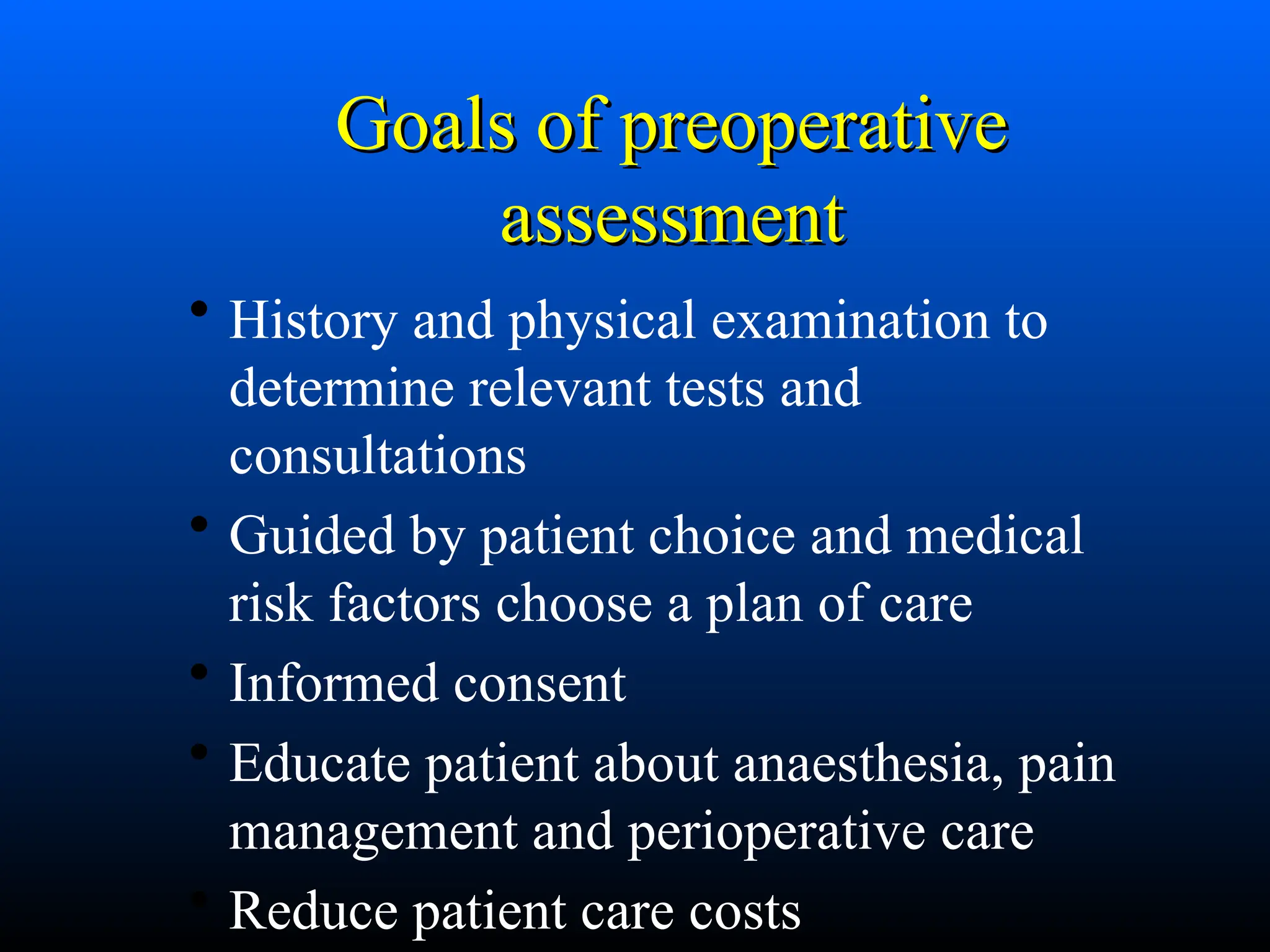
• History and physical examination to
determine relevant tests and
consultations
• Guided by patient choice and medical
risk factors choose a plan of care
• Informed consent
• Educate patient about anaesthesia, pain
management and perioperative care
• Reduce patient care costs
5.
Mortality related toanaesthesia
Mortality related to anaesthesia
• One third of deaths are preventable
• Causes in order of frequency
– inadequate patient preparation
– inadequate postoperative management
– wrong choice of anaesthetic technique
– inadequate crisis management
6.
Predicting the DifficultAirway
• History
• General, Physical and regional Examination
• Specific test for assessment
7.
History and
History and
physicalare the
physical are the
most important
most important
assessors of
assessors of
disease and risk
disease and risk
Presenting complaint
Presenting complaint
Whydoes the patient need an operation now?
• Is it acute/chronic illness?
• Presenting symptoms?
e.g. anaemia, cachexia, pain, seizures etc
• What are the pathophysiological consequences?
e.g. thyroid mass
– Local - stridor, SVC obstruction
– Systemic - hypo/hyperthyroidism
10.
Associated medical conditions
Associatedmedical conditions
Given the presenting problems are there any other
conditions I am worried the patient could have?
• Bowel ca. - liver mets with abnormal LFTs,
abnormal coagulation, impaired drug metabolism
• Peripheral vascular disease - IHD, carotid disease,
HT, renal disease, COAD
11.
Other medical conditions
Othermedical conditions
Any other problems that may affect
perioperative morbidity and mortality?
• cardiac disease
• respiratory disease
• arthritis
• endocrine disease - diabetes, obesity etc
What is the patients functional capacity?
12.
Functional capacity
Functional capacity
•1 MET Can you dress yourself?
• 4 MET Can you climb a flight of stairs?
• 10 MET Can you participate in strenuous
activities
(swimming, tennis, football)
13.
Physical demand characteristicsof work
(1993 Leonard Matheson & Ministry of Labor)
Physical
Demand
Level
Occasional
0-33% work
day
Frequent
34-66% of
workday
Constant
67-100% of
workday
Typical Energy
Required
Sedentary 10 lbs Negligible Negligible 1.5 -2.1 METS
Light 20 lbs 10 lbs Negligible 2.2 – 3.5
METS
Medium 20-50 lbs 10-25 lbs 10 lbs 3.6 – 6.3
METS
Heavy 50-100 lbs 25-50 lbs 10-20 lbs 6.4 – 7.5
METS
Very
Heavy
Over 100
lbs
Over 50
lbs
Over 20 lbs Over 7.5
METS
ANAESTHETIC FACTORS
o Edema
oCompression or
perforation
o Pneumothorax
o Aspiration of gastric
contents
o Burns
o Bleeding
o Tracheal/oesophageal
stenosis
16.
Drug history
Drug history
Veryuseful, often forgotten
• Current medications
• ALLERGY
• Medic alert bracelets
• Smoking/alcohol history
• Other drugs of abuse!
17.
General, physical andregional
examination
i. Patency of nares : look for masses inside
nasal cavity
(e.g. polyps) deviated nasal septum, etc.
ii. Mouth opening of at least 2 large finger
breadths
iii. Teeth : Prominent upper incisors, or
canines
18.
iv. Palate :A high arched palate or a long, narrow
mouth
may present difficulty.
v. Assess patient’s ability to protrude the lower
jaw
beyond the upper incisors (Prognathism).
vi. Temporo-mandibular joint movement : It can
be
restricted ankylosis/fibrosis, tumors, etc.
vii. Measurement of submental space (hyomental/
Thyromental length > 6 cm)
19.
viii. Observation ofpatient’s neck : A short, thick
neck
ix. Presence of hoarse voice/stridor or previous
tracheostomy may suggest stenosis
x. Infections of airway (e.g. epiglottitis, abscess,
croup,
bronchitis, pneumonia).
xi. Physiologic conditions : Pregnancy and obesity
20.
Specific tests forassessment
Anatomical criteria
1. Relative to tongue/pharyngeal size
21.
Mallampati Score
Mallampati Score
Class I (easy)—visualization of the soft palate,
Class I (easy)—visualization of the soft palate,
fauces, uvula, and both anterior and posterior
fauces, uvula, and both anterior and posterior
pillars
pillars
Class II—visualization of the soft palate, fauces,
Class II—visualization of the soft palate, fauces,
and uvula
and uvula
Class III—visualization of the soft palate and the base
Class III—visualization of the soft palate and the base
of the uvula
of the uvula
Class IV (difficult)—the soft palate is not visible at all
Class IV (difficult)—the soft palate is not visible at all
Sensitivity: 44% - 81%
Specificity: 60% - 80%
Roughly corresponds to
Roughly corresponds to Cormack and Lehane’s
Cormack and Lehane’s
laryngoscopy views
laryngoscopy views
22.
Thyromental distance
Thyromental distance
Measurefrom upper edge of
Measure from upper edge of
thyroid cartilage to chin with
thyroid cartilage to chin with
the head fully extended.
the head fully extended.
– Normal is approx 7cm
Normal is approx 7cm
Relatively unreliable test unless combined with
Relatively unreliable test unless combined with
other tests.
other tests.
– Grade 3 or 4 Mallampati who also had a thyromental
Grade 3 or 4 Mallampati who also had a thyromental
distance of less than 7cm were likely to present
distance of less than 7cm were likely to present
difficulty with intubation.
difficulty with intubation.
» Sensitivity: 90.9% Specificity: 81.5%
Sensitivity: 90.9% Specificity: 81.5%
23.
Atlanto-occipital movement
Atlanto-occipital movement
The patient is asked to hold head erect, facing directly to the front, then
The patient is asked to hold head erect, facing directly to the front, then
he is asked to extend the head maximally and the examiner estimates the
he is asked to extend the head maximally and the examiner estimates the
angle traversed by the occlusal surface of upper teeth.
angle traversed by the occlusal surface of upper teeth.
– Visual assessment or using a goniometer.
Visual assessment or using a goniometer.
» Grade I >35 degrees
Grade I >35 degrees
» Grade II 22-34 degrees
Grade II 22-34 degrees
» Grade III 12–21 degrees
Grade III 12–21 degrees
» Grade IV <12 degrees
Grade IV <12 degrees
Assesses feasibility to make the optimal intubation position with
Assesses feasibility to make the optimal intubation position with
alignment of oral, pharyngeal and laryngeal axes into a straight line.
alignment of oral, pharyngeal and laryngeal axes into a straight line.
Limited A-O joint extension
Limited A-O joint extension
– Spondylosis, rheumatoid arthritis, halo-jacket fixation, and in patients with
Spondylosis, rheumatoid arthritis, halo-jacket fixation, and in patients with
symptoms indicating nerve compression with cervical extension.
symptoms indicating nerve compression with cervical extension.
24.
Further assessments
Further assessments
Sterno-mental distance
Sterno-mental distance
– Measured from the sternum to the tip of the mandible
Measured from the sternum to the tip of the mandible
with the head extended.
with the head extended.
» A sternomental distance of 12.5cm predicts a difficult
A sternomental distance of 12.5cm predicts a difficult
intubation.
intubation.
Mandibular protrusion
Mandibular protrusion
– If the patient is able to protrude the lower teeth beyond
If the patient is able to protrude the lower teeth beyond
the upper incisors intubation is usually straightforward
the upper incisors intubation is usually straightforward
– If the patient cannot get the upper and lower incisors
If the patient cannot get the upper and lower incisors
into alignment intubation is likely to be difficult.
into alignment intubation is likely to be difficult.
25.
Dr. Binnions LemonLaw: An easy way to
remember multiple tests…
• Look externally.
• Evaluate the 3-3-2 rule.
• Mallampati.
• Obstruction?
• Neck mobility.
26.
L: Look Externally
•Obesity or very small.
• Short Muscular neck
• Large breasts
• Prominent Upper Incisors (Buck Teeth)
• Receding Jaw (Dentures)
• Burns
• Facial Trauma
• Stridor
• Macroglossia
27.
E-Evaluate the 3-3-2rule
27
3 fingers fit in mouth
3 fingers fit from mentum
to hyoid cartilage
2 fingers fit from the floor
of the mouth to the top of
the thyroid cartilage
28.
M- Mallampati classification
Class-1Class-11
Class-111 Class-1V
soft palate, fauces;
uvula, anterior and
the posterior pillars.
the soft palate, fauces
and uvula
soft palate and base of uvula Only hard palate
Predictors of difficultairway in
diabetics
Palm print
Grade 0 – All the phalangeal areas are visible.
Grade 1 – Deficiency in the interphalangeal areas
of the 4th and 5th digits.
Grade 2 – Deficiency in interphalangeal areas of
2nd to 5th digits.
Grade 3 – Only the tips of digits are seen.
34.
Prayer sign
Patient isasked to bring both the
palms together as ‘Namaste’ and sign is
categorized as–
Positive – When there is gap between palms.
Negative – When there is no gap between
palms.
35.
Assessment of paediatric
airway
History
complaints of snoring, apnoea, day time
somnolence, stridor, hoarse voice and prior surgery
or radiation treatment to face or neck
History of previous anaesthetic records with
attention being paid to history of oropharyngeal
injury, damage to teeth, awake tracheal intubation or
postponement of surgery following an anaesthetic.
36.
Physical examination
It shouldfocus on the anomalies of face, head, neck
and spine.
Evaluate size and shape of head, gross features of
the face; size and symmetry of the mandible,
presence of sub-mandibular pathology, size of
tongue, shape of palate, prominence of upper
incisors, range of motion of jaw, head and neck.
The presence of retractions (suprasternal/sternal/
infrasternal/ intercostal) should be sought for they
usually are signs of airway obstruction.
37.
Breath sounds– Crowing on inspiration is
indicative of extrathoracic airway obstruction
whereas, noise on exhalation is usually due to
intrathoracic lesions.
Noise on inspiration and expiration usually is due
to a lesion at thoracic inlet.
Obtaining blood gas and O2 saturation is
important to determine patient’s ability to
compensate for airway problems.
Transcutaneous CO2 determinations are very
helpful in infants and young children.
38.
Difficult airway
Difficult airway
ASAdefinition of difficult airway:
ASA definition of difficult airway:
“
“The clinical situation in which a
The clinical situation in which a
conventionally trained anaesthetist
conventionally trained anaesthetist
experiences difficulty with mask
experiences difficulty with mask
ventilation, difficulty with tracheal
ventilation, difficulty with tracheal
intubation or both.”
intubation or both.”
39.
Difficult ventilation
Difficult ventilation
Theinability of a trained anesthetist to
The inability of a trained anesthetist to
maintain the oxygen saturation > 90% using
maintain the oxygen saturation > 90% using
a face mask for ventilation and 100%
a face mask for ventilation and 100%
inspired oxygen, provided that the pre-
inspired oxygen, provided that the pre-
ventilation oxygen saturation level was
ventilation oxygen saturation level was
within the normal range.
within the normal range.
40.
Difficult intubation
Difficult intubation
Morethan 3 attempts
More than 3 attempts
Longer than 10 minutes
Longer than 10 minutes
Failure of optimal best attempt
Failure of optimal best attempt
41.
Predictors of difficultyto face
Predictors of difficulty to face
mask ventilate (OBESE)
mask ventilate (OBESE)
1.
1.The
The O
Obese (body mass index > 26
bese (body mass index > 26
kg/m2)
kg/m2)
2.
2.The
The B
Bearded
earded
3.
3.The
The E
Elderly (older than 55 y)
lderly (older than 55 y)
4.
4.The
The S
Snorers
norers
5.
5.The
The E
Edentulous
dentulous
42.
Prevalence
Prevalence
Difficult face mask
Difficultface mask
– 0.1% - 5%
0.1% - 5%
Difficult LMA
Difficult LMA
– 0.2% - 1%
0.2% - 1%
Difficult intubation
Difficult intubation
– 1-2% of normal surgical population
1-2% of normal surgical population
– 50% of rheumatic cervical disease
50% of rheumatic cervical disease
43.
Causes of difficult
Causesof difficult
airway
airway
Stiffness
Stiffness
– Arthritis of neck/jaw/larynx.
Arthritis of neck/jaw/larynx.
– Fixation devices
Fixation devices
– Scleroderma
Scleroderma
– Diabetes
Diabetes
Deformity
Deformity
– Cervical and craniofacial
Cervical and craniofacial
– Burns/trauma/infection
Burns/trauma/infection
Swelling
Swelling
– Infection/tumour/trauma/burns
Infection/tumour/trauma/burns
– Anaphylaxis/haematoma/acromegaly
Anaphylaxis/haematoma/acromegaly
Reflexes
Reflexes
– Cough/breathholding
Cough/breathholding
– Laryngospasm/salivation/regurgitation
Laryngospasm/salivation/regurgitation
Foreign body
Foreign body
Other – Pregnant/full stomach/VIP
Other – Pregnant/full stomach/VIP
44.
Wilson’s risk score
Wilson’srisk score
Score
Score
Weight
Weight 0=<90kg
0=<90kg
1=>90kg
1=>90kg
2=>110kg
2=>110kg
Head and
Head and
neck
neck
movement
movement
0=Above 90degrees
0=Above 90degrees
1=About 90degrees
1=About 90degrees
2=Below 90degrees
2=Below 90degrees
Jaw
Jaw
movement
movement
0=IG>5cm or SLux >0
0=IG>5cm or SLux >0
1=IG<5cm and SLux = 0
1=IG<5cm and SLux = 0
2=IG<5cm and SLux<0
2=IG<5cm and SLux<0
Receding
Receding
mandible
mandible
0=Normal
0=Normal
1=Moderate
1=Moderate
2=Severe
2=Severe
Buck teeth
Buck teeth 0=Normal
0=Normal
1=Moderate
1=Moderate
2=Severe
2=Severe
• Head movement assessed with
pencil taped to a patient’s forehead.
•IG = Interincisor gap measured
with mouth fully open.
•SLux = Maximal forward
protrusion of the lower incisors
beyond the upper incisors.
Intubation
Intubation
Equipment
Equipment
– TRAINEDASSISTANT
TRAINED ASSISTANT
– Laryngoscopes with a selection of blades
Laryngoscopes with a selection of blades
– Variety of endotracheal tubes
Variety of endotracheal tubes
– Introducers for endotracheal tubes (stylets or flexible bougies)
Introducers for endotracheal tubes (stylets or flexible bougies)
– Oral and nasal airways
Oral and nasal airways
– A cricothyroid puncture kit
A cricothyroid puncture kit
– Reliable suction equipment
Reliable suction equipment
– Laryngeal mask airways, sizes 3 AND 4
Laryngeal mask airways, sizes 3 AND 4
The safety of laryngoscopy can be increased by preoxygenating the patient prior to
The safety of laryngoscopy can be increased by preoxygenating the patient prior to
induction and attempts at intubation.
induction and attempts at intubation.
Intubation is attempted by optimal direct laryngoscopy;
Intubation is attempted by optimal direct laryngoscopy;
– optimal head and neck positioning
optimal head and neck positioning
– optimal muscle relaxation
optimal muscle relaxation
– optimal laryngoscope blade
optimal laryngoscope blade
– optimal external laryngeal manipulation
optimal external laryngeal manipulation
– optimal use of the bougie
optimal use of the bougie
After intubation correct placement of the tube should be confirmed by:
After intubation correct placement of the tube should be confirmed by:
– Observing the tube pass through the cords
Observing the tube pass through the cords
– Successful inflation of the chest on manual ventilation
Successful inflation of the chest on manual ventilation
– Auscultation over both lung fields in the axillae
Auscultation over both lung fields in the axillae
– Capnograph
Capnograph
– If in doubt – take it out
If in doubt – take it out
47.
Consider the meritsand feasibility
Awake Intubation vs Intubation after induction
of GA
Non-Invasive technique vs Invasive technique
for initial approach for initial approach
Preservation of spontaneous vs Ablation of spontaneous
Ventilation ventilation
47
48.
What are wegoing to do if we don’t get the
Tube?
• Plans “A”, “B” and “C”
• Know this answer before you tube.
49.
Plan “A”: (ALTERNATE)
•Different Length of blade
• Different Type of Blade
• Different Position
50.
Plan “B”: (BVMand BLIND INTUBATION
Techniques )
• Can you Ventilate with a BVM? (Consider
two person mask Ventilation)
• Combi-Tube?
• LMA an Option?
51.
What do wedo when faced with a
Can’t Intubate Can’t Ventilate
situation?
• Plan “C”: (CRIC) Needle, Surgical,
TFE catheter: preventthe ET tube form redundancy over
the guidewire decrease trauma, increase success rate
58.
References
References
Practice guidelinesfor management of the difficult airway: an updated
Practice guidelines for management of the difficult airway: an updated
report by the American Society of Anesthesiologists Task Force on
report by the American Society of Anesthesiologists Task Force on
Management of the Difficult Airway. Anesthesiology 2003; 98 (5):1269-
Management of the Difficult Airway. Anesthesiology 2003; 98 (5):1269-
77
77
Frerk CM. Predicting difficult intubation. Anaesthesia 1991; 46 (12):1005-
Frerk CM. Predicting difficult intubation. Anaesthesia 1991; 46 (12):1005-
8
8
Verghese C, Brimacombe JR
Verghese C, Brimacombe JR. Survey of laryngeal mask airway usage in
. Survey of laryngeal mask airway usage in
11,910 patients: safety and efficacy for conventional and nonconventional
11,910 patients: safety and efficacy for conventional and nonconventional
usage. Anesth Analg 1996; 82: 129–33
usage. Anesth Analg 1996; 82: 129–33
Gupta S, Sharma R, Jain D. Airway assessment – Predictors of a Difficult
Gupta S, Sharma R, Jain D. Airway assessment – Predictors of a Difficult
Airway. Indian Journal Of Anaesthetics 2005; 49(4) : 257 -262
Airway. Indian Journal Of Anaesthetics 2005; 49(4) : 257 -262
Wilson M, Spiegelhalter D, Robertson A, Predicting difficult intubation.
Wilson M, Spiegelhalter D, Robertson A, Predicting difficult intubation.
Br. J. Anaesth.
Br. J. Anaesth. (1988), 61, 211-216
(1988), 61, 211-216
The Difficult Airway Society Website:
The Difficult Airway Society Website: WWW.DAS.UK.COM
Reed M, Dunn M, McKeown D. Can an an airway assessment score
Reed M, Dunn M, McKeown D. Can an an airway assessment score
predict difficulty at intubation in the emergency department. Emerg Med J
predict difficulty at intubation in the emergency department. Emerg Med J
2005;22:99–102.
2005;22:99–102.
Editor's Notes
#21 This scoring system was first introduced in 1985 in the Canadian Anesthesia Society Journal based on the work of Mallampati. Place the patient in a seated position and have them hold head in a neutral position with mouth open wide and the tongue fully extended. MENTION MODIFIED -
#22 Thyromental distance
A short thyromental distance
equates with an anterior larynx that is at a more acute angle and also results in less space for the tongue to be compressed into by the laryngoscope blade.
This is a measurement taken from the thyroid notch to the tip of the jaw with the head extended. The normal distance is 6.5cm or greater and is dependant on a number of anatomical factors including the position of the larynx. If the distance is greater than 6.5cm, conventional intubation is usually possible. If it is less than 6cm intubation may be impossible [3].
By combining the modified Mallampati and thyromental distance, Frerk showed that patients who fulfilled the criteria of Grade 3 or 4 Mallampati who also had a thyromental distance of less than 7cm were likely to present difficulty with intubation [4]. Frerk suggests that using this combined approach should predict the majority of difficult intubations. A 7cm marker can be used (eg a cut off pencil or an appropriate number of examiners fingers) to determine whether the thyromental distance is greater that 7cm.
#23 Atlanto-Occipital Joint Distance
Atlantooccipital joint extension may be measured when the head is held erect and facing forward. The angle between the erect and extended planes of the occlusal surface of the upper teeth is measured and equals the degree of atlantooccipital joint extension. The "normal" amount of extension equals 35 degrees. Almost all extension of the head on the neck takes place at the atlantooccipital joint. The atlas or the first cervical vertebra is a ring of bone. It does not have a body or spine which would hamper the backward movement of the head. Therefore the greater the atlantooccipital distance in the neutral position, the greater degree of extension that is possible Conversely, if the occiput and the atlas are already in contact in the neutral position, no extension can take place at the atlantooccipital joint.
Because there is a wide variation in atlantooccipital joint distance in the population, it is important to assess head extension at the atlantooccipital joint. Additionally, limited A-O joint extension is present in certain pathological states such as spondylosis, rheumatoid arthritis, halo-jacket fixation, and in patients with symptoms indicating nerve compression with cervical extension. In these patients, it is even more important than usual to raise the occiput above the shoulders prior to laryngoscopy.
Check neck extension on to the chest. Limitation of neck extension (< 30 degrees) may interfere with the sniffing position and limit the laryngoscopic view
#46 Notes:
Intubation is attempted by optimal direct laryngoscopy and this has 5 components;
- optimal head and neck positioning
- optimal muscle relaxation
- optimal laryngoscope blade
- optimal external laryngeal manipulation
- optimal use of the bougie
A number of intubation attempts may be undertaken - to change the blade (long, straight McCoy etc), to use the bougie or to apply optimal external laryngeal manipulation. After 3-4 attempts at intubation, it is likely that the practitioner is repeating fruitless attempts and no further attempts should be made.
Correct positioning of the tube in the trachea (rather than oesophagus) should always be verified after intubation preferably by two out of the 3 best techniques of visual confirmation of the tube passing through the glottic aperture, six consecutive normal capnograph traces and inflation of the oesophageal detector device. No anaesthetist in the UK is ever expected to anaesthetise without using a working capnograph.
It is a deliberate act to stop attempts at direct laryngoscopy, announcing to your assistant 'Failed direct laryngoscopy'. This stops you having yet another attempt and alerts your assistant that you will be going on to Plan B. Start facemask ventilation and ask for a laryngeal mask. Ask for assistance. Go to plan B.