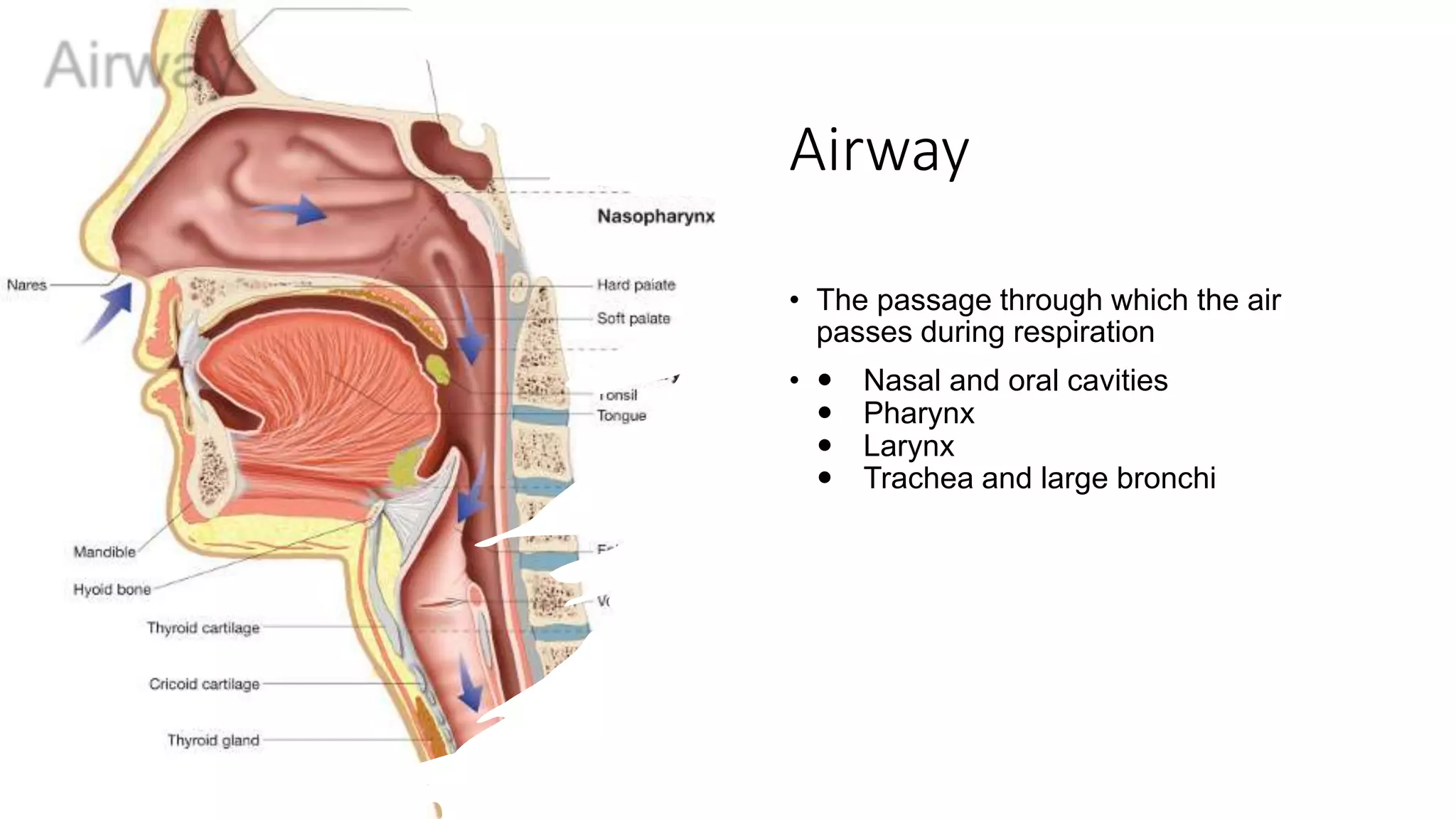
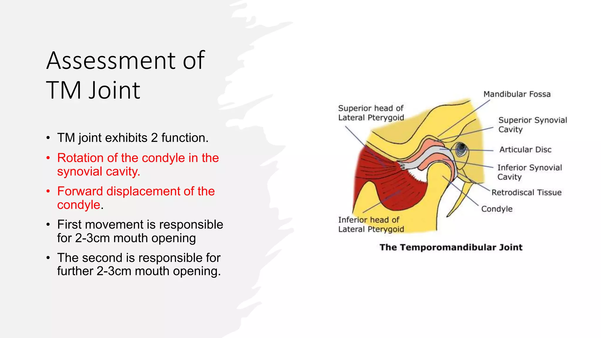
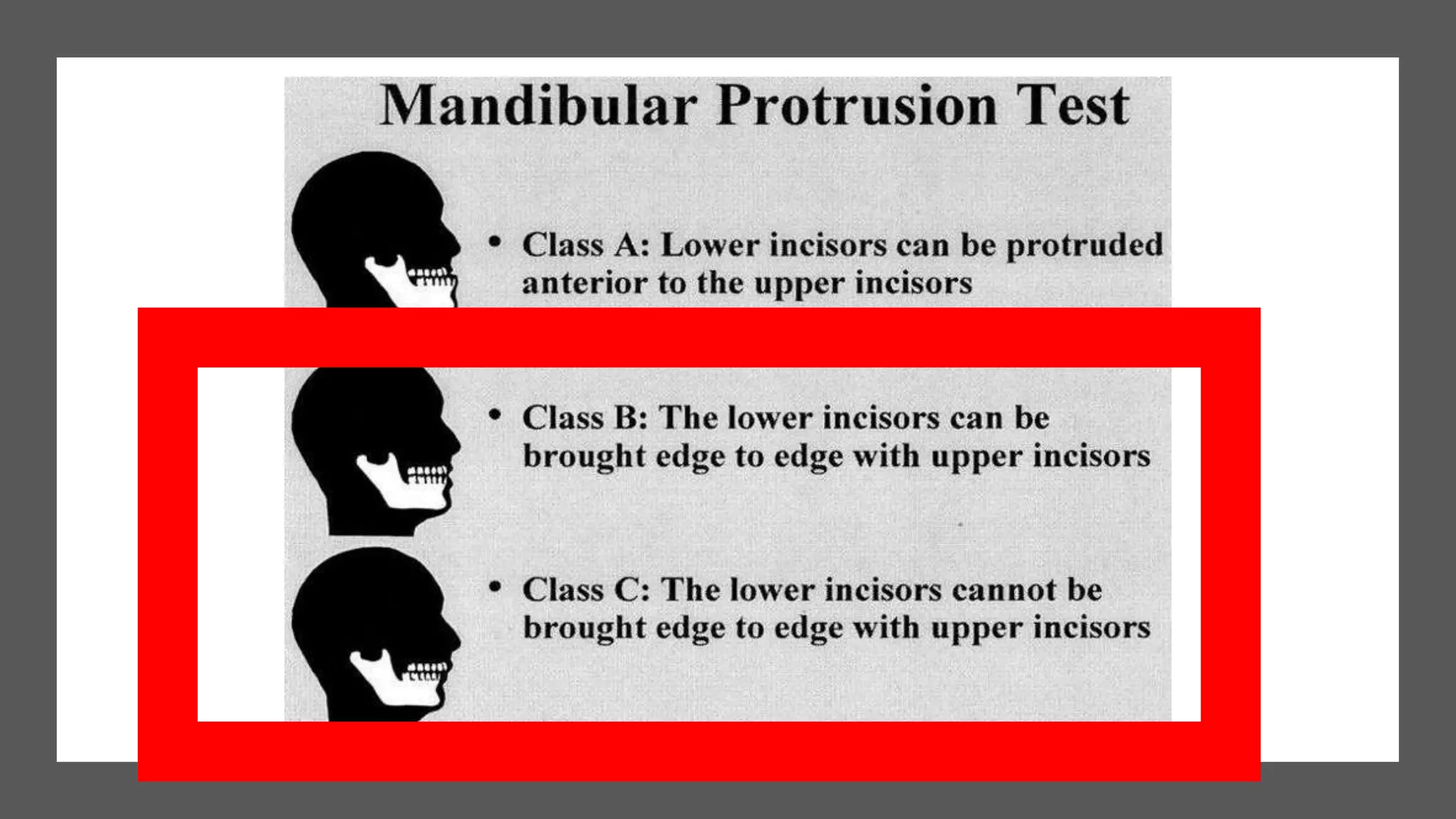
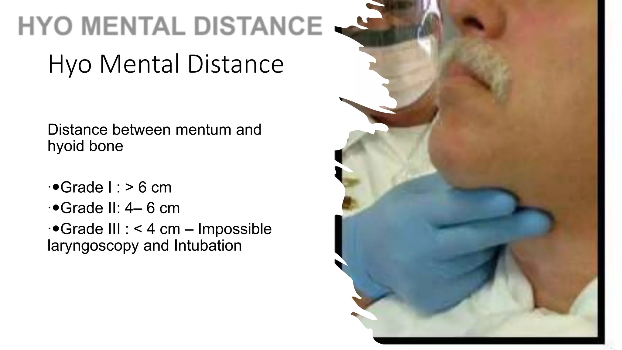
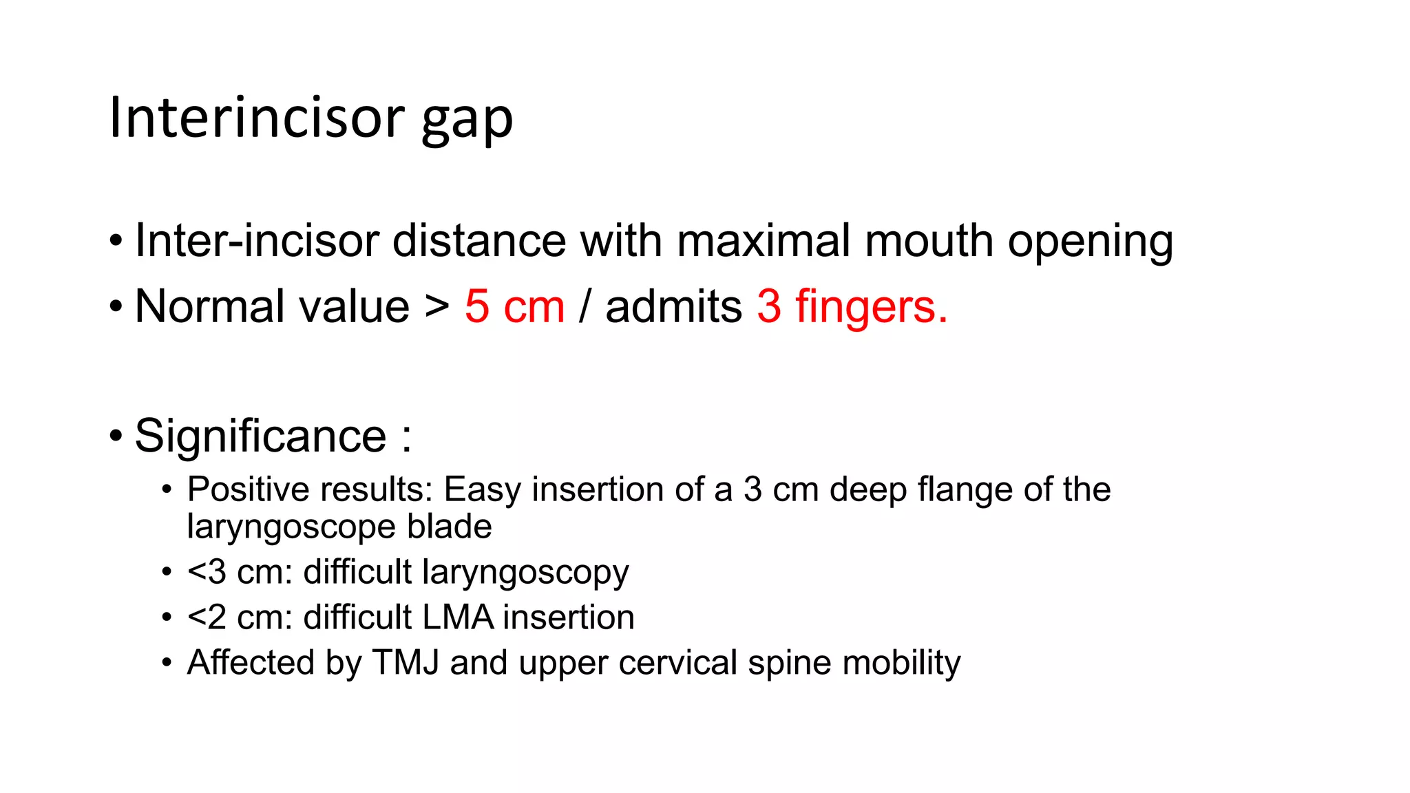
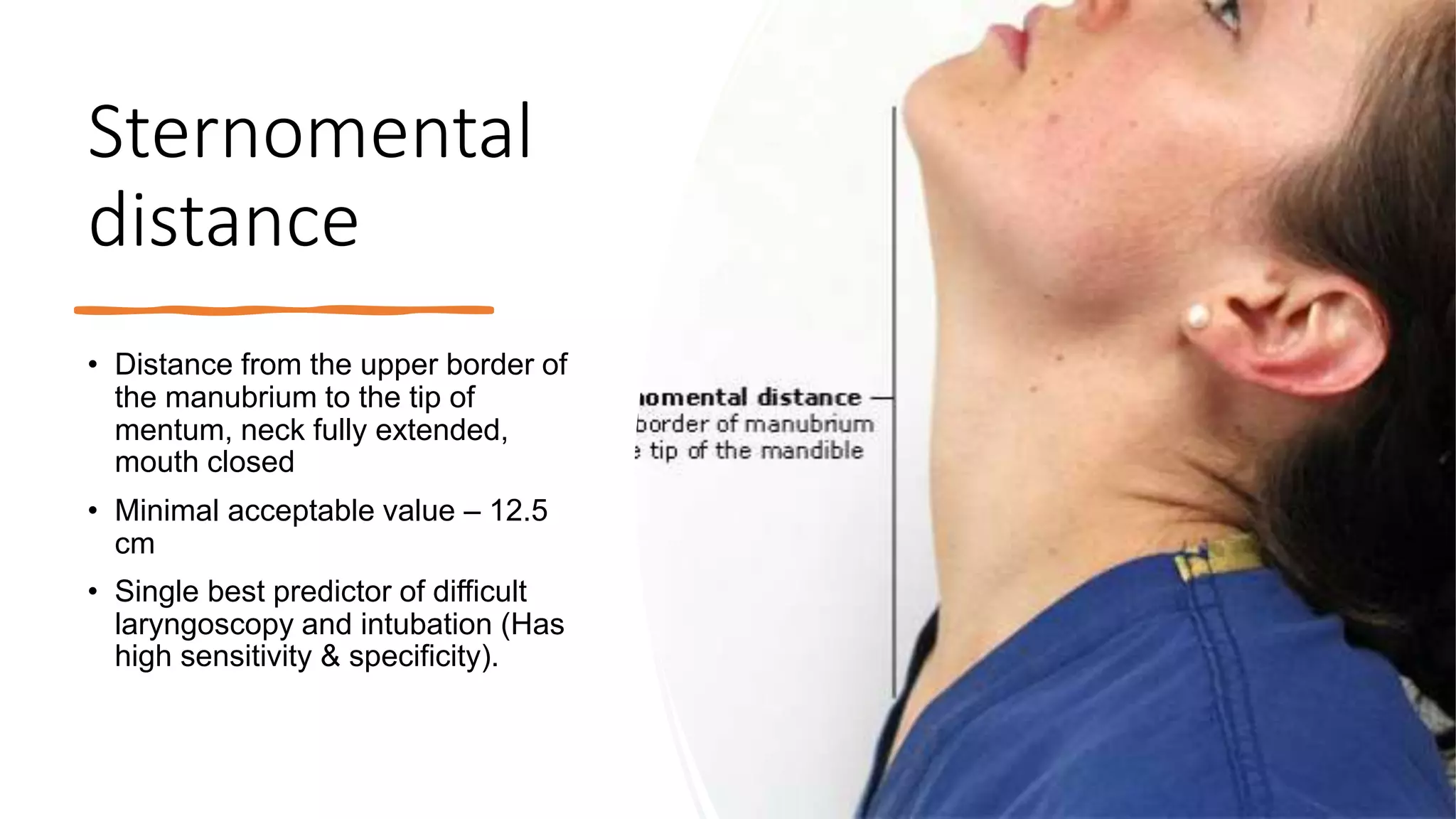
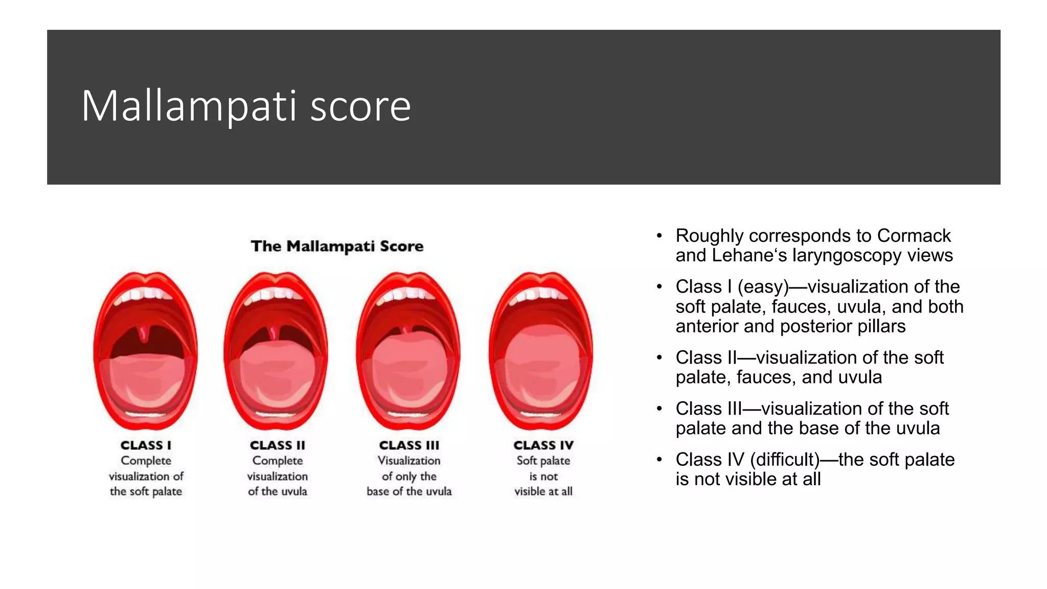
Airway Assessment
Mohamad Hasnol Hayat
Airway
• The passage through which the air
passes during respiration
•  Nasal and oral cavities
 Pharynx
 Larynx
 Trachea and large bronchi
Why need to assess
• Respiratory events are the most common anaesthetic related injuries,
following dental damage. Three main causes:
• Inadequate ventilation
• Oesophageal intubation
• Difficult tracheal intubation
• Difficult tracheal intubation accounts for 17% of the respiratory related
injuries and results in significant morbidity and mortality.
• Estimated that up to 28% of all anaesthetic related deaths are secondary
to the inability to mask ventilate or intubate.
• Prediction of the difficult airway allows time for proper selection of
equipment, technique and personnel experienced in difficult airways
Difficult airway
•ASA definition of difficult airway:
“The clinical situation in which a
conventionally trained anaesthetist
experiences difficulty with mask
ventilation, difficulty with tracheal
intubation or both.”
Difficult ventilation
• The inability of a trained anesthetist to
maintain the oxygen saturation > 90% using a
face mask for ventilation and 100% inspired
oxygen, provided that the pre-ventilation
oxygen saturation level was within the normal
range.
Difficult intubation
 More than 3 attempts
 Longer than 10 minutes
 Failure of optimal best attempt
Prevalence
• Difficult face mask ◦ 0.1% - 5%
• Difficult LMA ◦ 0.2% - 1%
• Difficult intubation
•1-2% of normal surgical population
•50% of rheumatic cervical disease
Components of airway examination
• Nostril patency
• Length of the upper incisors, alignment
• Condition of the teeth
• Relationship of the upper (maxillary) incisors to the lower (mandibular) incisors
• Ability to protrude or advance the lower (mandibular) incisors in front of the upper (maxillary) incisors
• Interincisor or intergum (if edentulous) distance
• Tongue size
• Visibility of the uvula e.g. mallampati
• Presence of heavy facial hair
• Compliance of the mandibular space
• Thyromental distance with the head in maximum extension
• Length of the neck
• Thickness or circumference of the neck
• Range of motion of the head and neck
• Cheek pad
Causes of difficult airway
• Stiffness
◦ Arthritis of neck/ jaw/ larynx. ◦ Fixation devices
◦ Scleroderma
◦ Diabetes
• Deformity
◦ Cervical and craniofacial
◦Burns/trauma/infection
• Swelling
◦ Infection/ tumour/ trauma/ burns
◦ Anaphylaxis/ haematoma/ acromegaly
• Reflexes
◦ Cough/ breathholding
◦ Laryngospasm/ salivation/ regurgitation
 Foreign body
 Other–Pregnant/ full stomach
Airway
assessment
• History
• Patient/notes/chart/medic-alert/spam letter
• Difficulty
• Surgery/burns
• Concurrentdisease
• Reflux/recentmeals
• General examination
• Do they just look difficult?
• Dentition (prominent upper incisors, receding chin)
• Distortion (edema, blood, vomits, tumor, infection)
• Disproportion (short chin-to-larynx distance, bull neck,
large tongue, small mouth)
• Dysmobility (TMJ and cervical spine)
• Massively obese or pregnant
• Beards +/- tubes
• Specific tests/indices
• Investigations.
• Nasoendoscopy
• X-ray, CT/MRI
• Flow volume loop
Predictors of difficulty to bag mask ventilate
• The Obese (body mass index > 35 kg/m2)
• The Bearded
• The Elderly (older than 60 y)
• The Snorers
• The Edentulous
(=BONES)
Predictors of
difficult
laryngoscopy
and intubation
• Individual indices
• Physical examination indices
• radiological indices
• advanced indices
• Group indices
• Wilson‘s score
• Benumof‘s analysis
• Saghei & safavi test
• Lemon assessment
• Arne‘s simplified score
• Magboul‘s 4 M‘s
Atlanto occipital movement
• The patient is asked to hold head erect, facing directly to
the front, then he is asked to extend the head maximally
and the examiner estimates the angle traversed by the
occlusal surface of upper teeth.
• Visual assessment or using a goniometer.
• Grade I >35 degrees
• Grade II 22-34 degrees
• Grade III 12–21 degrees
• Grade IV <12 degrees
• Assesses feasibility to make the optimal intubation position
with alignment of oral, pharyngeal and laryngeal axes into
a straight line.
• Limited A-O joint extension
• Spondylosis, rheumatoid arthritis, halo-jacket fixation,
and in patients with symptoms indicating nerve
compression with cervical extension.
Prayer sign
• A positive "prayer sign" can be elicited on
examination with the patient unable to
approximate the palmar surfaces of the
phalangeal joints while pressing their hands
together.
• Seen in diabetes
• This represents:- cervical spine immobility
and the potential for a difficult endotracheal
intubation.
Palm print test
• The palm and fingers of the dominant
hand of the patient is painted with black
writing ink using a brush.
• The patient then presses the hand firmly
against a white sheet of paper on a hard
surface. Scoring is done as:
• Grade 0 - All phalangeal areas
visible.
• Grade 1 - Deficiency in the inter-
phalangeal areas of 4th and/or
5thdigit.
• Grade2 - Deficiency in the inter-
phalangeal areas of 2nd to 5th digit.
• Grade 3 - Only the tips of digits
seen.
Assessment of
TM Joint
• TM joint exhibits 2 function.
• Rotation of the condyle in the
synovial cavity.
• Forward displacement of the
condyle.
• First movement is responsible
for 2-3cm mouth opening
• The second is responsible for
further 2-3cm mouth opening.
Subluxation of TMJ
• Index finger is placed in front of the
tragus & the thumb is placed in
front of the the lower part of the
mastoid process. patient is asked
to open his mouth as wide as
possible. Index finger in front of the
tragus can be intented in its space
and the thumb can feel the sliding
movement of the condyle as the
condyle of the mandible slides
forward.
Assessment of mandibular space
• Can be expressed as thyromental and hyomental
space.
• This space determines how easily the laryngeal and
pharyngeal axis will fall in line when the a-o joint is
extended.
Thyromental
Distance
• Measure from upper edge of thyroid
cartilage to chin with the head fully
extended.
• Normalis approx7cm.
• If the thyromental distance is short, <3
finger widths, the laryngeal axis makes a
more acute angle with the pharyngeal axis
and it will be difficult to achieve
alignment. Less space to displace the
tongue
• Limitation
• Little reliability in prediction
• Variation according to height, ethnicity
• Modification to improve the accuracy
• Ratio of height to thyromental distance (RHTMD)
• Useful bedside screening test
• RHTMD > 23.5 – very sensitive predictor of difficult
laryngoscopy
Hyo Mental Distance
Distance between mentum and
hyoid bone
Grade I : > 6 cm
Grade II: 4– 6 cm
Grade III : < 4 cm – Impossible
laryngoscopy and Intubation
Interincisor gap
• Inter-incisor distance with maximal mouth opening
• Normal value > 5 cm / admits 3 fingers.
• Significance :
• Positive results: Easy insertion of a 3 cm deep flange of the
laryngoscope blade
• <3 cm: difficult laryngoscopy
• <2 cm: difficult LMA insertion
• Affected by TMJ and upper cervical spine mobility
Sternomental
distance
• Distance from the upper border of
the manubrium to the tip of
mentum, neck fully extended,
mouth closed
• Minimal acceptable value – 12.5
cm
• Single best predictor of difficult
laryngoscopy and intubation (Has
high sensitivity & specificity).
Test for assessing adequacy of the oropharynx for
laryngoscopy and intubation
•Mallampati grading (samsoon and
young‘s modification)
•Narrowness of the palate
Mallampati score
• Roughly corresponds to Cormack
and Lehane‘s laryngoscopy views
• Class I (easy)—visualization of the
soft palate, fauces, uvula, and both
anterior and posterior pillars
• Class II—visualization of the soft
palate, fauces, and uvula
• Class III—visualization of the soft
palate and the base of the uvula
• Class IV (difficult)—the soft palate
is not visible at all
Significance of mallampati score
• Class III or IV: signifies that the angle between the base
of tongue and laryngeal inlet is more acute and not
conducive for easy laryngoscopy
• Limitations
• Poor interobserver reliability
• Limited accuracy
• Good predictor in pregnancy, obesity, acromegaly
CORMACK - LEHANE
Grading at direct
laryngoscopy
• Grade 1: Full exposure of
glottis (anterior + posterior
commissure)
• Grade 2: Anterior
commissure not visualised
• Grade 3: Epiglottis only
• Grade 4: No glottic structure
visible.
How to predict difficult placement
of supraglottic devices (RODS)
• Restricted mouth opening
• Obstruction of the upper airway
• Distrupted upper airway as following trauma,burn,caustic ingestion .
• Stiff lung (poor lung or thoracic compliance)
• Suggested by Hung and Murphy (Canadian journal of anesthesia 2004:10:963-8)
How to predict difficulty in creating surgical airway
(BANG)
•Bleeding tendency
•Agitated patient
•Neck scarring
•Growth or vascular abnormality in region of
surgical airway.
Reflection
• Airway assessment is a critical part .
• The difficult airway assessment must be performed prior
to ALL attempts.
• While this criteria helps identify difficult airways, it does
not guarantee an easy intubation—Be Prepared!
• Nothing is more expensive than the missed opportunity

airway assessment.pptx

  • 1.
  • 2.
    Airway • The passagethrough which the air passes during respiration •  Nasal and oral cavities  Pharynx  Larynx  Trachea and large bronchi
  • 3.
    Why need toassess • Respiratory events are the most common anaesthetic related injuries, following dental damage. Three main causes: • Inadequate ventilation • Oesophageal intubation • Difficult tracheal intubation • Difficult tracheal intubation accounts for 17% of the respiratory related injuries and results in significant morbidity and mortality. • Estimated that up to 28% of all anaesthetic related deaths are secondary to the inability to mask ventilate or intubate. • Prediction of the difficult airway allows time for proper selection of equipment, technique and personnel experienced in difficult airways
  • 4.
    Difficult airway •ASA definitionof difficult airway: “The clinical situation in which a conventionally trained anaesthetist experiences difficulty with mask ventilation, difficulty with tracheal intubation or both.”
  • 5.
    Difficult ventilation • Theinability of a trained anesthetist to maintain the oxygen saturation > 90% using a face mask for ventilation and 100% inspired oxygen, provided that the pre-ventilation oxygen saturation level was within the normal range.
  • 6.
    Difficult intubation  Morethan 3 attempts  Longer than 10 minutes  Failure of optimal best attempt
  • 7.
    Prevalence • Difficult facemask ◦ 0.1% - 5% • Difficult LMA ◦ 0.2% - 1% • Difficult intubation •1-2% of normal surgical population •50% of rheumatic cervical disease
  • 8.
    Components of airwayexamination • Nostril patency • Length of the upper incisors, alignment • Condition of the teeth • Relationship of the upper (maxillary) incisors to the lower (mandibular) incisors • Ability to protrude or advance the lower (mandibular) incisors in front of the upper (maxillary) incisors • Interincisor or intergum (if edentulous) distance • Tongue size • Visibility of the uvula e.g. mallampati • Presence of heavy facial hair • Compliance of the mandibular space • Thyromental distance with the head in maximum extension • Length of the neck • Thickness or circumference of the neck • Range of motion of the head and neck • Cheek pad
  • 9.
    Causes of difficultairway • Stiffness ◦ Arthritis of neck/ jaw/ larynx. ◦ Fixation devices ◦ Scleroderma ◦ Diabetes • Deformity ◦ Cervical and craniofacial ◦Burns/trauma/infection • Swelling ◦ Infection/ tumour/ trauma/ burns ◦ Anaphylaxis/ haematoma/ acromegaly • Reflexes ◦ Cough/ breathholding ◦ Laryngospasm/ salivation/ regurgitation  Foreign body  Other–Pregnant/ full stomach
  • 10.
    Airway assessment • History • Patient/notes/chart/medic-alert/spamletter • Difficulty • Surgery/burns • Concurrentdisease • Reflux/recentmeals • General examination • Do they just look difficult? • Dentition (prominent upper incisors, receding chin) • Distortion (edema, blood, vomits, tumor, infection) • Disproportion (short chin-to-larynx distance, bull neck, large tongue, small mouth) • Dysmobility (TMJ and cervical spine) • Massively obese or pregnant • Beards +/- tubes • Specific tests/indices • Investigations. • Nasoendoscopy • X-ray, CT/MRI • Flow volume loop
  • 11.
    Predictors of difficultyto bag mask ventilate • The Obese (body mass index > 35 kg/m2) • The Bearded • The Elderly (older than 60 y) • The Snorers • The Edentulous (=BONES)
  • 12.
    Predictors of difficult laryngoscopy and intubation •Individual indices • Physical examination indices • radiological indices • advanced indices • Group indices • Wilson‘s score • Benumof‘s analysis • Saghei & safavi test • Lemon assessment • Arne‘s simplified score • Magboul‘s 4 M‘s
  • 13.
    Atlanto occipital movement •The patient is asked to hold head erect, facing directly to the front, then he is asked to extend the head maximally and the examiner estimates the angle traversed by the occlusal surface of upper teeth. • Visual assessment or using a goniometer. • Grade I >35 degrees • Grade II 22-34 degrees • Grade III 12–21 degrees • Grade IV <12 degrees • Assesses feasibility to make the optimal intubation position with alignment of oral, pharyngeal and laryngeal axes into a straight line. • Limited A-O joint extension • Spondylosis, rheumatoid arthritis, halo-jacket fixation, and in patients with symptoms indicating nerve compression with cervical extension.
  • 14.
    Prayer sign • Apositive "prayer sign" can be elicited on examination with the patient unable to approximate the palmar surfaces of the phalangeal joints while pressing their hands together. • Seen in diabetes • This represents:- cervical spine immobility and the potential for a difficult endotracheal intubation.
  • 15.
    Palm print test •The palm and fingers of the dominant hand of the patient is painted with black writing ink using a brush. • The patient then presses the hand firmly against a white sheet of paper on a hard surface. Scoring is done as: • Grade 0 - All phalangeal areas visible. • Grade 1 - Deficiency in the inter- phalangeal areas of 4th and/or 5thdigit. • Grade2 - Deficiency in the inter- phalangeal areas of 2nd to 5th digit. • Grade 3 - Only the tips of digits seen.
  • 16.
    Assessment of TM Joint •TM joint exhibits 2 function. • Rotation of the condyle in the synovial cavity. • Forward displacement of the condyle. • First movement is responsible for 2-3cm mouth opening • The second is responsible for further 2-3cm mouth opening.
  • 17.
    Subluxation of TMJ •Index finger is placed in front of the tragus & the thumb is placed in front of the the lower part of the mastoid process. patient is asked to open his mouth as wide as possible. Index finger in front of the tragus can be intented in its space and the thumb can feel the sliding movement of the condyle as the condyle of the mandible slides forward.
  • 19.
    Assessment of mandibularspace • Can be expressed as thyromental and hyomental space. • This space determines how easily the laryngeal and pharyngeal axis will fall in line when the a-o joint is extended.
  • 20.
    Thyromental Distance • Measure fromupper edge of thyroid cartilage to chin with the head fully extended. • Normalis approx7cm. • If the thyromental distance is short, <3 finger widths, the laryngeal axis makes a more acute angle with the pharyngeal axis and it will be difficult to achieve alignment. Less space to displace the tongue
  • 21.
    • Limitation • Littlereliability in prediction • Variation according to height, ethnicity • Modification to improve the accuracy • Ratio of height to thyromental distance (RHTMD) • Useful bedside screening test • RHTMD > 23.5 – very sensitive predictor of difficult laryngoscopy
  • 22.
    Hyo Mental Distance Distancebetween mentum and hyoid bone Grade I : > 6 cm Grade II: 4– 6 cm Grade III : < 4 cm – Impossible laryngoscopy and Intubation
  • 23.
    Interincisor gap • Inter-incisordistance with maximal mouth opening • Normal value > 5 cm / admits 3 fingers. • Significance : • Positive results: Easy insertion of a 3 cm deep flange of the laryngoscope blade • <3 cm: difficult laryngoscopy • <2 cm: difficult LMA insertion • Affected by TMJ and upper cervical spine mobility
  • 24.
    Sternomental distance • Distance fromthe upper border of the manubrium to the tip of mentum, neck fully extended, mouth closed • Minimal acceptable value – 12.5 cm • Single best predictor of difficult laryngoscopy and intubation (Has high sensitivity & specificity).
  • 25.
    Test for assessingadequacy of the oropharynx for laryngoscopy and intubation •Mallampati grading (samsoon and young‘s modification) •Narrowness of the palate
  • 26.
    Mallampati score • Roughlycorresponds to Cormack and Lehane‘s laryngoscopy views • Class I (easy)—visualization of the soft palate, fauces, uvula, and both anterior and posterior pillars • Class II—visualization of the soft palate, fauces, and uvula • Class III—visualization of the soft palate and the base of the uvula • Class IV (difficult)—the soft palate is not visible at all
  • 27.
    Significance of mallampatiscore • Class III or IV: signifies that the angle between the base of tongue and laryngeal inlet is more acute and not conducive for easy laryngoscopy • Limitations • Poor interobserver reliability • Limited accuracy • Good predictor in pregnancy, obesity, acromegaly
  • 28.
    CORMACK - LEHANE Gradingat direct laryngoscopy • Grade 1: Full exposure of glottis (anterior + posterior commissure) • Grade 2: Anterior commissure not visualised • Grade 3: Epiglottis only • Grade 4: No glottic structure visible.
  • 30.
    How to predictdifficult placement of supraglottic devices (RODS) • Restricted mouth opening • Obstruction of the upper airway • Distrupted upper airway as following trauma,burn,caustic ingestion . • Stiff lung (poor lung or thoracic compliance) • Suggested by Hung and Murphy (Canadian journal of anesthesia 2004:10:963-8)
  • 31.
    How to predictdifficulty in creating surgical airway (BANG) •Bleeding tendency •Agitated patient •Neck scarring •Growth or vascular abnormality in region of surgical airway.
  • 36.
    Reflection • Airway assessmentis a critical part . • The difficult airway assessment must be performed prior to ALL attempts. • While this criteria helps identify difficult airways, it does not guarantee an easy intubation—Be Prepared! • Nothing is more expensive than the missed opportunity