The document discusses aeromicrobiology, which is the study of airborne microorganisms and their effects on human health and the environment. It defines aeromicrobiology and describes the various microbes that can be found in air, such as viruses, bacteria, fungi, and protozoa. The document also discusses how these microbes can be transmitted through the air and cause diseases in humans and other organisms. It provides examples of common airborne pathogens and the diseases they cause. Furthermore, the document discusses the sizes of airborne biological particles known as bioaerosols and different methods for sampling and analyzing bioaerosols in air, including various impactor and impinger sampling devices.
Air microbiology study of microbes suspended in air. Microflora of air depend on the location and environmental condition at particular place. There are different types of air trapping devices like Slit Sampler, Andersons samplers, Impingers etc. Air borne diseases mainly spread by droplet infection, contact with infected things . Air borne diseases are discussed and concluded with control of air borne microbes.
Air Microbiology. Aerobiology is defined as the study of life present in the air. Aeromicrobiology relates to the study of environmentally relevant microorganisms. ... In dry whether the microbial load of air is high while in wet weather the rain washes the microorganisms from the air.
Air is not a natural environment for microorganisms. Microorganisms present in air are liberated from various other sources. These various sources include soil, water, plant and animal surfaces and human beings.
Microbiology of Air
Aero-microbiology
Airborne diseases.
Sources of microorganisms in Air
Microbes in atmosphere
Bioaerosol
“Study of living microbes suspended in air”
Various layers present in the atmosphere at height of 1000km
Nearest to earth is troposphere
troposphere contains heavy load of microorganisms
Boundary layer responsible for transport of particles both short and long distances
Bio Aerosol
“particles release from terrestrial and marine ecosystem into the atmosphere they consist of both living and non living components
including organisms, dispersal method of organisms and excretion
air is not a natural environment for microorganisms. Physical & chemical parameters of air do not support the growth and multiplication of microorganisms. Microbes present in the troposphere are actually liberated into air from other sources like soil, water, plant & animal surfaces and human beings. Air acts mainly as a medium for dispersion and transmission of microorganisms. Several infectious diseases are transmitted through air.
Introduction
History
Definition
Aerobiological pathway
Fundamentals of Aerobiology
New techniques for advancing aerosol science and aerobiology
Airborne Diseases
Conclusion
A SEMINAR REPORT ON AIR MICROFLORA.
In addition to gases, dust particles and water vapour, air also contains microorganisms. There are vegetative cells and spores of bacteria, fungi and algae, viruses and protozoan cysts (Rintala et al., 2018).
Since air is often exposed to sunlight, it has a higher temperature and less moisture. So, if not protected from desiccation, most of these microbial forms will die. Air is mainly it transport or dispersal medium for microorganisms (Rintala et al., 2018).
They occur in relatively small numbers in air when compared with soil or water. The microflora of air can be studied under two headings outdoor and indoor microflora (Rintala et al., 2018).
The use of high efficient particulate air filters and immunization should be employed to control the spread of these airborne diseases. Obviously, the presence of a good ventilation system inside buildings eliminates to some extent the influence of indoor and outdoor sources. Proper ventilation helps to dilute the negative effects of indoor and outdoor air.
DENTAL BIOAEROSOL AS AN OCCUPATIONAL HAZARD IN A DENTIST’S WORKPLACE
NOTE : all this from my reading in some scientific website and articles
I hope that you enjoy and you benefit❤
Activity Relatedness of Environment and Distribution of Air borne Biocontamin...Premier Publishers
Airborne indoor and outdoor bacteria and fungi were assessed during the spring season using conventional methods to investigate the enumeration and identification of airborne micro-organisms taking into consideration anthropogenic variation of the environment. This was determined through air quality sampling using microbial air sampler (M.A.Q.S.II-90, OXOID, UK). The air samples were collected during the onset of harmattan in Nov 2017. four different zones located in Owerri local Govt, were chosen for the collection of airborne bacteria and fungi. These zones were Owerri downtown (Zone A), Imo State University (Zone B), Naze building material layout (Zone C) and the Open unoccupied area along Obinze Port Harcourt road (Zone D). were selected for air bio-pollutant measurement. Cultivation and total microscopic enumeration methods were employed for the sample analysis. Identification of isolates was done using conventional biochemical test and 16S Rrna. Twenty-six groups of bacteria and fungi, either of human or environmental origin were detected. Microbial count ranges from 20-2056 CFU/m-3 of air, with statistical significant variation (P<0.05) across locations which is proportional to human, other biological and physical activities. Environmental agents generally predominated while significantly higher counts were detected as the level of anthropogenic activities increases. Seven genera of fungi, mainly members of the genus Aspergillum, were isolated from all locations. Results correlated with data obtained from treatment centres establishes a link between the presence of these airborne bacteria and fungi and development of respiratory diseases.
For more details on our products and services, please feel free to visit us at: mold remediation, black mold, air quality testing, mold test & environmental testing.
Fundamental Principle Of Dental
I.A.Q.( Indoor Air Quality). Environmental Surfaces Contaminated patient care items and surfaces pose different degrees of risk for infection
transmission based on their location and potential to transmit pathogens. With regards to environmental surfaces, the latest precautionary dental guidelines also provide a better
understanding of how to discriminate between the two categories of environmental surfaces: clinical contact surfaces and housekeeping surfaces.
Indoor Air Quality -- The Basics And MoreMartyRayToo
This presentation covers the basics of Indoor Air Quality -- and much more. From actinomycetes to VOCs, we hope you find this primer on IAQ to be useful and informative. (c) 2002-2011 Michaels Engineering Inc.
Introduction to Basic Pharmaceutical MicrobiologyChittaranjan Das
Contains basic of pharmaceutical microbiology and major microflora in the cleanroom. Microorganisms like bacteria and fungi. Common microorganisms in the cleanroom and diseases they produce. Biofilm in the pharmaceutical cleanroom.
Seminar of U.V. Spectroscopy by SAMIR PANDASAMIR PANDA
Spectroscopy is a branch of science dealing the study of interaction of electromagnetic radiation with matter.
Ultraviolet-visible spectroscopy refers to absorption spectroscopy or reflect spectroscopy in the UV-VIS spectral region.
Ultraviolet-visible spectroscopy is an analytical method that can measure the amount of light received by the analyte.
This pdf is about the Schizophrenia.
For more details visit on YouTube; @SELF-EXPLANATORY;
https://www.youtube.com/channel/UCAiarMZDNhe1A3Rnpr_WkzA/videos
Thanks...!
(May 29th, 2024) Advancements in Intravital Microscopy- Insights for Preclini...Scintica Instrumentation
Intravital microscopy (IVM) is a powerful tool utilized to study cellular behavior over time and space in vivo. Much of our understanding of cell biology has been accomplished using various in vitro and ex vivo methods; however, these studies do not necessarily reflect the natural dynamics of biological processes. Unlike traditional cell culture or fixed tissue imaging, IVM allows for the ultra-fast high-resolution imaging of cellular processes over time and space and were studied in its natural environment. Real-time visualization of biological processes in the context of an intact organism helps maintain physiological relevance and provide insights into the progression of disease, response to treatments or developmental processes.
In this webinar we give an overview of advanced applications of the IVM system in preclinical research. IVIM technology is a provider of all-in-one intravital microscopy systems and solutions optimized for in vivo imaging of live animal models at sub-micron resolution. The system’s unique features and user-friendly software enables researchers to probe fast dynamic biological processes such as immune cell tracking, cell-cell interaction as well as vascularization and tumor metastasis with exceptional detail. This webinar will also give an overview of IVM being utilized in drug development, offering a view into the intricate interaction between drugs/nanoparticles and tissues in vivo and allows for the evaluation of therapeutic intervention in a variety of tissues and organs. This interdisciplinary collaboration continues to drive the advancements of novel therapeutic strategies.
Earliest Galaxies in the JADES Origins Field: Luminosity Function and Cosmic ...Sérgio Sacani
We characterize the earliest galaxy population in the JADES Origins Field (JOF), the deepest
imaging field observed with JWST. We make use of the ancillary Hubble optical images (5 filters
spanning 0.4−0.9µm) and novel JWST images with 14 filters spanning 0.8−5µm, including 7 mediumband filters, and reaching total exposure times of up to 46 hours per filter. We combine all our data
at > 2.3µm to construct an ultradeep image, reaching as deep as ≈ 31.4 AB mag in the stack and
30.3-31.0 AB mag (5σ, r = 0.1” circular aperture) in individual filters. We measure photometric
redshifts and use robust selection criteria to identify a sample of eight galaxy candidates at redshifts
z = 11.5 − 15. These objects show compact half-light radii of R1/2 ∼ 50 − 200pc, stellar masses of
M⋆ ∼ 107−108M⊙, and star-formation rates of SFR ∼ 0.1−1 M⊙ yr−1
. Our search finds no candidates
at 15 < z < 20, placing upper limits at these redshifts. We develop a forward modeling approach to
infer the properties of the evolving luminosity function without binning in redshift or luminosity that
marginalizes over the photometric redshift uncertainty of our candidate galaxies and incorporates the
impact of non-detections. We find a z = 12 luminosity function in good agreement with prior results,
and that the luminosity function normalization and UV luminosity density decline by a factor of ∼ 2.5
from z = 12 to z = 14. We discuss the possible implications of our results in the context of theoretical
models for evolution of the dark matter halo mass function.
Introduction:
RNA interference (RNAi) or Post-Transcriptional Gene Silencing (PTGS) is an important biological process for modulating eukaryotic gene expression.
It is highly conserved process of posttranscriptional gene silencing by which double stranded RNA (dsRNA) causes sequence-specific degradation of mRNA sequences.
dsRNA-induced gene silencing (RNAi) is reported in a wide range of eukaryotes ranging from worms, insects, mammals and plants.
This process mediates resistance to both endogenous parasitic and exogenous pathogenic nucleic acids, and regulates the expression of protein-coding genes.
What are small ncRNAs?
micro RNA (miRNA)
short interfering RNA (siRNA)
Properties of small non-coding RNA:
Involved in silencing mRNA transcripts.
Called “small” because they are usually only about 21-24 nucleotides long.
Synthesized by first cutting up longer precursor sequences (like the 61nt one that Lee discovered).
Silence an mRNA by base pairing with some sequence on the mRNA.
Discovery of siRNA?
The first small RNA:
In 1993 Rosalind Lee (Victor Ambros lab) was studying a non- coding gene in C. elegans, lin-4, that was involved in silencing of another gene, lin-14, at the appropriate time in the
development of the worm C. elegans.
Two small transcripts of lin-4 (22nt and 61nt) were found to be complementary to a sequence in the 3' UTR of lin-14.
Because lin-4 encoded no protein, she deduced that it must be these transcripts that are causing the silencing by RNA-RNA interactions.
Types of RNAi ( non coding RNA)
MiRNA
Length (23-25 nt)
Trans acting
Binds with target MRNA in mismatch
Translation inhibition
Si RNA
Length 21 nt.
Cis acting
Bind with target Mrna in perfect complementary sequence
Piwi-RNA
Length ; 25 to 36 nt.
Expressed in Germ Cells
Regulates trnasposomes activity
MECHANISM OF RNAI:
First the double-stranded RNA teams up with a protein complex named Dicer, which cuts the long RNA into short pieces.
Then another protein complex called RISC (RNA-induced silencing complex) discards one of the two RNA strands.
The RISC-docked, single-stranded RNA then pairs with the homologous mRNA and destroys it.
THE RISC COMPLEX:
RISC is large(>500kD) RNA multi- protein Binding complex which triggers MRNA degradation in response to MRNA
Unwinding of double stranded Si RNA by ATP independent Helicase
Active component of RISC is Ago proteins( ENDONUCLEASE) which cleave target MRNA.
DICER: endonuclease (RNase Family III)
Argonaute: Central Component of the RNA-Induced Silencing Complex (RISC)
One strand of the dsRNA produced by Dicer is retained in the RISC complex in association with Argonaute
ARGONAUTE PROTEIN :
1.PAZ(PIWI/Argonaute/ Zwille)- Recognition of target MRNA
2.PIWI (p-element induced wimpy Testis)- breaks Phosphodiester bond of mRNA.)RNAse H activity.
MiRNA:
The Double-stranded RNAs are naturally produced in eukaryotic cells during development, and they have a key role in regulating gene expression .
Nutraceutical market, scope and growth: Herbal drug technologyLokesh Patil
As consumer awareness of health and wellness rises, the nutraceutical market—which includes goods like functional meals, drinks, and dietary supplements that provide health advantages beyond basic nutrition—is growing significantly. As healthcare expenses rise, the population ages, and people want natural and preventative health solutions more and more, this industry is increasing quickly. Further driving market expansion are product formulation innovations and the use of cutting-edge technology for customized nutrition. With its worldwide reach, the nutraceutical industry is expected to keep growing and provide significant chances for research and investment in a number of categories, including vitamins, minerals, probiotics, and herbal supplements.
A brief information about the SCOP protein database used in bioinformatics.
The Structural Classification of Proteins (SCOP) database is a comprehensive and authoritative resource for the structural and evolutionary relationships of proteins. It provides a detailed and curated classification of protein structures, grouping them into families, superfamilies, and folds based on their structural and sequence similarities.
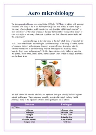
1. Aero microbiology
The term aeromicrobiology was coined in the 1930s by F.C.Meier in relation with a project
concerned with study of life in air. Aeromicrobiology has been defined in various ways as
“the study of aerosolisation, aerial transmission, and deposition of biological materials” or
more specifically as “the study of diseases that may be transmitted via respiratory route” or
even more aptly as “the study of airborne organisms and their effects on human health and
the environment”.
Aeromicrobiology in its wider sense is the study of all forms of microbial life
in air. To an environmental microbiologist, aeromicrobiology is “the study of various aspects
of intramural (indoor) and extramural (outdoor) aeromicrobiology in relation with the
airborne transmission of environmentally relevant microorganisms including viruses,
bacteria, fungi, yeasts and protozoans”. Besides these microbes, other biological materials
like pollens, insect debris, animal debris, animal danders (chief source of allergic disorders)
are also found in air.
It is well known that airborne microbes are important pathogens causing diseases in plants,
animals and humans. These pathogens spread by aeromicrobiological pathway (AMB
pathway). Some of the important airborne human pathogens are as follows:
Diseases Pathogens
(a)Viral:
Influenza Influenza virus
Chickenpox Varicella virus
Smallpox Variola virus
Measles Rubeola virus
German measles Rubella virus
Dengue fever Flavivirus
2. (b) Bacteria:
Diphtheria Corynebacterium diphtheriae
Pertussis (whooping cough) Bordetella pertusis
Meningitis (meningococcal) Neisseria meningitidis
Tuberculosis Mycobacterium tuberculosis
Klebsiella pneumonia Klebsiella pneumoniae
Serratia pneumonia Serratia marcescens
Q fever Coxiella burnetii
Pneumococcal Streptococcus
Pneumonia Pneumoniae
Sterp throat Streptococcus
Scarlet fever Pyogenes
(c) Fungal:
Aspergillosis Aspergillus fumigates
Blastomycosis Blastomyces dermatidis
Coccidioidomycosis Coccidiodes immitis
Cryptococcosis Cryptococcus neoformans
Histoplasmosis Histoplasma capsulatum
(d)Protozoal:
Pneumocytosis Pneumocytosis carinii
Dropletnuclei:
Airborne droplet nuclei develop when the fluid of pathogenic droplets (1-5 µm in
size; micrometre = one-thousandth of a millimetre) evaporates. They are so small and light
they may remain suspended in the air for several hours. Thus, they may also infect persons
entering a room which has been left by a patient long ago. Also, airborne droplet nuclei can
be widely dispersed by air currents. Tuberculosis, chickenpox, measles and possibly also
influenza may be transmitted this way.
3. When investigating the origins of droplet and airborne infections, there are several
well-known primary sources of infectious particles. Vomiting, toilet flushing, sneezing,
coughing, and talking. Moreover, toilet bowls, the water in them, and toilet seats may harbor
infectious particles after the initial flush, making additional aerosolization of infectious
particles possible with additional flushes for as long as 30 minutes after the initial flush.
Particle desiccation, discussed above, is important in this context. A single sneeze, for
example, generates as many as 40,000 large droplet particles; most will desiccate
immediately into small, infectious droplet nuclei, with 80% of the particles being smaller
than 100 μm.
Droplet nuclei released from various activities
The transmission of infectious diseases via airborne or droplet routes may also depend
on the frequency of the initiating activity. For example, while a single sneeze may produce
more total infectious particles than a cough, Couch et al. reported that coughing is more
frequent than sneezing during infection with Coxsackievirus A. This finding suggests that
coughing is a more likely method of airborne transmission for this disease than sneezing. As
coughing is also a common symptom of influenza infection, it may also contribute to the
airborne transmission of this pathogen.Finally, infectious individuals are not always the
immediate source of airborne infectious particles. Many people spend considerable time in
office buildings, for example, and as a result become exposed to airborne pathogens that
originate from nonhuman sources (e.g., molds, toxins produced by molds, pollen, pet dander,
and pest droppings). The health effects associated with naturally occurring indoor biological
air pollutants include disease, toxicoses, and hypersensitivity (i.e., allergic) diseases. In
addition, exposure to indoor biological air pollutants has been associated with “sick building
syndrome,”a set of nonspecific symptoms that may include upper-respiratory symptoms,
headaches, fatigue, and rash and“appear to be linked to time spent in a building, but no
specific illness or cause can be identified.”
Bioaerosols:
A bioaerosol is an aerosol comprising particles of variable biological origin.The
term bioaerosol is used to describe living airborne particles or those originating form
living organisms. This can be fungal spores, pollen grains, endotoxins, or particles of
animal dander. Bioaerosols are complex mixtures consisting of several components that
can stem from simple organic molecules (dimensions in the nanometer range), viruses,
4. bacteria and bacterial spores, mold spores and hyphae, pollen (with diameters as small
as 100 micrometers), and animal and plant debris (of various sizes).
Relative Sizes of Bioaerosols
Bioaerosols vary considerably in size. Their composition depends on several
factors including the type of microbe or toxin, type of particles they are associated with
(as mist or dust), and the gases in which the bioaerosol is suspended.In general,
bioaerosols range from less than 0.01µm to more than 100µm in diameter and are
classified on the basis of their size into different modes as follows:
Mode type Size range (µm in diameter)
Nuclei Fine <0.1
Accumulation particles 0.1 to 2.0
Coarse >2.00
Bioaerosols in the respirable size range (≤10µm) are of particular concern to human
health.The composition of bioaerosols can be liquid or solid or a mixture of the two and
should be thought of as microorganisms associated with airborne particles or as airborne
particles containing microorganisms. It is rare to find microbes not associated with dust
or mist or other airborne particles in the atmosphere.
Assessmentof Air Quality:
Airborne bacterial and fungal cells and spores may be present in droplets as
bioaerosols, as very small individual particles that stay suspended for long periods, or as
larger clumps and aggregates that settle rapidly onto surfaces. They can be an important
source of infection in medical facilities and can contaminate sensitive manufacturing
operations, but regular monitoring of airborne microorganisms is sometimes neglected.
Bioaerosols containing airborne microbes can be controlled at every point of spread
launching, transport and deposition by using different mechanisms which include ventilation,
filtration, biocidal agents and isolation.
5. Air Sampling:
Bioaerosol sampling provides useful data that can be used for a variety of
applications, such as (i)measuring the presence and concentration of airborne
biocontaminants; (ii)confirming dissemination from a biocontaminant source; (iii)evalution
of bioaerosol exposure and health risks; (iv)documentation of clearance to reoccupy a space,
following remediation efforts; and (v)compliance monitoring in industrial environments. The
objective of bioaerosol sampling is the efficient removal and collection of biological particles
from the air in a manner that does not affect the integrity of the sample. This depends on the
characteristics of the microorganisms and on the physical features of the sampling
instrument. The selection of a sampler depends on a number of factors, such as sampler
performance, expected bioaerosol concentration, and the analysis method.
Sampling Devices and Equipment:
Various methods have been developed for the collection of bioaerosols.One can
choose any of these sampling methods depending upon the site of collection, environmental
conditions of sampling and mobility and sampling efficiency. Different kinds of samplers
have been designed,from the very simple type, such as the Andersen six stage impaction
sampler.Several types of samplers are commonly used,which are based on different methods
of sampling-impingement, impaction, centrifugation, filtration and deposition.
Impingement is the trapping of airborne particles in a liquid matrix;
Impaction is the forced deposition of airborne particles on a solid surface; Centrifugation is
the mechanically forced deposition of airborne particles using increased forces of gravity;
Filtration is the trapping of airborne particles by size exclusion, and Deposistion is the
collection of airborne particles using only naturally-occuring deposition forces.
Impaction:
Impaction is the forced deposition of airborne particles usually on a solid agar surface.
The impaction method uses the inertia of particles to separate them from the air. The particles
are deposited onto a collection surface, usually on agar medium for culture based analysis or
an adhesive coated surface for microscopic analysis. The impaction process depends on the
inertial properties of the particles, such as size, density and velocity and on the physical
parameters of the impactor such as inlet nozzle dimensions and air flow pathway. The
impactor sampler draws air through an inlet nozzle dimensions and air flow pathway. The
impactor sampler draws air through an inlet nozzle toward a collection surface, and particles
with sufficient inertia impact while smaller particles remain in the air stream.
Impator samplers may be designed with multiple circular inlet mozzles; the sampler is
then referred to as a multiple-hole or sieve sampler.If there are several stages with
successively smaller nozzles, the sampler is referred to as a cascade impactor. For samplers
in which air is drawn through a single nozzle, the shape of the nozzle is usually rectangular
and the impactor is reffered to as a slit sampler. Some impactors utilize centrifugal
6. impaction, which also uses inertial forces to separate the particles from the air stream, but in a
radial geometry.
1.Multiple-hole impactor samplers:
The Andersen six-stage impactor sampler
The Andersen impactor sampler has been widely used for culturable bioaerosol
measurements. The sampler draws air at a flow rate of 28.3 1/min and is operated using an
electric vaccum pump. The Andersen six-stage impactor sampler consists of six stages with
decreasing nozzle diameter that collect progressively samller particles onto agar plates.The
general operating principle is that the air is sucked through the sampling pore and strikes agar
plates.There are six petridishes containing suitable growth medium, kept under sieves of
different pore size.Each sieve has 400 pores.The circular orfice at the top sucks air at flow
rate of 28.3 1/min. passing it through the sieve arranged in gradually decreasing order of pore
size. Air impacting on last petridish goes out.Larger particles are collected on the first layer,
and each successive stage collects smaller and smaller particles by increasing the flow
velocity and consequently the impaction potential.Particles of similar size impact on a given
petridish only. In this way spores/pollens of six dimension orders are impacted on agar
7. surface of individual petridish. The number of colony-forming units (CFU) that form on the
agar collection plates provides bioaerosol size distribution information.
Slit Impactors:
Rotorod intermittent sampler Burkard personal multiple hole sampler
Slit impactors are available for the measurement of either culturable or total airborne
microorgamisms.Many slit impactors have a moving collection surface that allows
enumeration of bioaerosol concentration over time. Slit impactors that deposit the bioaerosol
onto an agar surface for the estimation of culturable cells include the Mattson-Garvin air
sampler and the Slit-to-agar sampler. Slit impactors that collect particles onto an adhesive-
coated surface are generally used for the microscopic enumeration of fungal spores or pollen
grains. These include the Burkard spore traps and the personal air sampler, the Air-O-Cell
sampling cassette,the Allergenco air sampler and the Rotorod intermittent sampler.
Burkard personal slide sampler, battery operated, suitable for indoor environment is
10cm high having a rectangular orfice at the top. The microslide is coated with glycerine
jelly. The sampler sucks air at flow rate of 10.0 1/min. through the orfice. The particles get
impacted on the slide forming a band. Slide is mounted and scanned.
The Rotorod sampler has an intermittent rotating impactor, is power-operated and
ideal for both indoor and outdoor studies. It is light weight portable sampler. It has arm that
holds two cubical rods coated with silicon grease.Unexposed rods remain folded. Arm has a
shield at top for protection from rains. Exposure time can be regulated.The rods are mounted
on a grooved slide in a basic Fuschin stain and Scanned.
Liquid Impingement:
Liquid impingement samplers draw air through an inlet and particles are collected in a
liquid. The particle is removed from the air by inertial force and impaction into a swirling or
bubbling liquid. Aggregates of cells may be broken apart by the action of the collection fluid.
8. The collection of bioaerosol particles over a wide range of airborne particle concentrations,
the ability to divide the sample for multiple analyses, and the option of applying a variety of
analysis methods. A liquid sample can be concentrated by filtration or diluted by liquid
addition, depending on the concentration of collected microorganisms. In addition, several
culture media can be inoculated with aliquots of the collection medium for the culture of
groups of microorganisms with different nutrient requirements.
(i)The AGI-30 all glass impinge sampler; (ii)The SKC Biosampler; (iii)TheBurkardmulti-stage liquidimpinge
The AGI-30 all-glass impinge sampler is a widely used sampler that has a curved inlet tube
designed to simulate the nasal passage,making this sampler useful for studying the respirating
infection potential of bioaersols. The air is transmitted through a liquid medium where the
particles are trapped. The sampler is usually run at 12.5 1/min. flow rate at a height of 1.5 m,
the average human breathing height. It is easy to use, inexpensive, portable, easily sterilized
and has good bioefficiency. It is very efficient for particles in the range of 0.8 to 15µm. The
usual volume of the collection medium is 20 ml and duration of sampling approximately 20
minutes. Sampling media may be a simple one as 0.85% NaCl, or more complex as 1%
peptone. Enriched or defined growth media can also be used for specific microbes.
The Burkard multi-stage liquid impinger is a stainless steel sampler that collects
particles in three size fractions: > 10µm, 4 to 10µm and < 4µm. The BioSampler collects
particles by drawing air through three nozzles that are directed at an angle toward the inner
sampler wall and the liquid swirls upward on the inner wall of the sampler to remove
collected particles. Although other impingers are designed for use with water-based
collection fluids, the Biosamplers can be used with viscous collection fluids (e.g., heavy
white mineral oil) to minimize sampling stress and evaporation loss of the collection buffer.