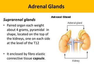
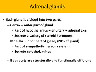
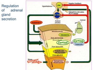
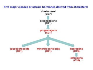
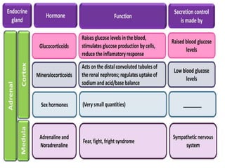
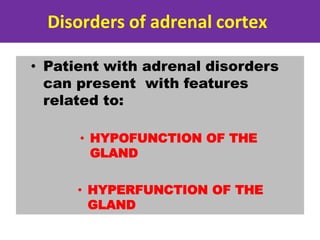
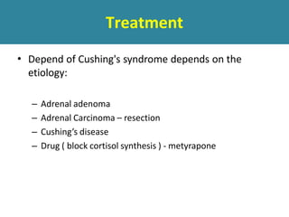
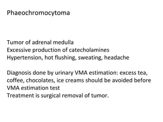
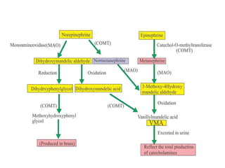
Adrenal Function Tests
Adrenal Glands
Suprarenal glands
• Paired organ each weight
about 4 grams, pyramidal in
shape, located on the top of
the kidneys, one on each side
at the level of the T12
• It enclosed by fibro elastic
connective tissue capsule.
Adrenal glands
• Each gland is divided into two parts:
– Cortex – outer part of gland
• Part of hypothalamus – pituitary – adrenal axis
• Secrete a variety of steroid hormones
– Medulla – inner part of gland, (20% of gland)
• Part of sympathetic nervous system
• Secrete catecholamines
– Both parts are structurally and functionally different
Adrenal cortex
• The large cortical cells are
arranged into three layers or
zones :
• zona glomerulosa,
• zona fasciculata,
• zona reticularis
Adrenal cortex
• Zona glomerulosa:
– Produce mineralocorticoids
– Mainly aldosterone
Hormones that help control the balance of
minerals (Na+ and K+) and water in the
blood
Aldosterone secretion
Adrenal cortex
• Zona fasciculata:
– Produce glucocorticoids
– Mainly cortisol and corticosterone
– The secretion of these cells is controled
by hypothalamic-pituitary axis via ACTH
Hormone that play a major role in
glucose metabolism as well as in
protein and lipid metabolism
Adrenal cortex
• Zona reticularis:
– The innermost layer of the adrenal
cortex, lying deep to the zona
faciculata and superficial to the
medulla.
– These cells produce androgens
Adrenal cortex
– The androgens produced includes
• Dehydroepiandrosterone (DHEA)
• Androstenedione
– Synthesized from cholesterol
• DHEA is further converted to DHEA-
sulfate via a sulfotransferase
Adrenal cortex
The androgens produced are released
into the blood stream and taken up in
the testis and ovaries to produce
testosterone and the estrogens
respectively.
Regulation
of adrenal
gland
secretion
ACTH
Cortisol
Cortisol
Disorders of adrenal cortex
• Patient with adrenal disorders
can present with features
related to:
• HYPOFUNCTION OF THE
GLAND
• HYPERFUNCTION OF THE
GLAND
DISORDERS OF ADRENAL
CORTEX
Adrenal Hypofunction
Adrenal insufficiency leads to a reduction in
the output of adrenal hormones
– glucocorticoids and/or mineralocorticoids
• Two types of adrenal insufficiency
• Primary insufficiency
• inability of the adrenal glands to produce enough
steroid hormones
• Secondary insufficiency
• inadequate pituitary or hypothalamic stimulation of the
adrenal glands
• Causes
– Glucocorticoid
treatment
– Autoimmune adrenalitis
– Tuberculosis
– Adrenalectomy
– Adrenal haemorrhage
Adrenal Hypofunction
Common
Adrenal Hypofunction
• Congenital causes:
Metabolic failure in hormone
production
• Congenital adrenal hyperplasia
e.g. 21-hydroxylase deficiency
• Progressive destruction of entire adrenal cortex ,
This is usually autoimmune based.
• Most likely the result of cytotoxic T lymphocytes,
Adrenal Hypofunction
Addison’s disease:
Addison’s disease: Clinical features
• Tiredness, generalized weakness, lethargy
• Anorexia, nausea, vomiting
• Hyponatremia
• Hyperkalemia ,Hypercalcemia
Dizziness and postural
hypotension
• Pigmentation
• Loss of body hair
Addison’s disease: clinical features
• hyperpigmentation
Addison’s disease: clinical features
• Hyperpigmentation
INVESTIGATIONS (HORMONAL)
• Plasma cortisol concentration
• ACTH stimulation test / Synacthen
test
• Measurement of plasma ACTH
• CRH stimulation test
• Plasma renin and aldosterone
levels
PLASMA ACTH MEASUREMENT
• To differentiate between primary
and secondary adrenal failure
• Primary insufficiency - ACTH
increased
• Secondary insufficiency - ACTH
decreased
CRH STIMULATION TEST
• To differentiate between secondary
adrenal insufficiency due to pituitary
or hypothalamic disease.
Pituitary disease – blunted or nil response
Hypothalamic lesions – positive response
PLASMA RENIN AND ALDOSTERONE
• Adrenal insufficiency
– Low aldosterone level with
high renin
Disorders of adrenal cortex
Adrenal Hyperfunction
• Cushing syndrome : High Cortisol
• Hyperaldosteronism : High aldosterone
• Pheochromocytoma : High catecholamine
.
Hyperaldosteronism
A medical condition where too much
aldosterone is produced by the adrenal
glands, which can lead to sodium retention
and potassium loss.
• Types:
– Primary hyperaldosteronism
– Secondary hyperaldosteronism
Primary hyperaldosteronism
Conn’s syndrome
Primary aldosteronism
CONN’S SYNDROME
•Characterized by autonomous excessive
production of aldosterone by adrenal glands
• Presents with hypertension, hypokalemia
• and renal K+ wasting
Conn’s Syndrome
• Causes:
– Adrenal adenoma
– Bilateral hypertrophy of zona
glomerulosa cells
– Adrenal carcinoma
Secondary aldosteronism
Increased adrenal aldosterone production
in response to non-pituitary, extra-adrenal
stimuli
• Commoner than primary aldosteronism
Secondary aldosteronism
– CCF
– Liver cirrhosis with ascitis
– Nephrotic Syndrome
Conn’s syndrome
• Clinical features:
– Hypertension : aldosterone induced Na retention
– Muscle weakness: Due to decrease K+
– Muscle paralysis: severe hypokalemia
– tetany and paraesthesia
INVESTIGATION
• Electrolyte & blood gasses:
– Hypernatremia
– Hypokalemia
– Alkalosis: Blood p H > 7.45
Plasma aldosterone : renin activity ratio high
Disorders of adrenal cortex
CUSHING’S SYNDROME
• Definition
• Clinical features
• Investigations
– Screening for Cushing’s syndrome
– Elucidation of the cause of Cushing’s syndrome
• Management
CUSHING’S SYNDROME
Adrenal cortex hyperfunction
• Any condition resulting from overproduction of
primarily glucocorticoid (cortisol)
• Mineralocorticoid and androgen may also be
excessive
Pseudo-Cushing’s syndrome
•
•
Appear cushingoid and have some biochemical
abnormalities of true Cushing’s disease
Causes
– Severe depression
– Alcoholism
– Obesity
– Polycystic ovarian syndrome
Etiology
• Excessive cortisol (ACTH dependent)~75%
– Pituitary disease
– Ectopic ACTH syndrome
• Malignancy - ( bronchus, thymus, pancreas, ovary )
– Ectopic CRH syndrome
– Exogenous ACTH administration
ACTH dependent causes
*Hypersecretion of ACTH and Cortisol is greater in ectopic ACTH syndrome
than Cushing Disease
Etiology
• Excessive cortisol (ACTH independent) ~25%
– Adrenal tumour
• Adenoma
• carcinoma
– Nodular hyperplasia
– Exogenous glucocorticoid administration
ACTH independent causes
Etiology
• Excess cortisol binding globulin
– Estrogen therapy : Osteoporosis, OCP
– Pregnancy
Clinical features
• Truncal obesity with deposition of adipose tissue in
characteristic site (moon face, buffalo hump)– exact
mechanism unknown
• Thinning of skin – catabolic response
• Purple striae – catabolic response
• Excessive bruising – catabolic response
Cont..
• Hirsutism ( esp adrenal carcinoma ) - ↑ adrenal
androgen
• Menstrual irregularities - ↑ adrenal androgen
• Skin pigmentation ( ACTH ↑ ) – melanocyte
stimulating activity
Cont..
• Hypertension – mineralocorticoid effect → sodium
retention
– metabolic alkalosis
• Glucose intolerance - ↑ hepatic gluconeogenesis and
insulin resistance
• Muscle weakness and wasting
Cont..
• Back pain
• Psychiatric disturbances
Laboratory investigations
 There are two diagnostic steps in the investigation
of patient suspected of having Cushing's
syndrome
Screening test
for identification of Cushing's syndrome.
the demonstration of high plasma cortisol level
 Identification of cause
1. Demonstration of increased cortisol
 Assessment of circadian rhythm in cortisol secretion
 24-Hour urinary free cortisol excretion
 Overnight / low dose dexamethasone suppression
test
Laboratory investigations
1. Assessment of circadian rhythm in cortisol
secretion.
 Measure 8 am and 11 pm serum cortisol
level
 Normal : Serum value at midnight is 50% less than value
at 8 am
 Cushing’s syndrome : rhythm is lost
 Pseudo-Cushing : normal circadian.
Laboratory investigations
2. Measuring 24-hour urinary free cortisol
> 100 microgram Diagnostic of
Cushing's syndrome
Laboratory investigations
3. Low dose Dexamethasone suppression test :
After injection of dexamethasone urinary and
plasma cortisol levels should fall
But in cushing’s syndrome there is no fall in cortisol
levels
• High dose Dexamethasone suppression test
• Normal individuals suppress plasma cortisol
• Patients with Cushing's syndrome fail to show
complete suppression of plasma cortisol levels.
This test is highly sensitive
2. Elucidation of the cause
• Plasma ACTH
– Low – adrenal causes
– Elevated
• Slight – pituitary dependent Cushing’s
• Gross – ectopic secretion of ACTH
Elucidation of the cause
• CRH Test
– Differentiate ectopic ACTH secretion and
Cushing’s disease.
– Cushing’s disease – plasma ACTH increases 50%
over baseline and cortisol increase by 20%
– Ectopic ACTH or adrenal tumour – no response
Elucidation of the cause
• Imaging
– CT scan of adrenal gland
– MRI of pituitary gland:
– CT scan/MRI of thorax & abdomen: ectopic ACTH
producing tumor
Treatment
• Depend of Cushing's syndrome depends on the
etiology:
– Adrenal adenoma
– Adrenal Carcinoma – resection
– Cushing’s disease
– Drug ( block cortisol synthesis ) - metyrapone
Tumor of adrenal medulla
Excessive production of catecholamines
Hypertension, hot flushing, sweating, headache
Diagnosis done by urinary VMA estimation: excess tea,
coffee, chocolates, ice creams should be avoided before
VMA estimation test
Treatment is surgical removal of tumor.
Phaeochromocytoma
adrenal function tests.pptx

adrenal function tests.pptx

  • 1.
  • 2.
    Adrenal Glands Suprarenal glands •Paired organ each weight about 4 grams, pyramidal in shape, located on the top of the kidneys, one on each side at the level of the T12 • It enclosed by fibro elastic connective tissue capsule.
  • 3.
    Adrenal glands • Eachgland is divided into two parts: – Cortex – outer part of gland • Part of hypothalamus – pituitary – adrenal axis • Secrete a variety of steroid hormones – Medulla – inner part of gland, (20% of gland) • Part of sympathetic nervous system • Secrete catecholamines – Both parts are structurally and functionally different
  • 4.
    Adrenal cortex • Thelarge cortical cells are arranged into three layers or zones : • zona glomerulosa, • zona fasciculata, • zona reticularis
  • 5.
    Adrenal cortex • Zonaglomerulosa: – Produce mineralocorticoids – Mainly aldosterone Hormones that help control the balance of minerals (Na+ and K+) and water in the blood
  • 6.
  • 7.
    Adrenal cortex • Zonafasciculata: – Produce glucocorticoids – Mainly cortisol and corticosterone – The secretion of these cells is controled by hypothalamic-pituitary axis via ACTH Hormone that play a major role in glucose metabolism as well as in protein and lipid metabolism
  • 8.
    Adrenal cortex • Zonareticularis: – The innermost layer of the adrenal cortex, lying deep to the zona faciculata and superficial to the medulla. – These cells produce androgens
  • 9.
    Adrenal cortex – Theandrogens produced includes • Dehydroepiandrosterone (DHEA) • Androstenedione – Synthesized from cholesterol • DHEA is further converted to DHEA- sulfate via a sulfotransferase
  • 10.
    Adrenal cortex The androgensproduced are released into the blood stream and taken up in the testis and ovaries to produce testosterone and the estrogens respectively.
  • 11.
  • 14.
    Disorders of adrenalcortex • Patient with adrenal disorders can present with features related to: • HYPOFUNCTION OF THE GLAND • HYPERFUNCTION OF THE GLAND
  • 15.
  • 16.
    Adrenal Hypofunction Adrenal insufficiencyleads to a reduction in the output of adrenal hormones – glucocorticoids and/or mineralocorticoids • Two types of adrenal insufficiency • Primary insufficiency • inability of the adrenal glands to produce enough steroid hormones • Secondary insufficiency • inadequate pituitary or hypothalamic stimulation of the adrenal glands
  • 17.
    • Causes – Glucocorticoid treatment –Autoimmune adrenalitis – Tuberculosis – Adrenalectomy – Adrenal haemorrhage Adrenal Hypofunction Common
  • 18.
    Adrenal Hypofunction • Congenitalcauses: Metabolic failure in hormone production • Congenital adrenal hyperplasia e.g. 21-hydroxylase deficiency
  • 19.
    • Progressive destructionof entire adrenal cortex , This is usually autoimmune based. • Most likely the result of cytotoxic T lymphocytes, Adrenal Hypofunction Addison’s disease:
  • 20.
    Addison’s disease: Clinicalfeatures • Tiredness, generalized weakness, lethargy • Anorexia, nausea, vomiting • Hyponatremia • Hyperkalemia ,Hypercalcemia Dizziness and postural hypotension • Pigmentation • Loss of body hair
  • 21.
    Addison’s disease: clinicalfeatures • hyperpigmentation
  • 22.
    Addison’s disease: clinicalfeatures • Hyperpigmentation
  • 23.
    INVESTIGATIONS (HORMONAL) • Plasmacortisol concentration • ACTH stimulation test / Synacthen test • Measurement of plasma ACTH • CRH stimulation test • Plasma renin and aldosterone levels
  • 24.
    PLASMA ACTH MEASUREMENT •To differentiate between primary and secondary adrenal failure • Primary insufficiency - ACTH increased • Secondary insufficiency - ACTH decreased
  • 25.
    CRH STIMULATION TEST •To differentiate between secondary adrenal insufficiency due to pituitary or hypothalamic disease. Pituitary disease – blunted or nil response Hypothalamic lesions – positive response
  • 26.
    PLASMA RENIN ANDALDOSTERONE • Adrenal insufficiency – Low aldosterone level with high renin
  • 27.
  • 28.
    Adrenal Hyperfunction • Cushingsyndrome : High Cortisol • Hyperaldosteronism : High aldosterone • Pheochromocytoma : High catecholamine .
  • 29.
    Hyperaldosteronism A medical conditionwhere too much aldosterone is produced by the adrenal glands, which can lead to sodium retention and potassium loss. • Types: – Primary hyperaldosteronism – Secondary hyperaldosteronism
  • 30.
  • 31.
    Primary aldosteronism CONN’S SYNDROME •Characterizedby autonomous excessive production of aldosterone by adrenal glands • Presents with hypertension, hypokalemia • and renal K+ wasting
  • 32.
    Conn’s Syndrome • Causes: –Adrenal adenoma – Bilateral hypertrophy of zona glomerulosa cells – Adrenal carcinoma
  • 33.
    Secondary aldosteronism Increased adrenalaldosterone production in response to non-pituitary, extra-adrenal stimuli • Commoner than primary aldosteronism
  • 34.
    Secondary aldosteronism – CCF –Liver cirrhosis with ascitis – Nephrotic Syndrome
  • 35.
    Conn’s syndrome • Clinicalfeatures: – Hypertension : aldosterone induced Na retention – Muscle weakness: Due to decrease K+ – Muscle paralysis: severe hypokalemia – tetany and paraesthesia
  • 36.
    INVESTIGATION • Electrolyte &blood gasses: – Hypernatremia – Hypokalemia – Alkalosis: Blood p H > 7.45 Plasma aldosterone : renin activity ratio high
  • 37.
  • 38.
    CUSHING’S SYNDROME • Definition •Clinical features • Investigations – Screening for Cushing’s syndrome – Elucidation of the cause of Cushing’s syndrome • Management
  • 39.
    CUSHING’S SYNDROME Adrenal cortexhyperfunction • Any condition resulting from overproduction of primarily glucocorticoid (cortisol) • Mineralocorticoid and androgen may also be excessive
  • 40.
    Pseudo-Cushing’s syndrome • • Appear cushingoidand have some biochemical abnormalities of true Cushing’s disease Causes – Severe depression – Alcoholism – Obesity – Polycystic ovarian syndrome
  • 41.
    Etiology • Excessive cortisol(ACTH dependent)~75% – Pituitary disease – Ectopic ACTH syndrome • Malignancy - ( bronchus, thymus, pancreas, ovary ) – Ectopic CRH syndrome – Exogenous ACTH administration
  • 42.
  • 43.
    ACTH dependent causes *Hypersecretionof ACTH and Cortisol is greater in ectopic ACTH syndrome than Cushing Disease
  • 44.
    Etiology • Excessive cortisol(ACTH independent) ~25% – Adrenal tumour • Adenoma • carcinoma – Nodular hyperplasia – Exogenous glucocorticoid administration
  • 45.
  • 46.
    Etiology • Excess cortisolbinding globulin – Estrogen therapy : Osteoporosis, OCP – Pregnancy
  • 47.
    Clinical features • Truncalobesity with deposition of adipose tissue in characteristic site (moon face, buffalo hump)– exact mechanism unknown • Thinning of skin – catabolic response • Purple striae – catabolic response • Excessive bruising – catabolic response
  • 48.
    Cont.. • Hirsutism (esp adrenal carcinoma ) - ↑ adrenal androgen • Menstrual irregularities - ↑ adrenal androgen • Skin pigmentation ( ACTH ↑ ) – melanocyte stimulating activity
  • 49.
    Cont.. • Hypertension –mineralocorticoid effect → sodium retention – metabolic alkalosis • Glucose intolerance - ↑ hepatic gluconeogenesis and insulin resistance • Muscle weakness and wasting
  • 50.
    Cont.. • Back pain •Psychiatric disturbances
  • 52.
    Laboratory investigations  Thereare two diagnostic steps in the investigation of patient suspected of having Cushing's syndrome Screening test for identification of Cushing's syndrome. the demonstration of high plasma cortisol level  Identification of cause
  • 53.
    1. Demonstration ofincreased cortisol  Assessment of circadian rhythm in cortisol secretion  24-Hour urinary free cortisol excretion  Overnight / low dose dexamethasone suppression test
  • 54.
    Laboratory investigations 1. Assessmentof circadian rhythm in cortisol secretion.  Measure 8 am and 11 pm serum cortisol level  Normal : Serum value at midnight is 50% less than value at 8 am  Cushing’s syndrome : rhythm is lost  Pseudo-Cushing : normal circadian.
  • 55.
    Laboratory investigations 2. Measuring24-hour urinary free cortisol > 100 microgram Diagnostic of Cushing's syndrome
  • 56.
    Laboratory investigations 3. Lowdose Dexamethasone suppression test : After injection of dexamethasone urinary and plasma cortisol levels should fall But in cushing’s syndrome there is no fall in cortisol levels
  • 57.
    • High doseDexamethasone suppression test • Normal individuals suppress plasma cortisol • Patients with Cushing's syndrome fail to show complete suppression of plasma cortisol levels. This test is highly sensitive
  • 58.
    2. Elucidation ofthe cause • Plasma ACTH – Low – adrenal causes – Elevated • Slight – pituitary dependent Cushing’s • Gross – ectopic secretion of ACTH
  • 59.
    Elucidation of thecause • CRH Test – Differentiate ectopic ACTH secretion and Cushing’s disease. – Cushing’s disease – plasma ACTH increases 50% over baseline and cortisol increase by 20% – Ectopic ACTH or adrenal tumour – no response
  • 60.
    Elucidation of thecause • Imaging – CT scan of adrenal gland – MRI of pituitary gland: – CT scan/MRI of thorax & abdomen: ectopic ACTH producing tumor
  • 61.
    Treatment • Depend ofCushing's syndrome depends on the etiology: – Adrenal adenoma – Adrenal Carcinoma – resection – Cushing’s disease – Drug ( block cortisol synthesis ) - metyrapone
  • 62.
    Tumor of adrenalmedulla Excessive production of catecholamines Hypertension, hot flushing, sweating, headache Diagnosis done by urinary VMA estimation: excess tea, coffee, chocolates, ice creams should be avoided before VMA estimation test Treatment is surgical removal of tumor. Phaeochromocytoma