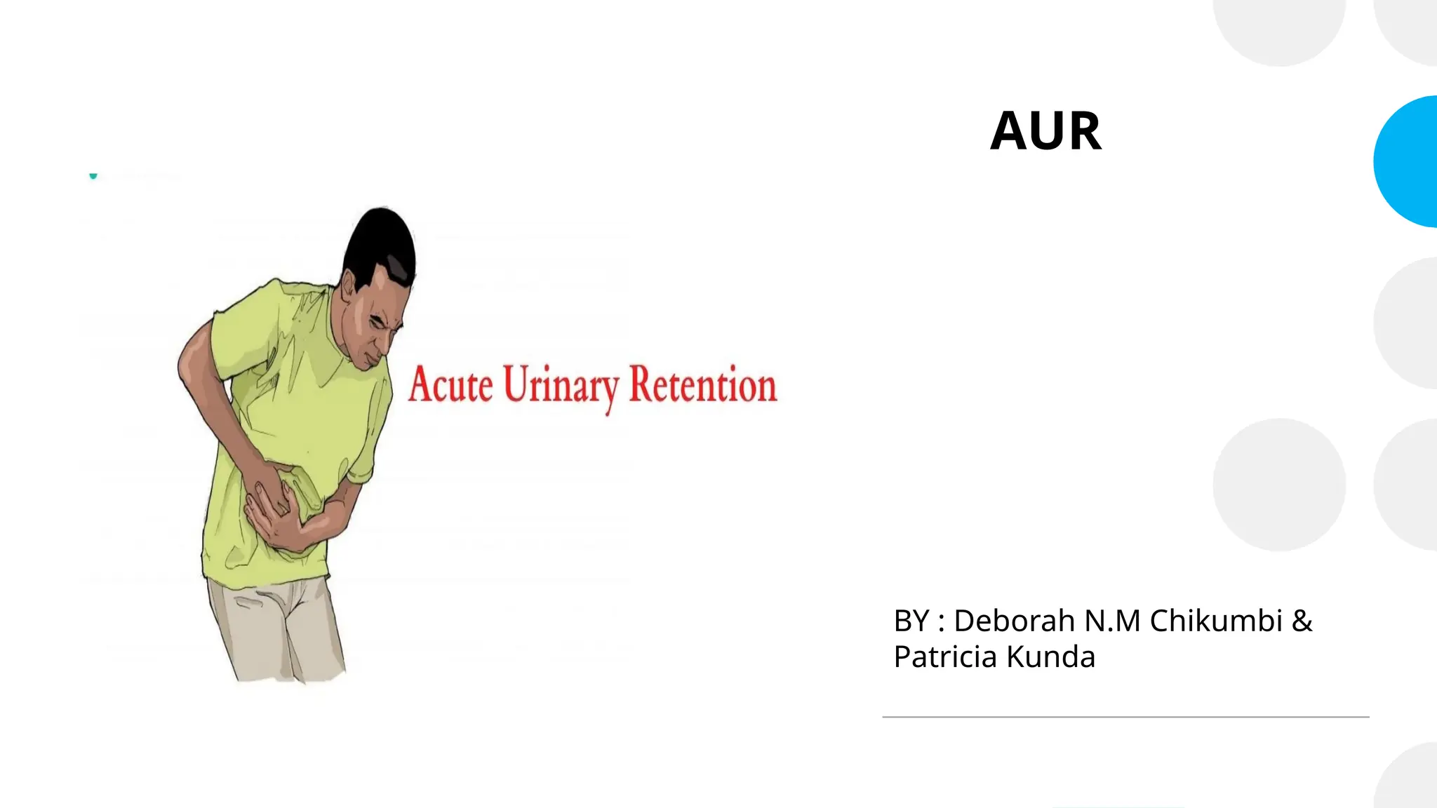
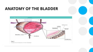
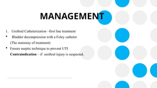
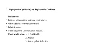
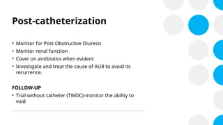
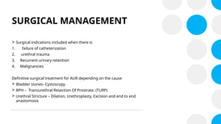
AUR
BY : Deborah N.M Chikumbi &
Patricia Kunda
OBJECTIVES
• Define Acute Urinary Retention and Know Its Etiology.
• To Learn Its Clinical Presentation Including How To Assess a patient.
• Learn the Approach and Manage of a patient.
DEFINITION
• Acute Urinary Retention is a term that refers to sudden onset of a failure to pass urine
when the urine production is normal inspite the desire to do so.
CLASSIFICATION
Urinary Retention can be classified in various ways:
1. History (duration of onset)
2. Symptoms (Presences or absences of pain)
3. The volume of residual urine.
CLASSIFICATION
The following classification is commonly used:
1.Acute Retention
Period of onset is short. Symptoms include severe pain and the volume of the urine
is <1000ml. Mostly between 300mls to 500mls.
2.Chronic Retention
Period of onset is Long. The patient presents in the outpatient department as a non
emergency. The symptoms are abdominal distention which is painless. The amount
of residual urine will exceed I liter ( >1000ml).
CLASSIFICATION
3.Acute on Chronic
Mixed usually the patient develops acute retention on pre-existing chronic
retention. This maybe precipitated by factors such as Travel, parties, bus drive ,
alcohol etc
ANATOMY OF THE BLADDER
• The appearance of the bladder varies depending on the amount of urine stored.
When full, it exhibits an oval shape, and when empty it is flattened by the
overlying bowel.
ANATOMY OF THE BLADDER
The parts of the bladder include:
• Apex – located superiorly, pointing towards the pubic symphysis. It is connected to the
umbilicus by the median umbilical ligament (a remnant of the urachus).
• Body – main part of the bladder, located between the apex and the fundus
• Fundus (or base) – located posteriorly. It is triangular-shaped, with the tip of the
triangle pointing backwards.
• Neck – formed by the convergence of the fundus and the two inferolateral surfaces. It
is continuous with the urethra.
ANATOMY OF THE BLADDER
PHYSIOLOGY OF MICTURITION
• Micturition is the process by which the urinary bladder becomes emptied when
it becomes filled.
It involves two steps:
• Firstly the bladder fills progressively until the tension in its wall raises above
threshold level.
• Secondly a nervous reflex known as the micturition reflex causes a conscious
desire to urinate.
PHYSIOLOGY OF MICTURITION
• The micturition reflex is an autonomic spinal cord reflex which can be facilitated or
inhibited by centers in the cerebral cortex of brain stem.
• Normal micturition has a central control in the pons that is known as the Barrington’s
Nucleus.
• It also has a spinal control centre called Onuf’s nucleus which is found at the area of S2-S4.
• When the bladder fills up to 300mls the Micturition reflex is triggered. This will result in
contracting of the bladder muscle which is known as the Detrusor muscle and the relaxing
of sphincter muscles automatically. Unless the Central control inhibits this process.
PHYSIOLOGY OF MICTURITION
• In Retention this process may fail. Mainly due to a physical obstruction in the
Urethral in 90% of case.
• It may also fail due to Micturition Reflex failure or more Broadly nervous
system diseases.
EPIDEMIOLOGY
• Acute Urinary Retention is more common in men that women.
• Men : Female ratio 13: 1
• This increase with age in men starting with incidences of 4.5 to 6.8 per 1,000
per person years to 300 per 1,000 persons years in men in their 80s.
• As compared to women who have 7 per 100,000 per year.
AETIOLOGY
• The aetiology of Urinary Retention is not well
known. However some risk factors have been
identified such as:
• Benign Prostatic Hypertrophy
• Constipation
• Urethral stricture
• Prostate cancer
• Post operative ( e.g haemorrhoidectomy and
fistulectomy)
• Neurological ( e.g spinal injuries, CVA, spinal cord
compression, general and epidural anasthesia)
RISK FACTORS
• Medications such as anticholinergic and sympathomimetics
• Genitourinary infections such as acute prostatitis, urethritis and perianal abscess
• Urethral trauma or injury or a blood clot.
• Blocked catheter
• Urinary stones
• Phimosis and paraphimosis
RISK FACTORS
• Prostate disease such as prostate infarction or infection can precipitate retention.
• Taking lots of fluid especially a large alcohol intake.
• No opportunity to void such as in long flights, trips, meetings, seminars, at parties and
conferences.
CLINICAL FEATURES
HISTORY
• Typically the patient will be an elderly male patient older than 50 years.
• Patients will present with acute suprapubic pain.
• Inability to micturate.
• Will have a history of BPE with a rising IPSS score or the Single Question
Nocturia Score.
• May have had a precipitating event such as travel, alcohol binge, trauma or
infection.
CLINICAL FEATURES
• Any associated fevers, rigors, or lethargy may suggest an infective cause.
PHYSICAL EXAMINATION
• The examination can be divided into the General and Specific examination.
General Examination:
• The age of the patient.
• Habitus, gaiting and the use of crutches.
• General features of wasting, anaemia, uremia frost and fever may suggest urinary
infection.
PHYSICAL EXAMINATION
Specific Examination:
Begins with the abdominal examination then the examination of the perineum and lastly the
DRE.
1.Abdominal Examination:
• Focuses on the examination of the kidneys and bladder.
• ballottement of the Kidneys to examine whether they are enlarged.
• Examine the extent of the bladder distention by palpation and percussion.
• If the extent is below the umbilicus this is Acute Urinary Retention.
• If it is above the Umbilicus this Chronic Urinary Retention.
PHYSICAL EXAMINATION
PHYSICAL EXAMINATION.
2. Perineal Examination:
• Examine the genital region with a focus on look for thickening of the
Urethral, the prepuce, signs of fistulation or sinuses, signifying previous STI
or Stricture.
• Next examine the Scrotum and Testis. In trauma there may be a butterfly
hematoma.
PHYSICAL EXAMINATION
3. Digital Rectal Examination (DRE):
• It is important to perform a DRE especially in older patients, to assess for any prostate
enlargement or constipation.
• Examine the Prostate, fell its size, consistency, extent, mucosal attachment as well as the
nature of the median groove.
• In trauma a high riding prostate and initial hematuria may be seen.
• Assess the anal winking reflex and the sphincter tone. To ensure that the retention is not due
to neurological impairment such as multiple sclerosis, transverse myelitis or spinal cord
injury. If the anal winking reflex is absent perform a neurological examination.
PHYSICAL EXAMINATION
• Assess the anal winking reflex and the sphincter tone. To ensure that the retention is not
due to neurological impairment such as multiple sclerosis, transverse myelitis or spinal
cord injury.
• If the anal winking reflex is absent perform a neurological examination.
INVESTIGATIONS
• General tests – PSA, urinalysis, E &U&CR, FBC and Urine
MCS
SPECIFIC TESTS
• Ultrasound of the bladder, pelvic and prostate
• Renal tract ultrasound
• KUB X-Ray
• Urethrogram
• Prostatic biopsy
• Cystoscopy
Diagnosis
• Diagnosis is clinical usually made after a proper
history and physical examination
MANAGEMENT
GENERAL
• Manage pain
• Treat the infection if indicated
SPECIFIC
- Per urethral catheterisation
- Suprapubic
- Manage post obstruction diuresis
- Identify the primary cause
MANAGEMENT
1. Urethral Catheterization –first line treatment
 Bladder decompression with a Foley catheter
(The mainstay of treatment)
 Ensure aseptic technique to prevent UTI
Contraindication – if urethral injury is suspected.
2. Suprapubic Cystostomy or Suprapubic Catheter.
Indications
• Patients with urethral stricture or strictures
• When urethral catheterization fails
• Pelvic trauma
• when long term Catheterization needed.
Contraindications – 1. CA Bladder
2. Ascites
3. Active pelvic infection
Post-catheterization
• Monitor for Post Obstructive Diuresis
• Monitor renal function
• Cover on antibiotics when evident
• Investigate and treat the cause of AUR to avoid its
recurrence.
FOLLOW-UP
• Trial without catheter (TWOC)-monitor the ability to
void
SURGICAL MANAGEMENT
 Surgical indications included when there is
1. failure of catheterization
2. urethral trauma
3. Recurrent urinary retention
4. Malignancies
Definitive surgical treatment for AUR depending on the cause
 Bladder stones- Cystoscopy.
 BPH – Transurethral Resection Of Prostrate. (TURP)
 Urethral Stricture – Dilation, Urethroplasty, Excision and end to end
anastomosis
COMPLICATIONS
• Urinary tract infections
• hematuria
• Bladder hypertrophy
• Post-Obstructive Diuresis
• Urethral strictures
• Bladder stones
• Hydronephrosis
• Urolithiasis
• Reduced quality of life

ACUTE URINARY RETENTION PRESENTATION 1.pptx

  • 1.
    AUR BY : DeborahN.M Chikumbi & Patricia Kunda
  • 2.
    OBJECTIVES • Define AcuteUrinary Retention and Know Its Etiology. • To Learn Its Clinical Presentation Including How To Assess a patient. • Learn the Approach and Manage of a patient.
  • 3.
    DEFINITION • Acute UrinaryRetention is a term that refers to sudden onset of a failure to pass urine when the urine production is normal inspite the desire to do so.
  • 4.
    CLASSIFICATION Urinary Retention canbe classified in various ways: 1. History (duration of onset) 2. Symptoms (Presences or absences of pain) 3. The volume of residual urine.
  • 5.
    CLASSIFICATION The following classificationis commonly used: 1.Acute Retention Period of onset is short. Symptoms include severe pain and the volume of the urine is <1000ml. Mostly between 300mls to 500mls. 2.Chronic Retention Period of onset is Long. The patient presents in the outpatient department as a non emergency. The symptoms are abdominal distention which is painless. The amount of residual urine will exceed I liter ( >1000ml).
  • 6.
    CLASSIFICATION 3.Acute on Chronic Mixedusually the patient develops acute retention on pre-existing chronic retention. This maybe precipitated by factors such as Travel, parties, bus drive , alcohol etc
  • 7.
    ANATOMY OF THEBLADDER • The appearance of the bladder varies depending on the amount of urine stored. When full, it exhibits an oval shape, and when empty it is flattened by the overlying bowel.
  • 8.
    ANATOMY OF THEBLADDER The parts of the bladder include: • Apex – located superiorly, pointing towards the pubic symphysis. It is connected to the umbilicus by the median umbilical ligament (a remnant of the urachus). • Body – main part of the bladder, located between the apex and the fundus • Fundus (or base) – located posteriorly. It is triangular-shaped, with the tip of the triangle pointing backwards. • Neck – formed by the convergence of the fundus and the two inferolateral surfaces. It is continuous with the urethra.
  • 9.
  • 10.
    PHYSIOLOGY OF MICTURITION •Micturition is the process by which the urinary bladder becomes emptied when it becomes filled. It involves two steps: • Firstly the bladder fills progressively until the tension in its wall raises above threshold level. • Secondly a nervous reflex known as the micturition reflex causes a conscious desire to urinate.
  • 11.
    PHYSIOLOGY OF MICTURITION •The micturition reflex is an autonomic spinal cord reflex which can be facilitated or inhibited by centers in the cerebral cortex of brain stem. • Normal micturition has a central control in the pons that is known as the Barrington’s Nucleus. • It also has a spinal control centre called Onuf’s nucleus which is found at the area of S2-S4. • When the bladder fills up to 300mls the Micturition reflex is triggered. This will result in contracting of the bladder muscle which is known as the Detrusor muscle and the relaxing of sphincter muscles automatically. Unless the Central control inhibits this process.
  • 12.
  • 13.
    PHYSIOLOGY OF MICTURITION •In Retention this process may fail. Mainly due to a physical obstruction in the Urethral in 90% of case. • It may also fail due to Micturition Reflex failure or more Broadly nervous system diseases.
  • 14.
    EPIDEMIOLOGY • Acute UrinaryRetention is more common in men that women. • Men : Female ratio 13: 1 • This increase with age in men starting with incidences of 4.5 to 6.8 per 1,000 per person years to 300 per 1,000 persons years in men in their 80s. • As compared to women who have 7 per 100,000 per year.
  • 15.
    AETIOLOGY • The aetiologyof Urinary Retention is not well known. However some risk factors have been identified such as: • Benign Prostatic Hypertrophy • Constipation • Urethral stricture • Prostate cancer • Post operative ( e.g haemorrhoidectomy and fistulectomy) • Neurological ( e.g spinal injuries, CVA, spinal cord compression, general and epidural anasthesia)
  • 16.
    RISK FACTORS • Medicationssuch as anticholinergic and sympathomimetics • Genitourinary infections such as acute prostatitis, urethritis and perianal abscess • Urethral trauma or injury or a blood clot. • Blocked catheter • Urinary stones • Phimosis and paraphimosis
  • 17.
    RISK FACTORS • Prostatedisease such as prostate infarction or infection can precipitate retention. • Taking lots of fluid especially a large alcohol intake. • No opportunity to void such as in long flights, trips, meetings, seminars, at parties and conferences.
  • 18.
    CLINICAL FEATURES HISTORY • Typicallythe patient will be an elderly male patient older than 50 years. • Patients will present with acute suprapubic pain. • Inability to micturate. • Will have a history of BPE with a rising IPSS score or the Single Question Nocturia Score. • May have had a precipitating event such as travel, alcohol binge, trauma or infection.
  • 19.
    CLINICAL FEATURES • Anyassociated fevers, rigors, or lethargy may suggest an infective cause.
  • 20.
    PHYSICAL EXAMINATION • Theexamination can be divided into the General and Specific examination. General Examination: • The age of the patient. • Habitus, gaiting and the use of crutches. • General features of wasting, anaemia, uremia frost and fever may suggest urinary infection.
  • 21.
    PHYSICAL EXAMINATION Specific Examination: Beginswith the abdominal examination then the examination of the perineum and lastly the DRE. 1.Abdominal Examination: • Focuses on the examination of the kidneys and bladder. • ballottement of the Kidneys to examine whether they are enlarged. • Examine the extent of the bladder distention by palpation and percussion. • If the extent is below the umbilicus this is Acute Urinary Retention. • If it is above the Umbilicus this Chronic Urinary Retention.
  • 22.
  • 23.
    PHYSICAL EXAMINATION. 2. PerinealExamination: • Examine the genital region with a focus on look for thickening of the Urethral, the prepuce, signs of fistulation or sinuses, signifying previous STI or Stricture. • Next examine the Scrotum and Testis. In trauma there may be a butterfly hematoma.
  • 24.
    PHYSICAL EXAMINATION 3. DigitalRectal Examination (DRE): • It is important to perform a DRE especially in older patients, to assess for any prostate enlargement or constipation. • Examine the Prostate, fell its size, consistency, extent, mucosal attachment as well as the nature of the median groove. • In trauma a high riding prostate and initial hematuria may be seen. • Assess the anal winking reflex and the sphincter tone. To ensure that the retention is not due to neurological impairment such as multiple sclerosis, transverse myelitis or spinal cord injury. If the anal winking reflex is absent perform a neurological examination.
  • 25.
    PHYSICAL EXAMINATION • Assessthe anal winking reflex and the sphincter tone. To ensure that the retention is not due to neurological impairment such as multiple sclerosis, transverse myelitis or spinal cord injury. • If the anal winking reflex is absent perform a neurological examination.
  • 26.
    INVESTIGATIONS • General tests– PSA, urinalysis, E &U&CR, FBC and Urine MCS SPECIFIC TESTS • Ultrasound of the bladder, pelvic and prostate • Renal tract ultrasound • KUB X-Ray • Urethrogram • Prostatic biopsy • Cystoscopy
  • 27.
    Diagnosis • Diagnosis isclinical usually made after a proper history and physical examination
  • 28.
    MANAGEMENT GENERAL • Manage pain •Treat the infection if indicated SPECIFIC - Per urethral catheterisation - Suprapubic - Manage post obstruction diuresis - Identify the primary cause
  • 29.
    MANAGEMENT 1. Urethral Catheterization–first line treatment  Bladder decompression with a Foley catheter (The mainstay of treatment)  Ensure aseptic technique to prevent UTI Contraindication – if urethral injury is suspected.
  • 30.
    2. Suprapubic Cystostomyor Suprapubic Catheter. Indications • Patients with urethral stricture or strictures • When urethral catheterization fails • Pelvic trauma • when long term Catheterization needed. Contraindications – 1. CA Bladder 2. Ascites 3. Active pelvic infection
  • 31.
    Post-catheterization • Monitor forPost Obstructive Diuresis • Monitor renal function • Cover on antibiotics when evident • Investigate and treat the cause of AUR to avoid its recurrence. FOLLOW-UP • Trial without catheter (TWOC)-monitor the ability to void
  • 32.
    SURGICAL MANAGEMENT  Surgicalindications included when there is 1. failure of catheterization 2. urethral trauma 3. Recurrent urinary retention 4. Malignancies Definitive surgical treatment for AUR depending on the cause  Bladder stones- Cystoscopy.  BPH – Transurethral Resection Of Prostrate. (TURP)  Urethral Stricture – Dilation, Urethroplasty, Excision and end to end anastomosis
  • 33.
    COMPLICATIONS • Urinary tractinfections • hematuria • Bladder hypertrophy • Post-Obstructive Diuresis • Urethral strictures • Bladder stones • Hydronephrosis • Urolithiasis • Reduced quality of life