Downloaded 75 times


![BLOOD TEST:
Acompletebloodcount(CBC)demonstratesleukocytosis(whitebloodcell
[WBC]counthigherthan12,000/µL)withthedifferentialbeingshifted
towardthesegmentedpolymorphonuclear(PMN)cells.Leukocytosismay
representinflammationorinfection.
AC-reactiveprotein(CRP)valuecanbeobtained24-48hoursafter
presentationtoprovidesomeindicationofprognosis.Higherlevelshave
beenshowntocorrelatewithapropensitytowardorganfailure.ACRPvalue
indoublefigures(ie,≥10mg/dL)stronglyindicatesseverepancreatitis.CRP
isanacute-phasereactantthatisnotspecificforpancreatitis.](https://image.slidesharecdn.com/pancreatitisraj-150708175937-lva1-app6891/75/ACUTE-PANCREATITIS-33-2048.jpg)
![LIVER ENZYMES :
Determine alkaline phosphatase, total bilirubin, aspartate aminotransferase (AST), and
alanine aminotransferase (ALT) levels to search for evidence of gallstone pancreatitis. An
ALT level higher than 150 U/L suggests gallstone pancreatitis and a more fulminant disease
course.
Obtain measurements for blood urea nitrogen (BUN), creatinine, and electrolytes; a great
disturbance in the electrolyte balance is usually found, secondary to third spacing of fluids.
Measure blood glucose level because it may be elevated from B-cell injury in the pancreas.
Measure calcium, cholesterol, and triglyceride levels to search for an etiology of
pancreatitis (eg, hypercalcemia or hyperlipidemia) or complications of pancreatitis (eg,
hypocalcemia resulting from saponification of fats in the retroperitoneum). However, be
aware that baseline serum triglyceride levels can be falsely lowered during an episode of
acute pancreatitis.
Elevated serum amylase and lipase levels, in combination with severe abdominal pain,
often trigger the initial diagnosis of acute pancreatitis.
Serum lipase rises 4 to 8 hours from the onset of symptoms and normalizes within 7 to 14
days after treatment.
Serum amylase may be normal (in 10% of cases) for cases of acute or chronic pancreatitis
(depleted acinar cell mass) and hypertriglyceridemia.
Reasons for false positive elevated serum amylase include salivary gland disease (elevated
salivary amylase), bowel obstruction, infarction, cholecystitis, and a perforated ulcer.
If the lipase level is about 2.5 to 3 times that of amylase, it is an indication of pancreatitis
due to alcohol.[9]
Decreased serum calcium](https://image.slidesharecdn.com/pancreatitisraj-150708175937-lva1-app6891/75/ACUTE-PANCREATITIS-34-2048.jpg)


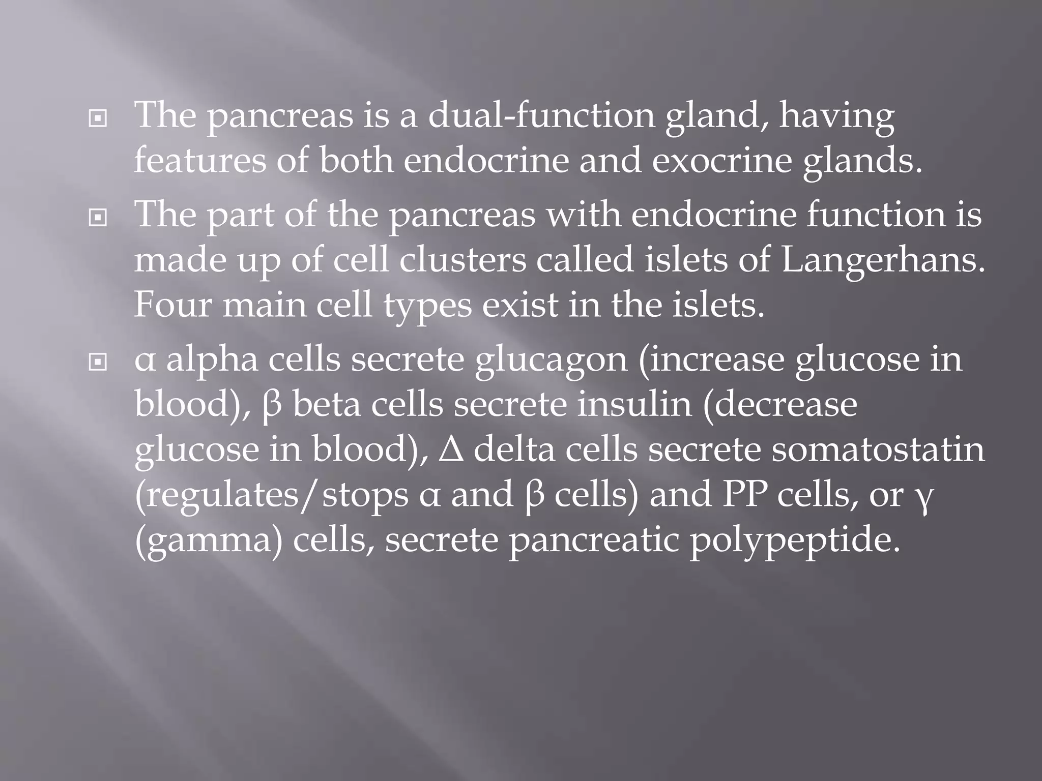
The document provides an in-depth overview of acute pancreatitis, including its anatomy, causes, symptoms, diagnosis, treatment, and complications. Key points include the dual function of the pancreas, the major etiological factors for acute pancreatitis, and diagnostic criteria using blood tests, ultrasounds, and CT scans. It also discusses both conservative and surgical treatment options along with dietary recommendations for managing the condition.


