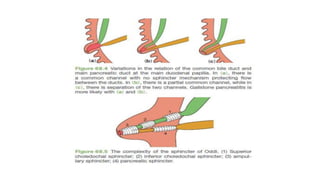
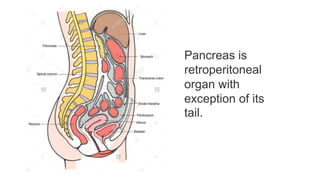
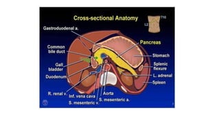
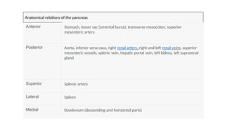
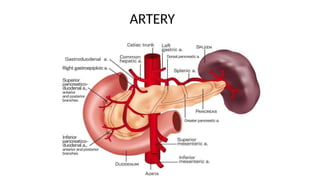
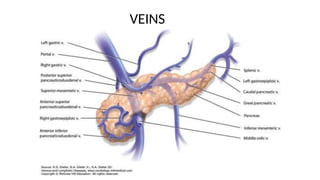
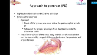
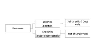
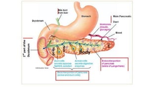
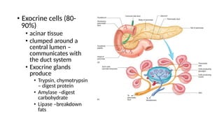
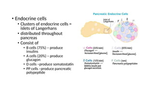
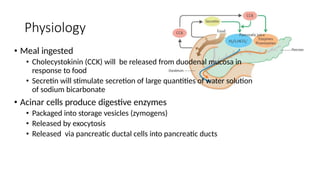
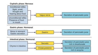
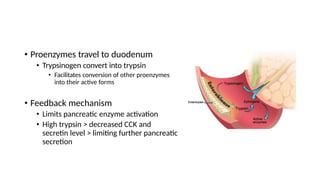
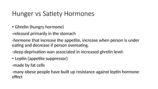
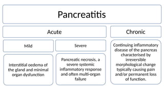
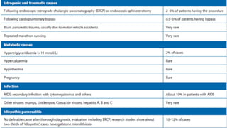
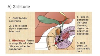
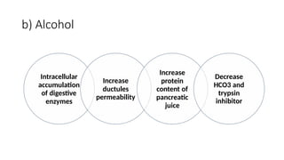
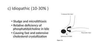
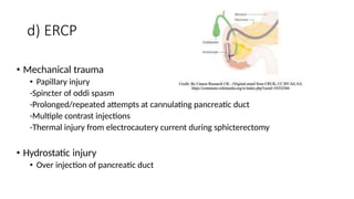
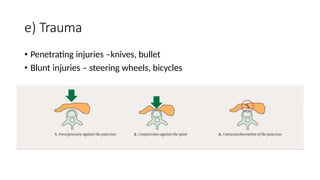
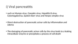
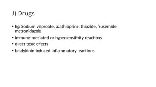
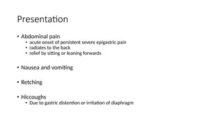
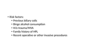
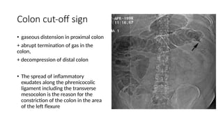
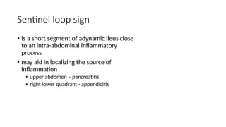
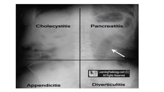
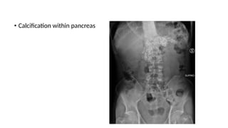
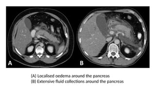
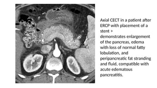
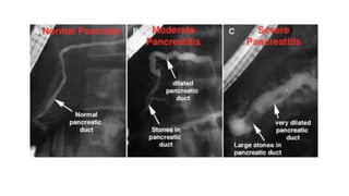
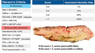
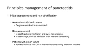
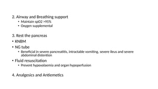
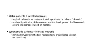
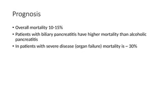
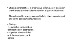
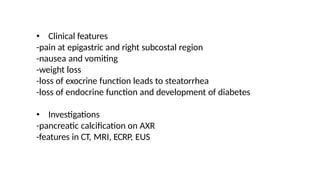
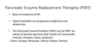
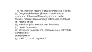
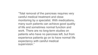
ACUTE PANCREATITIS
AHMAD IRFAN SYAKIR BIN KAMALUDDIN
Outline
• Diagnosis
• Anatomy
• Investigation
• Severity assessment
• Complication
• Management
• Prognosis
• Physiology
• Pathophysiology
• Causes
• Presentation
• Classification
• Acute pancreatitis
• Chronic pancreatitis
• Exocrine Pancreatic Insuffiency
ANATOMY
• Derived from Greek:
• ‘pan’ (all) and ‘kreas’ (flesh)
• Retroperitonium organ
• Positioned at the level of the transpyloric plane (L1)
• Weighs ~100g and 14-20cm long
• Divided into
• Head
• Body
• Tail
• The head
- lies within the curve of the duodenum, body of the second lumbar
vertebra and the vena cava.
• The neck
-aorta and superior mesenteric vessel behind of the neck
-the superior mesenteric vein joins the splenic vein to form the portal
vein.
• The tip of the pancreatic tail extends up to the splenic hilum
Duct system
• Main pancreatic duct (Wirsung
duct)
• Accessory duct (Santorini duct)
• Pancreatic duct joins CBD to
form ampulla of Vater
Pancreas is
retroperitoneal
organ with
exception of its
tail.
Anatomical relations of the pancreas
Anterior Stomach, lesser sac (omental bursa), transverse mesocolon, superior
mesenteric artery
Posterior Aorta, inferior vena cava, right renal artery, right and left renal veins, superior
mesenteric vessels, splenic vein, hepatic portal vein, left kidney, left suprarenal
gland
Superior
Lateral
Medial
Splenic artery
Spleen
Duodenum (descending and horizontal parts)
ARTERY
VEINS
RESECTIONAL ANATOMY OF PANCREAS
PHYSIOLOGY
Acinar cells & Duct
cells
Exocrine
(digestion)
Pancrease
Endocrine
Islet of Langerhans
(glucose homeostasis)
• Exocrine cells (80-
90%)
• acinar tissue
• clumped around a
central lumen –
communicates with
the duct system
• Exocrine glands
produce
• Trypsin, chymotrypsin
– digest protein
• Amylase –digest
carbohydrate
• Lipase –breakdown
fats
• Endocrine cells
• Clusters of endocrine cells =
islets of Langerhans
• distributed throughout
pancreas
• Consist of
• B cells (75%) – produce
insulins
• A cells (20%) – produce
glucagon
• D cells –produce somatostatin
• PP cells –produce pancreatic
polypeptide
Physiology
• Meal ingested
• Cholecystokinin (CCK) will be released from duodenal mucosa in
response to food
• Secretin will stimulate secretion of large quantities of water solution
of sodium bicarbonate
• Acinar cells produce digestive enzymes
• Packaged into storage vesicles (zymogens)
• Released by exocytosis
• Released via pancreatic ductal cells into pancreatic ducts
• Proenzymes travel to duodenum
• Trypsinogen convert into trypsin
• Facilitates conversion of other proenzymes
into their active forms
• Feedback mechanism
• Limits pancreatic enzyme activation
• High trypsin > decreased CCK and
secretin level > limiting further pancreatic
secretion
Hunger vs Satiety Hormones
• Ghrelin (hungry hormone)
-released primarily in the stomach
-hormone that increase the appetite, increase when person is under
eating and decrease if person overeating.
-sleep deprivation wan associated in increased ghrelin level.
• Leptin (appetite suppressor)
-made by fat cells
-many obese people have built up resistance against leptin hormone
effect
PANCREATITIS
• is inflammation of the gland parenchyma of the pancreas.
• Divided into acute, which is an emergency and chronic which is
prolonged resulting from development of fibrosis In pancreas.
• incidence : 5-30 cases per 100,000
• overall case fatality rate : ~ 5%
• Can occur at any age, with a peak in young men and older women.
Pancreatitis
Acute Chronic
Continuing inflammatory
disease of the pancreas
characterised by
Severe
Mild
Pancreatic necrosis, a
severe systemic
inflammatory response
and often multi-organ
failure
irreversible
morphological change
typically causing pain
and/or permanent loss
of function.
Interstitial oedema of
the gland and minimal
organ dysfunction
1. ACUTE PANCREATITIS
• An acute condition presenting with abdominal pain and usually
associated with raised pancreatic enzyme levels as a result of
pancreatic inflammation.
• Mechanism of injury
-premature activation of pancreatic enzymes within the pancreas,
leading to process of auto digestion.
a)Acinar cells injury
b)Impair secretion of zymogen
c)Delay of enzymatic secretions
CAUSES
• Common causes:
• Gallstone 50-70%
• Alcohol 25%
• ‘I GET SMASHED’
A) Gallstone
b) Alcohol
Increase
protein
content of
pancreatic
juice
Intracellular
accumulation
of digestive
enzymes
Decrease
HCO3 and
trypsin
Increase
ductules
permeability
inhibitor
c) Idiopathic (10-30% )
• Sludge and microlithiasis
• Relative deficiency of
phosphatidylcholine in bile
• Causing fast and extensive
cholesterol crystallization
d) ERCP
• Mechanical trauma
• Papillary injury
-Spincter of oddi spasm
-Prolonged/repeated attempts at cannulating pancreatic duct
-Multiple contrast injections
-Thermal injury from electrocautery current during sphicterectomy
• Hydrostatic injury
• Over injection of pancreatic duct
e) Trauma
• Penetrating injuries –knives, bullet
• Blunt injuries – steering wheels, bicycles
f) Steroids
• Corticosteroids might obstruct small pancreatic ductules by leading to
increased viscosity of pancreatic secretions, resulting in pancreatic
changes.
• These changes included reduced basophilia, vacuolization of acini,
peripancreatic fat necrosis, and hyperplasia of the islets of
Langerhans
g) Hypercalcaemia
• It is postulated that hypercalcemia leads to accelerated
intrapancreatic conversion of trypsinogen to trypsin, which causes the
pancreatic damage
• Can cause formation of pancreatic calculi and by modifying pancreatic
secretion, may lead to protein plug formation.
h) Hyperlipidemia
• TG > 1000mg/dL, a level at which chylomicrons are present
• Chylomicrons are triglyceride-rich lipoprotein particles.
• These are large enough to occlude the pancreatic capillaries leading
to ischemia and subsequent acinar structural alteration and release of
pancreatic lipase.
i) Viral pancreatitis
• such as Mumps virus, Coxsakie virus, Hepatitis B virus,
Cytomegalovirus, Epstein-Barr virus and Herpes simplex virus
• Direct destruction of pancreatic acinar cells by inflammation and
edema
• The damaging of pancreatic acinar cells by the virus leads to a leaking
intracellular enzyme or precipitates a process of cell death
J) Drugs
• Eg: Sodium valproate, azathioprine, thiazide, frusemide,
metronidazole
• immune-mediated or hypersensitivity reactions
• direct toxic effects
• bradykinin-induced inflammatory reactions
PRESENTATION
Presentation
• Abdominal pain
• acute onset of persistent severe epigastric pain
• radiates to the back
• relief by sitting or leaning forwards
• Nausea and vomiting
• Retching
• Hiccoughs
• Due to gastric distention or irritation of diaphragm
• Risk factors:
• Previous biliary colic
• Binge alcohol consumption
• H/o trauma/MVA
• Family history of HPL
• Recent operative or other invasive procedures
Physical examination:
• Appearance: well or gravely ill –profound shock, toxicity, confusion
• Tachypnoea, tachycardia, hypotension
• Mild icterus
• Pleural effusion, pulmonary edema
• Abdominal tenderness, distention, guarding
• Ascites
• Severe necrotizing pancreatitis
• Cullen sign
• Grey Turner sign
• Erythematous skin nodules
• due to focal subcutaneous fat
necrosis
• <1cm, on extensor skin surface
• polyarthritis
CLASSIFICATION
DIAGNOSIS
Diagnosis
• Requires at least 2 features:
1. Characteristic abdominal pain
2. Biochemical evidence of pancreatitis
• Amylase or lipase >3x above reference range
3. Characteristic imaging findings
• Amylase
• Not specific, Short half life
• Rises within few hours of pancreatic
damage
• Declines over the next 4-8 days
• Lipase
• More sensitive and specific to
pancrease
• Longer half-life, remains high for 12
days
• Does not indicate whether the disease is
mild, moderate or severe
Investigation Findings
FBC • Leucocytosis
• Hemoconcentration due to fluid sequestration
• Low Hct due to dehydration or hemorrhage
R P, e • electrolyte imbalance secondary to third spacing of fluids
• Hypocalcaemia due to saponification of fats in retroperitoneum
• Hypercalcaemia causing pancreatitis
• TRO renal failure
LFT • ALP, TB, AST, ALT
• ALT>150U/L –suggests gallstone pancreatitis
CRP • Indication of prognosis
• Higher level correlates with a propensity towards organ failure
FLP Hyperlipidaemia as the cause
Higher in severe pancreatitis
LDH
ABG
IgG4
To monitor oxygenation and acid-base status
Autoimmune pancreatitis
IMAGING
Investigation Findings
AXR • Sentinel loop –localized ileus of small intestine
• Colon cut-off sign –spasm of the descending
colon
• Gallstone, pancreatic calcification
CXR • Pleural effusion
• Hemidiaphragm elevation
• Basal atelectasis
• Pulmonary edema –suggestive of ARDS
• Air under diaphragm
- TRO Perforated viscus
Colon cut-off sign
• gaseous distension in proximal colon
+ abrupt termination of gas in the
colon,
+ decompression of distal colon
• The spread of inflammatory
exudates along the phrenicocolic
ligament including the transverse
mesocolon is the reason for the
constriction of the colon in the area
of the left flexure
Sentinel loop sign
• is a short segment of adynamic ileus close
to an intra-abdominal inflammatory
process
• may aid in localizing the source of
inflammation
• upper abdomen – pancreatitis
• right lower quadrant - appendicitis
• Calcification within pancreas
Investigation Findings
USG Abdomen Detect gallstones
Identify area of necrosis –hypoechoic regions
EUS Visualize pancrease and biliary tract
(Endoscopic Detect microlithiasis and periampullary lesions
ultrasound)
Transverse ultrasound demonstrates
diffuse enlargement of the pancreas ,
which appears abnormally hypoechoic
Investigation
MRCP
Findings
• Non invasive image of biliary and pancreatic ducts
ERCP • Pancreatitis secondary to chelodecholithiasis
• Biliary pancreatitis with worsening jaundice and clinical
deterioration despite max support therapy
CT • Pancreatic tumour
• Severe pancreatitis
• To assess complications
• Prognosis –Balthazar
CT scan
•Indications:
• Diagnostic uncertainty
• Severe pancreatitis – to distinguish interstitial from
necrotizing pancreatitis (Balthazar criteria)
• Organ failure, signs of sepsis or progressive clinical
deterioration
• Localised complication is suspected – fluid collection,
pseudocyst, pseudoaneurysm
(A) Localised oedema around the pancreas
(B) Extensive fluid collections around the pancreas
Axial CECT in a patient after
ERCP with placement of a
stent >
demonstrates enlargement
of the pancreas, edema
with loss of normal fatty
lobulation, and
peripancreatic fat stranding
and fluid, compatible with
acute edematous
pancreatitis.
SEVERITY ASSESSMENT
Assessment of severity
•Ranson criteria
•Glasgow criteria
•APACHE II score
>10mmol/L
<2.0 mmol/L
If the score ≥ 3, severe pancreatitis likely.
If the score < 3, severe pancreatitis is unlikely
If the score ≥ 3, severe
pancreatitis likely.
If the score < 3, severe
pancreatitis is unlikely
Measure the severity of disease for adult patients admitted to intensive care units
MANAGEMENTS
Principles management of pancreatitis
1. Initial assessment and risk stratification
• Assess hemodynamic status
• Begin resuscitation as needed
• Risk assessment
• to stratify patients into higher- and lower-risk categories
• to assist triage, such as admission to an intensive care setting
• Patients with organ failure
• Admit to intensive care unit or intermediary care setting whenever possible
2. Airway and Breathing support
• Maintain spO2 >95%
• Oxygen supplemental
3. Rest the pancreas
• KNBM
• NG tube
• Beneficial in severe pancreatitis, intractable vomiting, severe ileus and severe
abdominal distention
• Fluid resuscitation
• Prevent hypovolaemia and organ hypoperfusion
4. Analgesics and Antiemetics
4. Blood products in acute pancreatitis
• hemorrhagic pancreatitis – transfuse to HCT level of 30%
• If coagulopathic and bleeding – transfuse FFP and platelets
5. Correct electrolyte abnormalities
• Hypocalcaemia
• Hypokalaemia
• Hypomagnesemia
Nutrition
• Mild AP
• oral feedings can be started immediately if there is no nausea and vomiting,
and abdominal pain has resolved.
• initiation of feeding with a low-fat solid diet appears as safe as a clear liquid
diet.
• Severe AP
• enteral nutrition is recommended to prevent infectious complications.
• Parenteral nutrition should be avoided unless the enteral route is not
available, not tolerated, or not meeting caloric requirements.
Antibiotics
• prophylactic antibiotics in severe acute pancreatitis is not
recommended
• antibiotics in sterile necrosis to prevent the development of infected
necrosis is not recommended
• Infected necrosis should be considered in patients with
• pancreatic or extrapancreatic necrosis
• who deteriorate or fail to improve after 7–10 days of hospitalization.
• antibiotics known to penetrate pancreatic necrosis
• carbapenems, quinolones, and metronidazole
• Treat U/L causes
• Gallstones – early lap cholecystectomy
• Urgent ERCP
• Within 72H
• In pt with gallstones, causing jaundice, cholangitis or dilated CBD
• Reduce incidence of infective complication
COMPLICATIONS
IEP in a 43-year-old man. Axial contrast-
enhanced CT image shows peripancreatic
inflammation (black arrow) and a
homogeneous fluid-attenuation
collection in the left anterior pararenal
space (white arrow), a finding that is
consistent with APFC.
Figure 1 Drawings illustrate pancreatic necrosis (a), peripancreatic necrosis (b), and combined pancreatic-
peripancreatic necrosis (c).
Figure 4b Pancreatic necrosis in a 65-year-old man. (a) Axial contrast material–enhanced CT image obtained 2
days after the onset of acute abdominal pain shows peripancreatic fluid and stranding (arrows) and normal-
appearing pancreatic parenchyma. (b) Axial contrast-enhanced CT image obtained 5 days later owing to the
patient’s worsening clinical condition reveals an ill-defined hypoattenuating region in the body of the pancreas
(*), a finding that suggests pancreatic necrosis. Peripancreatic fluid and stranding (arrows) are also seen.
Figure 21a Axial contrast-enhanced CT image acquired 4 weeks after the onset of disease in the same
patient as in Figure 20a shows a well-defined, homogeneous peripancreatic collection around the tail
of the pancreas (arrows), a finding that is compatible with a pseudocyst.
Local complications
• Acute fluid collection
• Sterile and infected pancreatic necrosis
• Pancreatic abscess
• Pancreatic ascites
• Pancreatic effusion
• Haemorrhage
• Portal or splenic vein thrombosis
• pseudocyst
Systemic Complication
• Occurs within days of onset
• Disseminated intravascular coagulation (DIC)
• Acute respiratory distress syndrome (ARDS)
• Hypocalcaemia
• Fat necrosis from released lipases
• results in the release of free fatty acids
• react with serum calcium to form chalky deposits in fatty tissue
• Hyperglycaemia
• Secondary to destruction of islets of Langerhans
• subsequent disturbances to insulin metabolism
Surgery in Acute Pancreatitis
• Mild AP + cholelithiasis
• cholecystectomy should be performed before discharge
• to prevent a recurrence of AP
• necrotizing biliary AP
• cholecystectomy is to be deferred until active inflammation subsides and fluid
collections resolve or stabilize
• to prevent infection
• Asymptomatic pseudocysts + pancreatic and/or extrapancreatic necrosis
• do not warrant intervention regardless of size, location, and/or extension
• stable patients + infected necrosis
• surgical, radiologic, or endoscopic drainage should be delayed (>4 weeks)
• to allow liquefication of the contents and the development of a fibrous wall
around the necrosis (walled-off necrosis)
• symptomatic patients + infected necrosis
• minimally invasive methods of necrosectomy are preferred to open
necrosectomy
Prognosis
• Overall mortality 10-15%
• Patients with biliary pancreatitis have higher mortality than alcoholic
pancreatitis
• In patients with severe disease (organ failure) mortality is ~ 30%
CHRONIC PANCREATITIS
• Chronic pancreatitis is a progressive Inflammatory disease in
which there is irreversible destruction of pancreatic tissues.
• Characterized by severe pain and in later stage, exocrine and
endocrine pancreatic insufficiency.
• Etiology:
-high alcohol consumption
-pancreatic duct obstruction
-congenital abnormalities
-autoimmune pancreatitis
-others
• Clinical features
-pain at epigastric and right subcostal region
-nausea and vomiting
-weight loss
-loss of exocrine function leads to steatorrhea
-loss of endocrine function and development of diabetes
• Investigations
-pancreatic calcification on AXR
-features in CT, MRI, ECRP, EUS
Treatments
A) Medical treatments
• Treat the addiction
-stop alcohol and tobacco
• Alleviate abdominal pains
-eliminate obstructive factors
• Nutritional and pharmacological measures
-diet low in fat and high in protein and carbohydates
-pancreatic enzyme supplementation with meals
-correct malabsorptions of fat soluble vitamins
• Treat diabetes mellitus
b) Endoscopics or Surgical interventions
-relieve obstruction of the pancreatic duct, bile duct or the
duodenum, or in dealing with complications.
• Endoscopic pancreatic spincterotomy
-for papillary stenosis, high spicter pressure, pancreatic ductal
pressure
• Pancreatoduodenectomy (Beger procedure)
-mass in the head of pancrease
• Longitudinal pancreatojejunostomy (Frey procedure)
-if duct is markedly dilated
Exocrine Pancreatic Insuffiency
• Deficiency of the exocrine pancreatic enzyme, resulting in
the inability to digest food properly, or maldigestion.
• Amylase, lipase, protease
• Management
-lifestyle modifications: ex- avoiding fatty foods, limitation of
alcohol intake, consumption of well-balanced diet
-vitamin supplementation (A, D, E, K)
-Pancreatic Enzyme Replacement Theraphy (PERT)
Pancreatic Enzyme Replacement Theraphy (PERT)
• Basis of treatment of EPI
• Typical indication are progressive weight loss and
steatorrhea.
• The Pancreatic Enzyme Products (PEPs) use for PERT are
extract of porcine pancreas that contain all 3 pancreatic
enzymes (amylase, lipase, protease)
- Creon, Zenpap, Pancreaze, Ultresa Viokace, Pertzye
NON-ALCOHOLIC FAT TY PANCREAS DISEASE (NAFPD)
ABSTRACT
• Fat accumulation in the pancreas, defined as fatty pancreas, is usually an
incidental finding during transabdominal ultrasound examination.
• Fatty pancreas without any significant alcohol consumption is defined as non-
alcoholic fatty pancreas disease
• Its clinical impact is still largely unknown, hypothetically the disease progression
could lead to chronic pancreatitis and possibly pancreatic cancer development.
• Metabolic problems such as diabetes, central obesity, fatty liver, and
dyslipidaemia have been considered important risk factors related to non-
alcoholic fatty pancreas disease and pancreatic cancer
Endoscopic ultrasound image showing bright
hyperechoic of pancreas parenchyma.
The risk induction factors of steatopancreatitis include:
(a) Congenital diseases (Shwachman-Diamond
syndrome, Johanson-Blizzard syndrome, cystic
fibrosis, heterozygous carboxyl-ester lipase mutation)
(b) Alcohol abuse
(c) Infections (viral infection with Reovirus)
(d) Hemochromatosis
(e) Medicines (rosiglitazone, corticosteroids, octreotide,
gemcitabine)
(f) Malnutrition
(g) NAFLD, chronic hepatitis B
CAN YOU LIVE WITHOUT THE PANCREAS?
By Professor Hemant Kocher
MBBS, MS, MD, FRCS
Professor of Liver and Pancreas Surgery
“Total removal of the pancreas requires very
careful medical treatment and close
monitoring by a specialist. With medications,
many such patients can achieve good quality
of life and sometimes normal function and
work. There are no long-term studies on
patients who have no pancreas left, but from
experience patients go on to have normal life
expectancy with careful medical
supervision.”
References:
• American Gastroenterological Association Institute Guideline on Initial
Management of Acute Pancreatitis
https://www.gastrojournal.org/article/S0016-5085(18)30076-
3/fulltext#secsectitle0025
• Acute Pancreatitis Medication
https://emedicine.medscape.com/article/181364-medication#showall
• Bailey & Love
• RadioGraphics. Necrotizing Pancreatitis: Diagnosis, Imaging, and
Intervention
https://pubs.rsna.org/doi/full/10.1148/rg.345130012
• https://epomedicine.com/clinical-cases/acute-pancreatitis-case-
discussion/
References:
• https://www.cureus.com/articles/36219-steroid-induced-
pancreatitis-a-challenging-diagnosis
• https://www.ijem.in/article.asp?issn=2230-
8210;year=2013;volume=17;issue=5;spage=799;epage=805;aulast=K
ota
• https://www.ncbi.nlm.nih.gov/pmc/articles/PMC4919820/#:~:text=It
%20is%20postulated%20that%20hypercalcemia,6%2C%207%2C%208
%5D.
References:
https://www.topdoctors.co.uk/medical-articles/can-you-live-
without-the-pancreas#
https://www.emjreviews.com/gastroenterology/article/non-alcoholic-fatty-
pancreas-disease-pancreatic-cancer-and-impact-of-endoscopic-ultrasound-
examination-on-screening-and-
surveillance/#:~:text=Fat%20accumulation%20in%20the%20pancreas,non
%2Dalcoholic%20fatty%20pancreas%20disease.
Anatomy and Histology of the Pancreas
Daniel Longnecker, MD
Department of Pathology, Geisel School of Medicine at
Dartmouth, Lebanon, NH
https://www.slideshare.net/HappyKagathara/3-cmeasa-
ahmedabad-january-2015
THANK YOU

acutepancreatitisclinicalpresentation.ppt

  • 1.
    ACUTE PANCREATITIS AHMAD IRFANSYAKIR BIN KAMALUDDIN
  • 2.
    Outline • Diagnosis • Anatomy •Investigation • Severity assessment • Complication • Management • Prognosis • Physiology • Pathophysiology • Causes • Presentation • Classification • Acute pancreatitis • Chronic pancreatitis • Exocrine Pancreatic Insuffiency
  • 3.
  • 4.
    • Derived fromGreek: • ‘pan’ (all) and ‘kreas’ (flesh) • Retroperitonium organ • Positioned at the level of the transpyloric plane (L1) • Weighs ~100g and 14-20cm long • Divided into • Head • Body • Tail
  • 9.
    • The head -lies within the curve of the duodenum, body of the second lumbar vertebra and the vena cava. • The neck -aorta and superior mesenteric vessel behind of the neck -the superior mesenteric vein joins the splenic vein to form the portal vein. • The tip of the pancreatic tail extends up to the splenic hilum
  • 10.
    Duct system • Mainpancreatic duct (Wirsung duct) • Accessory duct (Santorini duct) • Pancreatic duct joins CBD to form ampulla of Vater
  • 12.
  • 14.
    Anatomical relations ofthe pancreas Anterior Stomach, lesser sac (omental bursa), transverse mesocolon, superior mesenteric artery Posterior Aorta, inferior vena cava, right renal artery, right and left renal veins, superior mesenteric vessels, splenic vein, hepatic portal vein, left kidney, left suprarenal gland Superior Lateral Medial Splenic artery Spleen Duodenum (descending and horizontal parts)
  • 15.
  • 16.
  • 17.
  • 23.
  • 24.
    Acinar cells &Duct cells Exocrine (digestion) Pancrease Endocrine Islet of Langerhans (glucose homeostasis)
  • 26.
    • Exocrine cells(80- 90%) • acinar tissue • clumped around a central lumen – communicates with the duct system • Exocrine glands produce • Trypsin, chymotrypsin – digest protein • Amylase –digest carbohydrate • Lipase –breakdown fats
  • 27.
    • Endocrine cells •Clusters of endocrine cells = islets of Langerhans • distributed throughout pancreas • Consist of • B cells (75%) – produce insulins • A cells (20%) – produce glucagon • D cells –produce somatostatin • PP cells –produce pancreatic polypeptide
  • 28.
    Physiology • Meal ingested •Cholecystokinin (CCK) will be released from duodenal mucosa in response to food • Secretin will stimulate secretion of large quantities of water solution of sodium bicarbonate • Acinar cells produce digestive enzymes • Packaged into storage vesicles (zymogens) • Released by exocytosis • Released via pancreatic ductal cells into pancreatic ducts
  • 30.
    • Proenzymes travelto duodenum • Trypsinogen convert into trypsin • Facilitates conversion of other proenzymes into their active forms • Feedback mechanism • Limits pancreatic enzyme activation • High trypsin > decreased CCK and secretin level > limiting further pancreatic secretion
  • 31.
    Hunger vs SatietyHormones • Ghrelin (hungry hormone) -released primarily in the stomach -hormone that increase the appetite, increase when person is under eating and decrease if person overeating. -sleep deprivation wan associated in increased ghrelin level. • Leptin (appetite suppressor) -made by fat cells -many obese people have built up resistance against leptin hormone effect
  • 32.
    PANCREATITIS • is inflammationof the gland parenchyma of the pancreas. • Divided into acute, which is an emergency and chronic which is prolonged resulting from development of fibrosis In pancreas. • incidence : 5-30 cases per 100,000 • overall case fatality rate : ~ 5% • Can occur at any age, with a peak in young men and older women.
  • 33.
    Pancreatitis Acute Chronic Continuing inflammatory diseaseof the pancreas characterised by Severe Mild Pancreatic necrosis, a severe systemic inflammatory response and often multi-organ failure irreversible morphological change typically causing pain and/or permanent loss of function. Interstitial oedema of the gland and minimal organ dysfunction
  • 34.
  • 35.
    • An acutecondition presenting with abdominal pain and usually associated with raised pancreatic enzyme levels as a result of pancreatic inflammation. • Mechanism of injury -premature activation of pancreatic enzymes within the pancreas, leading to process of auto digestion. a)Acinar cells injury b)Impair secretion of zymogen c)Delay of enzymatic secretions
  • 36.
  • 41.
    • Common causes: •Gallstone 50-70% • Alcohol 25% • ‘I GET SMASHED’
  • 42.
  • 43.
    b) Alcohol Increase protein content of pancreatic juice Intracellular accumulation ofdigestive enzymes Decrease HCO3 and trypsin Increase ductules permeability inhibitor
  • 44.
    c) Idiopathic (10-30%) • Sludge and microlithiasis • Relative deficiency of phosphatidylcholine in bile • Causing fast and extensive cholesterol crystallization
  • 45.
    d) ERCP • Mechanicaltrauma • Papillary injury -Spincter of oddi spasm -Prolonged/repeated attempts at cannulating pancreatic duct -Multiple contrast injections -Thermal injury from electrocautery current during sphicterectomy • Hydrostatic injury • Over injection of pancreatic duct
  • 46.
    e) Trauma • Penetratinginjuries –knives, bullet • Blunt injuries – steering wheels, bicycles
  • 47.
    f) Steroids • Corticosteroidsmight obstruct small pancreatic ductules by leading to increased viscosity of pancreatic secretions, resulting in pancreatic changes. • These changes included reduced basophilia, vacuolization of acini, peripancreatic fat necrosis, and hyperplasia of the islets of Langerhans
  • 48.
    g) Hypercalcaemia • Itis postulated that hypercalcemia leads to accelerated intrapancreatic conversion of trypsinogen to trypsin, which causes the pancreatic damage • Can cause formation of pancreatic calculi and by modifying pancreatic secretion, may lead to protein plug formation.
  • 49.
    h) Hyperlipidemia • TG> 1000mg/dL, a level at which chylomicrons are present • Chylomicrons are triglyceride-rich lipoprotein particles. • These are large enough to occlude the pancreatic capillaries leading to ischemia and subsequent acinar structural alteration and release of pancreatic lipase.
  • 50.
    i) Viral pancreatitis •such as Mumps virus, Coxsakie virus, Hepatitis B virus, Cytomegalovirus, Epstein-Barr virus and Herpes simplex virus • Direct destruction of pancreatic acinar cells by inflammation and edema • The damaging of pancreatic acinar cells by the virus leads to a leaking intracellular enzyme or precipitates a process of cell death
  • 51.
    J) Drugs • Eg:Sodium valproate, azathioprine, thiazide, frusemide, metronidazole • immune-mediated or hypersensitivity reactions • direct toxic effects • bradykinin-induced inflammatory reactions
  • 52.
  • 53.
    Presentation • Abdominal pain •acute onset of persistent severe epigastric pain • radiates to the back • relief by sitting or leaning forwards • Nausea and vomiting • Retching • Hiccoughs • Due to gastric distention or irritation of diaphragm
  • 54.
    • Risk factors: •Previous biliary colic • Binge alcohol consumption • H/o trauma/MVA • Family history of HPL • Recent operative or other invasive procedures
  • 55.
    Physical examination: • Appearance:well or gravely ill –profound shock, toxicity, confusion • Tachypnoea, tachycardia, hypotension • Mild icterus • Pleural effusion, pulmonary edema • Abdominal tenderness, distention, guarding • Ascites
  • 56.
    • Severe necrotizingpancreatitis • Cullen sign • Grey Turner sign • Erythematous skin nodules • due to focal subcutaneous fat necrosis • <1cm, on extensor skin surface • polyarthritis
  • 57.
  • 59.
  • 60.
    Diagnosis • Requires atleast 2 features: 1. Characteristic abdominal pain 2. Biochemical evidence of pancreatitis • Amylase or lipase >3x above reference range 3. Characteristic imaging findings
  • 61.
    • Amylase • Notspecific, Short half life • Rises within few hours of pancreatic damage • Declines over the next 4-8 days • Lipase • More sensitive and specific to pancrease • Longer half-life, remains high for 12 days • Does not indicate whether the disease is mild, moderate or severe
  • 63.
    Investigation Findings FBC •Leucocytosis • Hemoconcentration due to fluid sequestration • Low Hct due to dehydration or hemorrhage R P, e • electrolyte imbalance secondary to third spacing of fluids • Hypocalcaemia due to saponification of fats in retroperitoneum • Hypercalcaemia causing pancreatitis • TRO renal failure LFT • ALP, TB, AST, ALT • ALT>150U/L –suggests gallstone pancreatitis CRP • Indication of prognosis • Higher level correlates with a propensity towards organ failure FLP Hyperlipidaemia as the cause Higher in severe pancreatitis LDH ABG IgG4 To monitor oxygenation and acid-base status Autoimmune pancreatitis
  • 64.
  • 65.
    Investigation Findings AXR •Sentinel loop –localized ileus of small intestine • Colon cut-off sign –spasm of the descending colon • Gallstone, pancreatic calcification CXR • Pleural effusion • Hemidiaphragm elevation • Basal atelectasis • Pulmonary edema –suggestive of ARDS • Air under diaphragm - TRO Perforated viscus
  • 66.
    Colon cut-off sign •gaseous distension in proximal colon + abrupt termination of gas in the colon, + decompression of distal colon • The spread of inflammatory exudates along the phrenicocolic ligament including the transverse mesocolon is the reason for the constriction of the colon in the area of the left flexure
  • 67.
    Sentinel loop sign •is a short segment of adynamic ileus close to an intra-abdominal inflammatory process • may aid in localizing the source of inflammation • upper abdomen – pancreatitis • right lower quadrant - appendicitis
  • 69.
  • 70.
    Investigation Findings USG AbdomenDetect gallstones Identify area of necrosis –hypoechoic regions EUS Visualize pancrease and biliary tract (Endoscopic Detect microlithiasis and periampullary lesions ultrasound)
  • 73.
    Transverse ultrasound demonstrates diffuseenlargement of the pancreas , which appears abnormally hypoechoic
  • 77.
    Investigation MRCP Findings • Non invasiveimage of biliary and pancreatic ducts ERCP • Pancreatitis secondary to chelodecholithiasis • Biliary pancreatitis with worsening jaundice and clinical deterioration despite max support therapy CT • Pancreatic tumour • Severe pancreatitis • To assess complications • Prognosis –Balthazar
  • 78.
    CT scan •Indications: • Diagnosticuncertainty • Severe pancreatitis – to distinguish interstitial from necrotizing pancreatitis (Balthazar criteria) • Organ failure, signs of sepsis or progressive clinical deterioration • Localised complication is suspected – fluid collection, pseudocyst, pseudoaneurysm
  • 79.
    (A) Localised oedemaaround the pancreas (B) Extensive fluid collections around the pancreas
  • 80.
    Axial CECT ina patient after ERCP with placement of a stent > demonstrates enlargement of the pancreas, edema with loss of normal fatty lobulation, and peripancreatic fat stranding and fluid, compatible with acute edematous pancreatitis.
  • 82.
  • 83.
    Assessment of severity •Ransoncriteria •Glasgow criteria •APACHE II score
  • 84.
    >10mmol/L <2.0 mmol/L If thescore ≥ 3, severe pancreatitis likely. If the score < 3, severe pancreatitis is unlikely
  • 85.
    If the score≥ 3, severe pancreatitis likely. If the score < 3, severe pancreatitis is unlikely
  • 86.
    Measure the severityof disease for adult patients admitted to intensive care units
  • 87.
  • 88.
    Principles management ofpancreatitis 1. Initial assessment and risk stratification • Assess hemodynamic status • Begin resuscitation as needed • Risk assessment • to stratify patients into higher- and lower-risk categories • to assist triage, such as admission to an intensive care setting • Patients with organ failure • Admit to intensive care unit or intermediary care setting whenever possible
  • 89.
    2. Airway andBreathing support • Maintain spO2 >95% • Oxygen supplemental 3. Rest the pancreas • KNBM • NG tube • Beneficial in severe pancreatitis, intractable vomiting, severe ileus and severe abdominal distention • Fluid resuscitation • Prevent hypovolaemia and organ hypoperfusion 4. Analgesics and Antiemetics
  • 90.
    4. Blood productsin acute pancreatitis • hemorrhagic pancreatitis – transfuse to HCT level of 30% • If coagulopathic and bleeding – transfuse FFP and platelets 5. Correct electrolyte abnormalities • Hypocalcaemia • Hypokalaemia • Hypomagnesemia
  • 91.
    Nutrition • Mild AP •oral feedings can be started immediately if there is no nausea and vomiting, and abdominal pain has resolved. • initiation of feeding with a low-fat solid diet appears as safe as a clear liquid diet. • Severe AP • enteral nutrition is recommended to prevent infectious complications. • Parenteral nutrition should be avoided unless the enteral route is not available, not tolerated, or not meeting caloric requirements.
  • 92.
    Antibiotics • prophylactic antibioticsin severe acute pancreatitis is not recommended • antibiotics in sterile necrosis to prevent the development of infected necrosis is not recommended • Infected necrosis should be considered in patients with • pancreatic or extrapancreatic necrosis • who deteriorate or fail to improve after 7–10 days of hospitalization. • antibiotics known to penetrate pancreatic necrosis • carbapenems, quinolones, and metronidazole
  • 93.
    • Treat U/Lcauses • Gallstones – early lap cholecystectomy • Urgent ERCP • Within 72H • In pt with gallstones, causing jaundice, cholangitis or dilated CBD • Reduce incidence of infective complication
  • 94.
  • 97.
    IEP in a43-year-old man. Axial contrast- enhanced CT image shows peripancreatic inflammation (black arrow) and a homogeneous fluid-attenuation collection in the left anterior pararenal space (white arrow), a finding that is consistent with APFC.
  • 98.
    Figure 1 Drawingsillustrate pancreatic necrosis (a), peripancreatic necrosis (b), and combined pancreatic- peripancreatic necrosis (c).
  • 99.
    Figure 4b Pancreaticnecrosis in a 65-year-old man. (a) Axial contrast material–enhanced CT image obtained 2 days after the onset of acute abdominal pain shows peripancreatic fluid and stranding (arrows) and normal- appearing pancreatic parenchyma. (b) Axial contrast-enhanced CT image obtained 5 days later owing to the patient’s worsening clinical condition reveals an ill-defined hypoattenuating region in the body of the pancreas (*), a finding that suggests pancreatic necrosis. Peripancreatic fluid and stranding (arrows) are also seen.
  • 100.
    Figure 21a Axialcontrast-enhanced CT image acquired 4 weeks after the onset of disease in the same patient as in Figure 20a shows a well-defined, homogeneous peripancreatic collection around the tail of the pancreas (arrows), a finding that is compatible with a pseudocyst.
  • 101.
    Local complications • Acutefluid collection • Sterile and infected pancreatic necrosis • Pancreatic abscess • Pancreatic ascites • Pancreatic effusion • Haemorrhage • Portal or splenic vein thrombosis • pseudocyst
  • 102.
    Systemic Complication • Occurswithin days of onset • Disseminated intravascular coagulation (DIC) • Acute respiratory distress syndrome (ARDS) • Hypocalcaemia • Fat necrosis from released lipases • results in the release of free fatty acids • react with serum calcium to form chalky deposits in fatty tissue • Hyperglycaemia • Secondary to destruction of islets of Langerhans • subsequent disturbances to insulin metabolism
  • 103.
    Surgery in AcutePancreatitis • Mild AP + cholelithiasis • cholecystectomy should be performed before discharge • to prevent a recurrence of AP • necrotizing biliary AP • cholecystectomy is to be deferred until active inflammation subsides and fluid collections resolve or stabilize • to prevent infection • Asymptomatic pseudocysts + pancreatic and/or extrapancreatic necrosis • do not warrant intervention regardless of size, location, and/or extension
  • 104.
    • stable patients+ infected necrosis • surgical, radiologic, or endoscopic drainage should be delayed (>4 weeks) • to allow liquefication of the contents and the development of a fibrous wall around the necrosis (walled-off necrosis) • symptomatic patients + infected necrosis • minimally invasive methods of necrosectomy are preferred to open necrosectomy
  • 105.
    Prognosis • Overall mortality10-15% • Patients with biliary pancreatitis have higher mortality than alcoholic pancreatitis • In patients with severe disease (organ failure) mortality is ~ 30%
  • 106.
  • 107.
    • Chronic pancreatitisis a progressive Inflammatory disease in which there is irreversible destruction of pancreatic tissues. • Characterized by severe pain and in later stage, exocrine and endocrine pancreatic insufficiency. • Etiology: -high alcohol consumption -pancreatic duct obstruction -congenital abnormalities -autoimmune pancreatitis -others
  • 108.
    • Clinical features -painat epigastric and right subcostal region -nausea and vomiting -weight loss -loss of exocrine function leads to steatorrhea -loss of endocrine function and development of diabetes • Investigations -pancreatic calcification on AXR -features in CT, MRI, ECRP, EUS
  • 109.
    Treatments A) Medical treatments •Treat the addiction -stop alcohol and tobacco • Alleviate abdominal pains -eliminate obstructive factors • Nutritional and pharmacological measures -diet low in fat and high in protein and carbohydates -pancreatic enzyme supplementation with meals -correct malabsorptions of fat soluble vitamins • Treat diabetes mellitus
  • 110.
    b) Endoscopics orSurgical interventions -relieve obstruction of the pancreatic duct, bile duct or the duodenum, or in dealing with complications. • Endoscopic pancreatic spincterotomy -for papillary stenosis, high spicter pressure, pancreatic ductal pressure • Pancreatoduodenectomy (Beger procedure) -mass in the head of pancrease • Longitudinal pancreatojejunostomy (Frey procedure) -if duct is markedly dilated
  • 112.
    Exocrine Pancreatic Insuffiency •Deficiency of the exocrine pancreatic enzyme, resulting in the inability to digest food properly, or maldigestion. • Amylase, lipase, protease • Management -lifestyle modifications: ex- avoiding fatty foods, limitation of alcohol intake, consumption of well-balanced diet -vitamin supplementation (A, D, E, K) -Pancreatic Enzyme Replacement Theraphy (PERT)
  • 113.
    Pancreatic Enzyme ReplacementTheraphy (PERT) • Basis of treatment of EPI • Typical indication are progressive weight loss and steatorrhea. • The Pancreatic Enzyme Products (PEPs) use for PERT are extract of porcine pancreas that contain all 3 pancreatic enzymes (amylase, lipase, protease) - Creon, Zenpap, Pancreaze, Ultresa Viokace, Pertzye
  • 114.
    NON-ALCOHOLIC FAT TYPANCREAS DISEASE (NAFPD)
  • 115.
    ABSTRACT • Fat accumulationin the pancreas, defined as fatty pancreas, is usually an incidental finding during transabdominal ultrasound examination. • Fatty pancreas without any significant alcohol consumption is defined as non- alcoholic fatty pancreas disease • Its clinical impact is still largely unknown, hypothetically the disease progression could lead to chronic pancreatitis and possibly pancreatic cancer development. • Metabolic problems such as diabetes, central obesity, fatty liver, and dyslipidaemia have been considered important risk factors related to non- alcoholic fatty pancreas disease and pancreatic cancer
  • 116.
    Endoscopic ultrasound imageshowing bright hyperechoic of pancreas parenchyma.
  • 117.
    The risk inductionfactors of steatopancreatitis include: (a) Congenital diseases (Shwachman-Diamond syndrome, Johanson-Blizzard syndrome, cystic fibrosis, heterozygous carboxyl-ester lipase mutation) (b) Alcohol abuse (c) Infections (viral infection with Reovirus) (d) Hemochromatosis (e) Medicines (rosiglitazone, corticosteroids, octreotide, gemcitabine) (f) Malnutrition (g) NAFLD, chronic hepatitis B
  • 118.
    CAN YOU LIVEWITHOUT THE PANCREAS? By Professor Hemant Kocher MBBS, MS, MD, FRCS Professor of Liver and Pancreas Surgery
  • 119.
    “Total removal ofthe pancreas requires very careful medical treatment and close monitoring by a specialist. With medications, many such patients can achieve good quality of life and sometimes normal function and work. There are no long-term studies on patients who have no pancreas left, but from experience patients go on to have normal life expectancy with careful medical supervision.”
  • 120.
    References: • American GastroenterologicalAssociation Institute Guideline on Initial Management of Acute Pancreatitis https://www.gastrojournal.org/article/S0016-5085(18)30076- 3/fulltext#secsectitle0025 • Acute Pancreatitis Medication https://emedicine.medscape.com/article/181364-medication#showall • Bailey & Love • RadioGraphics. Necrotizing Pancreatitis: Diagnosis, Imaging, and Intervention https://pubs.rsna.org/doi/full/10.1148/rg.345130012 • https://epomedicine.com/clinical-cases/acute-pancreatitis-case- discussion/
  • 121.
  • 122.
  • 123.