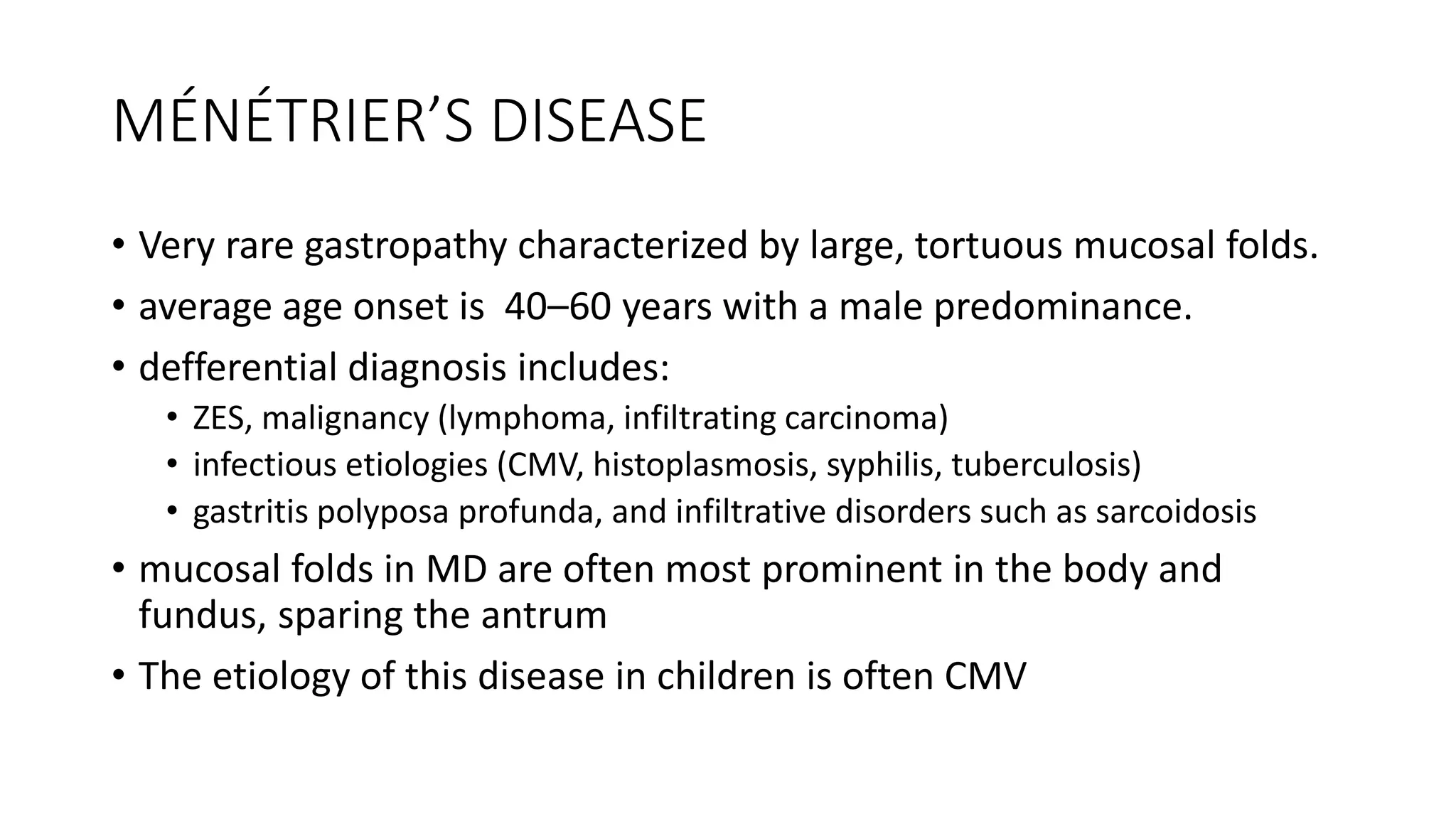
Gastritis
MD.Kiyumars Karimi
Definitions
• Gastritis is histologically documented inflammation of the gastric
mucosa
• Not interchangeable with dyspepsia
• Correlations between histologic findings, clinical picture and
endoscopic findings of gastric mucosa is poor.
• There is no typical clinical manifestations for gastritis.
Classifications of gastritis
I. Acute gastritis
A. Acute H.pylori infecton
B. Other acute infections gastridies
• Bacterial (other than H.pylori)
• H.Heilmani
• Phlegmonous
• Mycobacterial
• Syphlitic
• Viral
• Parsitic
• Fungal
II. Chronic atrophic gastritis
A. Type A: Autoimmune, body-predominant
B. Type B: H. pylori–related, antral-predominant
C. Indeterminate
III. Uncommon forms of gastritis
A. Lymphocytic
B. Eosinophilic
C. Crohn’s disease
D. Sarcoidosis
E. Isolated granulomatous gastritis
F. Russell body gastritis
Acute Gastritis
• The most common causes are infectious.
• H. pylori induced gastritis
• Presents with sudden onset of epigastric pain, nausea, vomiting and marked infiltrate of
neutrophils with edema and hyperemia in mucosal histologic studies.
• evolve into one of chronic gastritis in case not treated.
• phlegmonous gastritis
• Elderly individuals, alcoholics, and AIDS patients may be affected.
• Potential iatrogenic causes include:
• polypectomy
• mucosal injection with India ink
• Organisms associated with this entity include streptococci, staphylococci, Escherichia coli, Proteus,
and Haemophilus species
• Failure of supportive measures and antibiotics may result in gastrectomy
• Other types of infectious gastritis
• may occur in immunocompromised individuals
• Examples include herpetic (herpes simplex) or CMV gastritis
Chronic gastritis
• Identified by lymphocytic and plasma cell infiltration
• Patchy distribution of inflammation, initially involving superficial and glandular
portions of the gastric mucosa.
• may progress to more severe glandular destruction, with atrophy and metaplasia.
• According to histologic charachteristic classified in to:
• Superfacial atrophic changes
• Gastric atrophy
• The final stage of chronic gastritis is gastric atrophy.
• according to the predominant site of involvement chronic gastritis is classified in
to:
• Type A or body predominant form (autoimmune)
• Type B or antral predominant form (H.pylori-related)
• AB gastritis has been used to refer to a mixed antral/body picture.
Type A gastritis
• less common form involves primarily the fundus and body, with antral
sparing.
• This form of gastritis has been associated with pernicious anemia.
• Antibodies to parietal cells have been detected in >90% of patients
with pernicious anemia and in up to 50% of patients with type A
gastritis.
• The parietal cell antibody is directed against H+,K+-ATPase.
• up to 20% of individuals over age 60 and ~20% of patients with vitiligo
and Addison’s disease have these antibodies.
Cont…
• Anti-IF antibodies are more specific than parietal cell antibodies for type A
gastritis, being present in ~40% of patients with pernicious anemia.
• Parietal cell is targeted in this form of gastritis, and achlorhydria results.
• Parietal cells are the source of IF, the lack of which will lead to vitamin B12
deficiency
• Gastric acid plays an important role in feedback inhibition of gastrin
release.
• Gastrin levels can be markedly elevated (>500 pg/mL) in patients with
pernicious anemia.
• gastrin trophic effects can cause ECL cell hyperplasia with frank
development of gastric carcinoid tumors
Type B gastritis
• More common form of chronic gastritis.
• H. pylori infection is the cause of this entity.
• “antral-predominant,” is likely a misnomer in view of studies.
• conversion to a pangastritis is time-dependent and estimated to require 15–
20 years.
• Increases with age, being present in up to 100% of persons over age
70.
• Histology improves after H. pylori eradication
• The number of H. pylori organisms decreases dramatically with
progression to gastric atrophy
Cont…
• Multifocal atrophic gastritis, gastric atrophy with subsequent metaplasia,
has been observed in this form of gastritis
• may ultimately lead to development of gastric adenocarcinoma
• H. pylori infection is now considered an independent risk factor for gastric
cancer.
• Seropositivity for H. pylori is associated with a three to six fold increased
risk of gastric cancer.
• H. pylori is also associated with development of a low-grade B cell
lymphoma, gastric MALT lymphoma
• T cell stimulation leads to production of cytokines that promote the B cell
tumor.
• Such tumor should be followed by EUS every 2–3 months
Treatment of chronic gastritis
• Treatment is aimed at the sequelae and not the underlying inflammation
• Patients with pernicious anemia will require parenteral vitamin B12
supplementation.
• Eradication of H. pylori is often recommended even if PUD or alow-grade
MALT lymphoma is not present.
Miscellaneous Forms of Gastritis
• Lymphocytic gastritis
• Characterized histologically by intense
infiltration of the surface epithelium with
lymphocytes
• infiltrative process is primarily in the body of the
stomach
• Etiology is unknown
• No specific symptoms suggest lymphocytic
gastritis.
• H. pylori probably plays no significant role in
lymphocytic gastritis.
Cont…
• Eisoniphilic gastritis
• Marked eosinophilic infiltration involving any layer of the stomach
• Affected individuals have circulating eosinophilia with clinical manifestation of
systemic allergy.
• Involvment range from isolated gastric disease to diffuse gastroenteritits
• Treatment with glucocorticoids has been successful
• Granulomatous gastritits
• may be associated with several systemic disorder
• Gastric involvement has been observed in Crohn’s disease.
• Several rare infectious processes can lead to granulomatous gastritis,
including histoplasmosis, candidiasis, syphilis, and tuberculosis
Cont…
• Russell body gastritis (RBG)
• mucosal lesion of unknown etiology that has a pseudotumoral endoscopic
appearance
• Histologically, defined by the presence of numerous plasma cells containing
Russell bodies (RBs) that express kappa and lambda light chains.
• Only 10 cases have been reported, and 7 of these have been associated with
H. pylori infection.
• lesion can be confused with a neoplastic process
• There have been cases resolved by eradicating of H.pylori
MÉNÉTRIER’S DISEASE
• Very rare gastropathy characterized by large, tortuous mucosal folds.
• average age onset is 40–60 years with a male predominance.
• defferential diagnosis includes:
• ZES, malignancy (lymphoma, infiltrating carcinoma)
• infectious etiologies (CMV, histoplasmosis, syphilis, tuberculosis)
• gastritis polyposa profunda, and infiltrative disorders such as sarcoidosis
• mucosal folds in MD are often most prominent in the body and
fundus, sparing the antrum
• The etiology of this disease in children is often CMV
Cont…
• Overexpression of TGF-α has been demonstrated in patients with MD.
• overexpression of TGF-α in turn results in overstimulation EGFR pathway and
increased proliferation of mucus cells, resulting in the observed foveolar
hyperplasia.
• clinical presentation:
• Epigastric pain, nausea, vomiting, anorexia, peripheral edema, and weight loss
• Occult GIbleeding may occur
• 100% of patients develop a protein-losing gastropathy
• Gastric acid secretion is usually reduced
• Diagnosis
• Radiography
• Endoscopic methods.
Treatment of MD
• Varying result with:
• anticholinergic agents prostaglandins, PPIs, prednisone, somatostatin
analogues (octreotide) and H2 receptor antagonists
• Ulcers should be treated with a standard approach
• EGF inhibitory antibody
• Cetuximab is now considered the first-line treatment for MD
Refernces
• Kasper, D. L., Fauci, A. S., Hauser, S. L., Longo, D. L. 1., Jameson, J. L., &
Loscalzo, J. (2018). Harrison's principles of internal medicine (20th
edition.). New York: McGraw Hill Education
• a LANGE medical book. 2017. CURRENT. Medical Diagnosis.
& Treatment.

Gastritis

  • 1.
  • 2.
    Definitions • Gastritis ishistologically documented inflammation of the gastric mucosa • Not interchangeable with dyspepsia • Correlations between histologic findings, clinical picture and endoscopic findings of gastric mucosa is poor. • There is no typical clinical manifestations for gastritis.
  • 3.
    Classifications of gastritis I.Acute gastritis A. Acute H.pylori infecton B. Other acute infections gastridies • Bacterial (other than H.pylori) • H.Heilmani • Phlegmonous • Mycobacterial • Syphlitic • Viral • Parsitic • Fungal II. Chronic atrophic gastritis A. Type A: Autoimmune, body-predominant B. Type B: H. pylori–related, antral-predominant C. Indeterminate III. Uncommon forms of gastritis A. Lymphocytic B. Eosinophilic C. Crohn’s disease D. Sarcoidosis E. Isolated granulomatous gastritis F. Russell body gastritis
  • 4.
    Acute Gastritis • Themost common causes are infectious. • H. pylori induced gastritis • Presents with sudden onset of epigastric pain, nausea, vomiting and marked infiltrate of neutrophils with edema and hyperemia in mucosal histologic studies. • evolve into one of chronic gastritis in case not treated. • phlegmonous gastritis • Elderly individuals, alcoholics, and AIDS patients may be affected. • Potential iatrogenic causes include: • polypectomy • mucosal injection with India ink • Organisms associated with this entity include streptococci, staphylococci, Escherichia coli, Proteus, and Haemophilus species • Failure of supportive measures and antibiotics may result in gastrectomy • Other types of infectious gastritis • may occur in immunocompromised individuals • Examples include herpetic (herpes simplex) or CMV gastritis
  • 5.
    Chronic gastritis • Identifiedby lymphocytic and plasma cell infiltration • Patchy distribution of inflammation, initially involving superficial and glandular portions of the gastric mucosa. • may progress to more severe glandular destruction, with atrophy and metaplasia. • According to histologic charachteristic classified in to: • Superfacial atrophic changes • Gastric atrophy • The final stage of chronic gastritis is gastric atrophy. • according to the predominant site of involvement chronic gastritis is classified in to: • Type A or body predominant form (autoimmune) • Type B or antral predominant form (H.pylori-related) • AB gastritis has been used to refer to a mixed antral/body picture.
  • 8.
    Type A gastritis •less common form involves primarily the fundus and body, with antral sparing. • This form of gastritis has been associated with pernicious anemia. • Antibodies to parietal cells have been detected in >90% of patients with pernicious anemia and in up to 50% of patients with type A gastritis. • The parietal cell antibody is directed against H+,K+-ATPase. • up to 20% of individuals over age 60 and ~20% of patients with vitiligo and Addison’s disease have these antibodies.
  • 9.
    Cont… • Anti-IF antibodiesare more specific than parietal cell antibodies for type A gastritis, being present in ~40% of patients with pernicious anemia. • Parietal cell is targeted in this form of gastritis, and achlorhydria results. • Parietal cells are the source of IF, the lack of which will lead to vitamin B12 deficiency • Gastric acid plays an important role in feedback inhibition of gastrin release. • Gastrin levels can be markedly elevated (>500 pg/mL) in patients with pernicious anemia. • gastrin trophic effects can cause ECL cell hyperplasia with frank development of gastric carcinoid tumors
  • 10.
    Type B gastritis •More common form of chronic gastritis. • H. pylori infection is the cause of this entity. • “antral-predominant,” is likely a misnomer in view of studies. • conversion to a pangastritis is time-dependent and estimated to require 15– 20 years. • Increases with age, being present in up to 100% of persons over age 70. • Histology improves after H. pylori eradication • The number of H. pylori organisms decreases dramatically with progression to gastric atrophy
  • 11.
    Cont… • Multifocal atrophicgastritis, gastric atrophy with subsequent metaplasia, has been observed in this form of gastritis • may ultimately lead to development of gastric adenocarcinoma • H. pylori infection is now considered an independent risk factor for gastric cancer. • Seropositivity for H. pylori is associated with a three to six fold increased risk of gastric cancer. • H. pylori is also associated with development of a low-grade B cell lymphoma, gastric MALT lymphoma • T cell stimulation leads to production of cytokines that promote the B cell tumor. • Such tumor should be followed by EUS every 2–3 months
  • 12.
    Treatment of chronicgastritis • Treatment is aimed at the sequelae and not the underlying inflammation • Patients with pernicious anemia will require parenteral vitamin B12 supplementation. • Eradication of H. pylori is often recommended even if PUD or alow-grade MALT lymphoma is not present.
  • 13.
    Miscellaneous Forms ofGastritis • Lymphocytic gastritis • Characterized histologically by intense infiltration of the surface epithelium with lymphocytes • infiltrative process is primarily in the body of the stomach • Etiology is unknown • No specific symptoms suggest lymphocytic gastritis. • H. pylori probably plays no significant role in lymphocytic gastritis.
  • 14.
    Cont… • Eisoniphilic gastritis •Marked eosinophilic infiltration involving any layer of the stomach • Affected individuals have circulating eosinophilia with clinical manifestation of systemic allergy. • Involvment range from isolated gastric disease to diffuse gastroenteritits • Treatment with glucocorticoids has been successful • Granulomatous gastritits • may be associated with several systemic disorder • Gastric involvement has been observed in Crohn’s disease. • Several rare infectious processes can lead to granulomatous gastritis, including histoplasmosis, candidiasis, syphilis, and tuberculosis
  • 16.
    Cont… • Russell bodygastritis (RBG) • mucosal lesion of unknown etiology that has a pseudotumoral endoscopic appearance • Histologically, defined by the presence of numerous plasma cells containing Russell bodies (RBs) that express kappa and lambda light chains. • Only 10 cases have been reported, and 7 of these have been associated with H. pylori infection. • lesion can be confused with a neoplastic process • There have been cases resolved by eradicating of H.pylori
  • 17.
    MÉNÉTRIER’S DISEASE • Veryrare gastropathy characterized by large, tortuous mucosal folds. • average age onset is 40–60 years with a male predominance. • defferential diagnosis includes: • ZES, malignancy (lymphoma, infiltrating carcinoma) • infectious etiologies (CMV, histoplasmosis, syphilis, tuberculosis) • gastritis polyposa profunda, and infiltrative disorders such as sarcoidosis • mucosal folds in MD are often most prominent in the body and fundus, sparing the antrum • The etiology of this disease in children is often CMV
  • 18.
    Cont… • Overexpression ofTGF-α has been demonstrated in patients with MD. • overexpression of TGF-α in turn results in overstimulation EGFR pathway and increased proliferation of mucus cells, resulting in the observed foveolar hyperplasia. • clinical presentation: • Epigastric pain, nausea, vomiting, anorexia, peripheral edema, and weight loss • Occult GIbleeding may occur • 100% of patients develop a protein-losing gastropathy • Gastric acid secretion is usually reduced • Diagnosis • Radiography • Endoscopic methods.
  • 19.
    Treatment of MD •Varying result with: • anticholinergic agents prostaglandins, PPIs, prednisone, somatostatin analogues (octreotide) and H2 receptor antagonists • Ulcers should be treated with a standard approach • EGF inhibitory antibody • Cetuximab is now considered the first-line treatment for MD
  • 20.
    Refernces • Kasper, D.L., Fauci, A. S., Hauser, S. L., Longo, D. L. 1., Jameson, J. L., & Loscalzo, J. (2018). Harrison's principles of internal medicine (20th edition.). New York: McGraw Hill Education • a LANGE medical book. 2017. CURRENT. Medical Diagnosis. & Treatment.