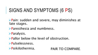
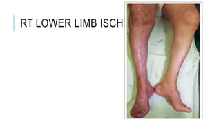
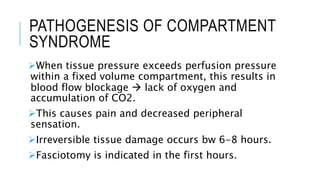
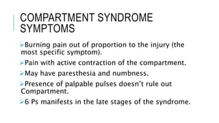
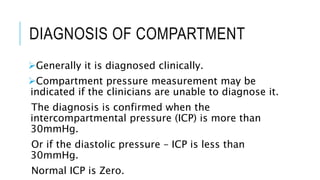
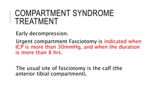
ACUTE ISCHEMIA Hamzeh Yacoub
ACUTE ISCHEMIA OVERVIEW
Occurs when arterial blood supply is suddenly
interrupted.
Most commonly due to embolization but can also
be due to thrombosis.
No time for collaterals to form.
More common in lower limbs.
Pain is due to ischemic muscles and nerves.
Irreversible changes within hours (Emergency).
SIGNS AND SYMPTOMS (6 PS)
Pain: sudden and severe, may diminishes at
late stages.
Paresthesia and numbness.
Paralysis.
Pallor below the level of obstruction.
Pulselessness.
Poikilothermia. PAIR TO COMPARE.
RT LOWER LIMB ISCHEMIA
ACUTE LIMB ISCHEMIA CAUSES
It can be caused by either Embolism or
Thrombus in situ, or maybe due to trauma.
Embolism cause may happen from:
Cardiac origin (85%).
Non-cardiac origin (15%).
CARDIAC CAUSES OF EMBOLISM
I. Atrial thrombus due to Atrial fibrillation
(irregularly irregular pulse).
II. Ventricular thrombus after MI, ventricles may
not contract properly. Or due to ventricular
aneurism.
III. Debris from damaged valves: as in Rheumatic
heart diseases.
IV. Venous embolism to right heart then causes PE.
But if remnant foramen ovale (hole bw right and
left sides), embolus can move from right to left
then to peripheral circulation (paradoxical
embolism).
NON CARDIAC ORIGIN EMBOLI
Atherosclerotic plaque debris (mainly
iatrogenic).
Aneurism can cause thrombi, most commonly
Aorta and the popliteal arteries.
THROMBOSIS CAUSES
Less common than emboli causes.
Mainly in patients with history of peripheral
artery disease.
ACUTE LIMB ISCHEMIA DIAGNOSIS
Clinically.
The best initial test is doppler ultrasound.
Normal healthy artery produces a triphasic signal,
biphasic signals are often normal but may present
an early disease state. Monophasic signals are
abnormal and signify a severe reduction in blood
flow. Most patients with acute ischemia have
absent doppler signals.
CTA of abdomen, pelvis, and limbs is the gold
standard as it can be performed quickly.
TREATMENT OF ACUTE ISCHEMIA
IV heparin unless contraindicated.
IV hydration to help optimize perfusion
through collateral vessels.
Urine alkalinization to protect kidneys from
myoglobinuria.
Position the affected limb in appropriate way
to ensure enough blood stream.
EMBOLECTOMY
The procedure of choice in case of
macroembolism in large artery such as common
femoral artery.
Performed by introducing a balloon-tipped
catheter through transverse Arteriotomy at the
occlusion level.
Inflation of the balloon and withdraw the catheter
to extract the thrombus.
Procedure is repeated until bleeding occurs.
Called Fogarty balloon catheter
EMBOLECTOMY
CONTRAINDICATIONS
Post embolectomy operation, the patient should
continue heparin therapy for long-term.
So, heparin is contraindicated for cerebral vascular
hemmorhages, subdural hematoma, recent trauma,
recent stroke patients...
THROMBOLYSIS
Performed when ischemia is not so severe, intra-
arterial thrombolysis is performed.
Narrow catheter is passed into the occluded
vessel and embedded with the clot.
tPA is infused through the catheter.
Contraindications: recent stroke, pregnancy
(pregnancy category C).
COMPARTMENT SYNDROME
Painful emergency condition, occurs when
tissue pressure exceeds perfusion pressure.
Resulting in ischemia and necrosis of muscles.
Lower extremities are more affected than
upper ex.
PATHOGENESIS OF COMPARTMENT
SYNDROME
When tissue pressure exceeds perfusion pressure
within a fixed volume compartment, this results in
blood flow blockage  lack of oxygen and
accumulation of CO2.
This causes pain and decreased peripheral
sensation.
Irreversible tissue damage occurs bw 6-8 hours.
Fasciotomy is indicated in the first hours.
COMPARTMENT SYNDROME
SYMPTOMS
Burning pain out of proportion to the injury (the
most specific symptom).
Pain with active contraction of the compartment.
May have paresthesia and numbness.
Presence of palpable pulses doesn’t rule out
Compartment.
6 Ps manifests in the late stages of the syndrome.
COMPARTMENT SYNDROME RISK
FACTORS
The most common cause is fractures (eg. Tibia
fracture).
Soft tissue injury (eg. Burns).
Constrictive dressing (eg. Splints, casts)
Penetrating wounds.
DIAGNOSIS OF COMPARTMENT
Generally it is diagnosed clinically.
Compartment pressure measurement may be
indicated if the clinicians are unable to diagnose it.
The diagnosis is confirmed when the
intercompartmental pressure (ICP) is more than
30mmHg.
Or if the diastolic pressure – ICP is less than
30mmHg.
Normal ICP is Zero.
COMPARTMENT SYNDROME
TREATMENT
Early decompression.
Urgent compartment Fasciotomy is indicated when
ICP is more than 30mmHg, and when the duration
is more than 8 hrs.
The usual site of fasciotomy is the calf (the
anterior tibial compartment).

Acute Limb ischemia

  • 1.
  • 2.
    ACUTE ISCHEMIA OVERVIEW Occurswhen arterial blood supply is suddenly interrupted. Most commonly due to embolization but can also be due to thrombosis. No time for collaterals to form. More common in lower limbs. Pain is due to ischemic muscles and nerves. Irreversible changes within hours (Emergency).
  • 3.
    SIGNS AND SYMPTOMS(6 PS) Pain: sudden and severe, may diminishes at late stages. Paresthesia and numbness. Paralysis. Pallor below the level of obstruction. Pulselessness. Poikilothermia. PAIR TO COMPARE.
  • 4.
    RT LOWER LIMBISCHEMIA
  • 5.
    ACUTE LIMB ISCHEMIACAUSES It can be caused by either Embolism or Thrombus in situ, or maybe due to trauma. Embolism cause may happen from: Cardiac origin (85%). Non-cardiac origin (15%).
  • 6.
    CARDIAC CAUSES OFEMBOLISM I. Atrial thrombus due to Atrial fibrillation (irregularly irregular pulse). II. Ventricular thrombus after MI, ventricles may not contract properly. Or due to ventricular aneurism. III. Debris from damaged valves: as in Rheumatic heart diseases. IV. Venous embolism to right heart then causes PE. But if remnant foramen ovale (hole bw right and left sides), embolus can move from right to left then to peripheral circulation (paradoxical embolism).
  • 7.
    NON CARDIAC ORIGINEMBOLI Atherosclerotic plaque debris (mainly iatrogenic). Aneurism can cause thrombi, most commonly Aorta and the popliteal arteries.
  • 8.
    THROMBOSIS CAUSES Less commonthan emboli causes. Mainly in patients with history of peripheral artery disease.
  • 10.
    ACUTE LIMB ISCHEMIADIAGNOSIS Clinically. The best initial test is doppler ultrasound. Normal healthy artery produces a triphasic signal, biphasic signals are often normal but may present an early disease state. Monophasic signals are abnormal and signify a severe reduction in blood flow. Most patients with acute ischemia have absent doppler signals. CTA of abdomen, pelvis, and limbs is the gold standard as it can be performed quickly.
  • 11.
    TREATMENT OF ACUTEISCHEMIA IV heparin unless contraindicated. IV hydration to help optimize perfusion through collateral vessels. Urine alkalinization to protect kidneys from myoglobinuria. Position the affected limb in appropriate way to ensure enough blood stream.
  • 12.
    EMBOLECTOMY The procedure ofchoice in case of macroembolism in large artery such as common femoral artery. Performed by introducing a balloon-tipped catheter through transverse Arteriotomy at the occlusion level. Inflation of the balloon and withdraw the catheter to extract the thrombus. Procedure is repeated until bleeding occurs. Called Fogarty balloon catheter
  • 13.
    EMBOLECTOMY CONTRAINDICATIONS Post embolectomy operation,the patient should continue heparin therapy for long-term. So, heparin is contraindicated for cerebral vascular hemmorhages, subdural hematoma, recent trauma, recent stroke patients...
  • 14.
    THROMBOLYSIS Performed when ischemiais not so severe, intra- arterial thrombolysis is performed. Narrow catheter is passed into the occluded vessel and embedded with the clot. tPA is infused through the catheter. Contraindications: recent stroke, pregnancy (pregnancy category C).
  • 16.
    COMPARTMENT SYNDROME Painful emergencycondition, occurs when tissue pressure exceeds perfusion pressure. Resulting in ischemia and necrosis of muscles. Lower extremities are more affected than upper ex.
  • 17.
    PATHOGENESIS OF COMPARTMENT SYNDROME Whentissue pressure exceeds perfusion pressure within a fixed volume compartment, this results in blood flow blockage  lack of oxygen and accumulation of CO2. This causes pain and decreased peripheral sensation. Irreversible tissue damage occurs bw 6-8 hours. Fasciotomy is indicated in the first hours.
  • 18.
    COMPARTMENT SYNDROME SYMPTOMS Burning painout of proportion to the injury (the most specific symptom). Pain with active contraction of the compartment. May have paresthesia and numbness. Presence of palpable pulses doesn’t rule out Compartment. 6 Ps manifests in the late stages of the syndrome.
  • 19.
    COMPARTMENT SYNDROME RISK FACTORS Themost common cause is fractures (eg. Tibia fracture). Soft tissue injury (eg. Burns). Constrictive dressing (eg. Splints, casts) Penetrating wounds.
  • 20.
    DIAGNOSIS OF COMPARTMENT Generallyit is diagnosed clinically. Compartment pressure measurement may be indicated if the clinicians are unable to diagnose it. The diagnosis is confirmed when the intercompartmental pressure (ICP) is more than 30mmHg. Or if the diastolic pressure – ICP is less than 30mmHg. Normal ICP is Zero.
  • 21.
    COMPARTMENT SYNDROME TREATMENT Early decompression. Urgentcompartment Fasciotomy is indicated when ICP is more than 30mmHg, and when the duration is more than 8 hrs. The usual site of fasciotomy is the calf (the anterior tibial compartment).