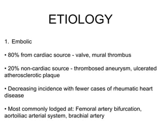
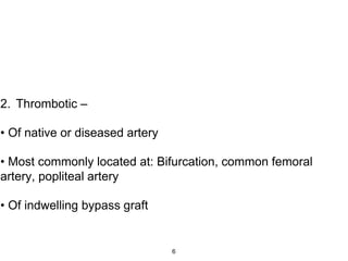
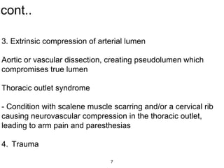
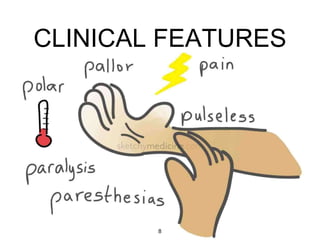
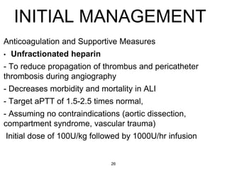
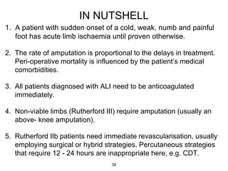
2. • Sudden loss of perfusion to the extremity/extremities of less than 14
days duration, resulting in variable ischemic clinical manifestations
and the potential risk of limb loss.
• Insufficient time for collaterals to compensate for loss of perfusion.
• A matter of a few hours can mean the difference between a major
amputation,limb salvage or death.
INTRODUCTION
2
3. Depends on:
—Degree of obstruction
—Site of occlusion
—Presence of Collaterals
—Affected Tissue
• Sluggish circulation distal to occlusion secondary
thrombosis occlusion of collaterals.
• Different tissue can tolerate ischemia at different rates.
PHYSIOLOGY
3
4. Cont.
• Biphasic injury pattern
1. Ischemic injury: Decreased arterial supply to a limb
causes ischemic injury by local hypoxemia.
2. Reperfusion injury: Return of arterial blood flow with
high oxygen content yields toxic oxygen free radicals, and
the
• re-circulation of muscle catabolic products and
inflammatory mediators. A patient at risk for a fatal
systemic reperfusion injury (multiple co-morbidities
and/or hemodynamically unstable) should instead
undergo an emergent amputation instead of
revascularization. 4
6. 2. Thrombotic –
• Of native or diseased artery
• Most commonly located at: Bifurcation, common femoral
artery, popliteal artery
• Of indwelling bypass graft
6
7. cont..
3. Extrinsic compression of arterial lumen
Aortic or vascular dissection, creating pseudolumen which
compromises true lumen
Thoracic outlet syndrome
- Condition with scalene muscle scarring and/or a cervical rib
causing neurovascular compression in the thoracic outlet,
leading to arm pain and paresthesias
4. Trauma
7
9. cont…
• Pain
—The severity of the initial symptoms depends on the
severity of ischemia and can range from incapacitating
pain to the sudden onset of mild claudication.
—More severe the ischemia, the faster the patient seeks
medical attention.
—Irreversible muscle necrosis occurs within 6 to 8 hours
if the condition is untreated.
9
10. Cont…
• Pallor
—The color of the skin reflects its vascular supply.
—Marble white skin is associated with acute total
ischemia.
—Slow capillary refill is a sign that at least a small
degree of distal flow is present and runoff vessels are
probably patent.
10
11. • Pulselessness
—Acute ischemia is associated with the loss of
peripheral pulses, which also helps define the level of
the occlusion. Palpable normal pulses in the
contralateral leg points toward embolism as the cause.
Cont…
11
13. Cont…
Paralysis: Loss of motor function indicates limb threatening
ischemia
Intrinsic foot muscles affected first followed by leg
muscles
Late irreversible ischemia indicated by loss of muscle
turgidity
13
15. EVALUATION
Following clinical assessment and classification, the anatomic
location of the arterial occlusion can be diagnosed with a high degree
of reliability.
• Aortic Occlusion:
—Paralysis of the legs is often the presenting feature
—patients are unwell, with mottled skin discoloration that often
extends above the inguinal ligament onto the lower abdomen
—No palpable extremity pulses.
—The kidneys are especially at risk, particularly if the aortic occlusion
is due to an aortic dissection.
—Successful revascularization restores the blood supply to a large
muscle mass, but the effects of ischemia-reperfusion may cause
further renal damage. 15
16. • Iliac Occlusion
—The femoral pulse is lost on the affected side, and mottling
usually extends to the inguinal level.
• Femoro-popliteal occlusion
—Most Common
—Severity depends on involvement of Profunda Femoris
Artery.
• Popliteal & Infra-popliteal Occlusion
—The calf muscles are ischemic with palpable femoral pulse.
Cont…
16
20. cont…
• Advantages:
- Noninvasive
- Fairly accurate for infrainguinal arterial occlusive disease,
especially of bypass grafts in this location
• Disadvantages:
- Suprainguinal arterial occlusions and distal run-off vessels not well-
visualized
- Calcifications of arterial walls (in diabetic patients, especially) can
create artifacts and obscure visualization.
• May be considered as a second-line approach to ALI imaging if
angiography is not possible for infrainguinal arterial occlusion.
20
22. DSA
• Advantages:
—Level and nature of occlusion
—During angiography, may be able
to immediately start therapeutic
intervention (intra-arterial
lysis)without losing time
• Disadvantages:
—Lack of collaterals and associated
spasm limits visualisation of more
distal vessels.
— incidence of contrast
nephrotoxicity
— Invasive
22
23. CT ANGIOGRAPHY
Differentiate Embolic & Thrombotic cause
if not clear clinically.
Conditions of vessels
Localizes level of Obstruction.
Visualises distal arterial tree and distal cut
off.
Disadvantages:
- Exposure to IV contrast –> If going to
angiography after CTA for intra-arterial
lysis of clot, patient will now receive a 2nd
contrast bolus, increasing the risk of renal
failure.
23
24. MR ANGIOGRAPHY
• Advantages:
- Less contrast load than angiography
- Non ionizing
• Disadvantages:
- Very time intensive and often unavailable
during weekend and night hours
-Can be used as an alternative imaging
modality for patients at high risk for contrast
angiography complications
Femoral-popliteal arterial system: sensitivity
92%, specificity 94%
Infra-popliteal arterial system: sensitivity 93%,
specificity = 71%24
26. INITIAL MANAGEMENT
Anticoagulation and Supportive Measures
• Unfractionated heparin
- To reduce propagation of thrombus and pericatheter
thrombosis during angiography
- Decreases morbidity and mortality in ALI
- Target aPTT of 1.5-2.5 times normal,
- Assuming no contraindications (aortic dissection,
compartment syndrome, vascular trauma)
Initial dose of 100U/kg followed by 1000U/hr infusion
26
27. cont…
• Hydration
—Patients are often relatively volume depleted, and
careful fluid resuscitation is necessary.
—The potential for myoglobinuria due to ischemia-
reperfusion, combined with the use of contrast agents
during diagnosis and treatment of ALI, increases the
risk of acute renal insufficiency.
• supplemental oxygen.
• intravenous analgesia.
27
28. ENDOVASCULAR TREATMENT
• Catheter Directed Thrombolysis
Viable to marginally threatened limb(class I & IIa)
Retrograde contralateral femoral approach preferred over Anterograde
ipsilateral Femoral
Multiple side hole 45/55cm 5F Glidecath catheter is used
Low dose Alteplase regimen: 1mg bolus followed by continuous infusion
of 0.5mg-1mg/hr for 12 hrs followed by arteriogram
High dose Alteplase regimen: 10mg bolus followed by 0.005mg/kg/hr
infusion for 6 hrs, max dose 4mg/hr.
Unfractioned heparin used 500U/hr to prevent peri sheath thrombosis
Monitoring of serum fibrinogen level 4 hrly, stop infusion if fibrinogen
drops below 100mg/dl, aPTT.
28
33. ENDARTERECTOMY
• The artery is opened
longitudinally at the site of
disease.
• The plaque is then separated
from the artery wall in the
direction of the arteriotomy &
removed
• The arteriotomy can be closed
primarily or with a patch.
33
35. • The reconstruction should be performed with preservation of
existing circulation.
• End to side anastomosis allows the maintenance of
anterograde flow in native vessel at proximal site.
• Distal anastomosis is placed in disease free segment distal
to obstruction to maintain retrograde flow through patent
branches.
• Align vessels without kink or twist.
• Arteriotomy measuring 1.2 - 2 times graft diameter created.
35
39. IN NUTSHELL
1. A patient with sudden onset of a cold, weak, numb and painful
foot has acute limb ischaemia until proven otherwise.
2. The rate of amputation is proportional to the delays in treatment.
Peri-operative mortality is influenced by the patient’s medical
comorbidities.
3. All patients diagnosed with ALI need to be anticoagulated
immediately.
4. Non-viable limbs (Rutherford III) require amputation (usually an
above- knee amputation).
5. Rutherford IIb patients need immediate revascularisation, usually
employing surgical or hybrid strategies. Percutaneous strategies
that require 12 - 24 hours are inappropriate here, e.g. CDT.
39