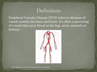
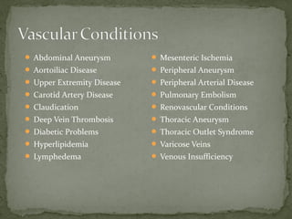
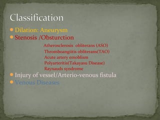
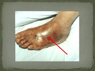
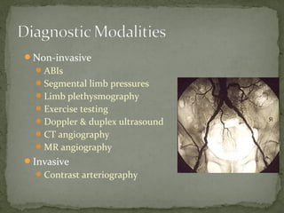
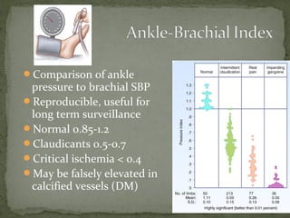
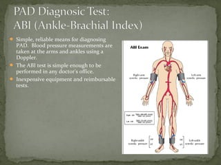
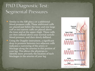
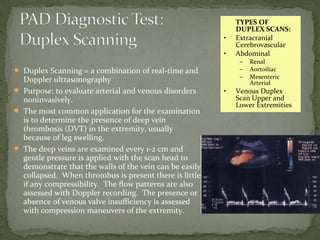
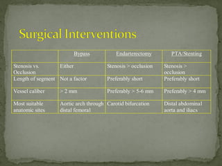
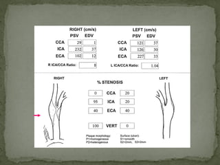
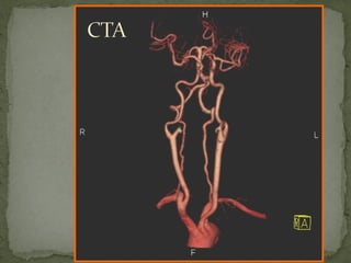
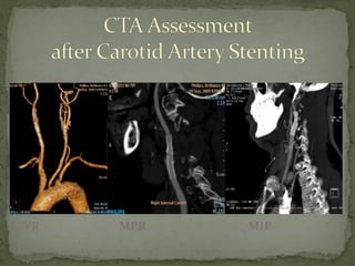
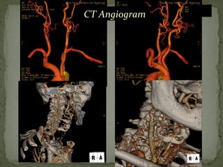
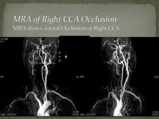
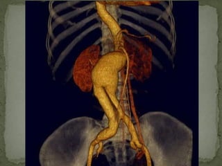
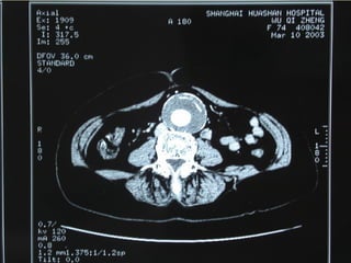
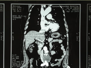
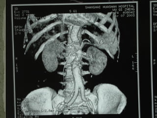
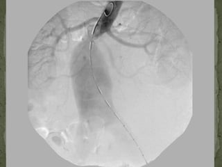
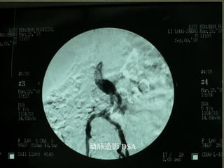
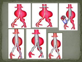
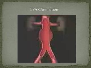
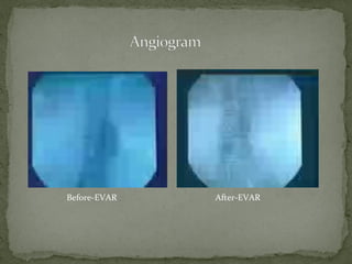
Prof. Bo Yu MD
Vascular Surgery Department ,Huashan Hospital
Fudan University
2013.12
Peripheral Vascular Disease (PVD) refers to diseases of
vessels outside the heart and brain. It's often a narrowing
of vessels that carry blood to the legs, arms, stomach or
kidneys.
* PVD definition is from the American Heart Association
 Abdominal Aneurysm
 Aortoiliac Disease
 Upper Extremity Disease
 Carotid Artery Disease
 Claudication
 Deep Vein Thrombosis
 Diabetic Problems
 Hyperlipidemia
 Lymphedema
 Mesenteric Ischemia
 Peripheral Aneurysm
 Peripheral Arterial Disease
 Pulmonary Embolism
 Renovascular Conditions
 Thoracic Aneurysm
 Thoracic Outlet Syndrome
 Varicose Veins
 Venous Insufficiency
Dilation: Aneurysm
Stenosis /Obsturction
Atherosclerosis obliterans (ASO)
Thromboangiitis obliterans(TAO)
Acute artery emoblism
Polyarteritis(Takayasu Disease)
Raynauds syndrome
Injury of vessel/Arterio-venous fistula
Venous Diseases
 Abdominal Aneurysm
 Aortoiliac Disease
 Upper Extremity Disease
 Carotid Artery Disease
 Claudication
 Deep Vein Thrombosis
 Diabetic Problems
 Hyperlipidemia
 Lymphedema
 Mesenteric Ischemia
 Peripheral Aneurysm
 Arterio-venous fistula
Pulmonary Embolism
 Renovascular Conditions
 Thoracic Aneurysm
 Thoracic Outlet Syndrome
 Varicose Veins
 Venous Insufficiency
NON-MODIFIABLE RISKS:
 Age. The risk of limb loss due to PAD increases with age. People 65 or older are two to three times
more likely to have an amputation.
 Gender. Men with PAD are twice as likely to undergo an amputation as women.
 Race/ethnicity. Some racial and ethnic groups have a higher risk of amputation (i.e., African
Americans, Latino Americans, and Native Americans). This is because they are at increased risk for
diabetes and cardiovascular disease.
 Family history of heart disease. A family history of cardiovascular disease is an indicator for risk at
developing PAD.
MODIFIABLE RISKS:
 Cigarette smoking. Smoking is a major risk factor for PAD. Smokers may have four times the risk of
PAD than nonsmokers.
 Obesity. People with a Body Mass Index (BMI) of 25 or higher are more likely to develop heart disease
and stroke even if they have no other risk factors.
 Diabetes mellitus. Having diabetes puts individuals at greater risk of developing PAD as well as other
cardiovascular diseases.
 Physical inactivity. Physical activity increases the distance that people with PAD can walk without
pain and also helps decrease the risk of heart attack or stroke. Supervised exercise programs are one of
the treatments for PAD patients.
 High blood cholesterol. High cholesterol contributes to the build-up of plaque in the arteries, which
can significantly reduce the blood's flow. This condition is known as atherosclerosis. Managing
cholesterol levels is essential to prevent or treat PAD.
 High blood pressure. When blood pressure remains high, the lining of the artery walls becomes
damaged. Many PAD patients also have high blood pressure.
 High levels of Homocysteine. This is an amino acid found in plasma (blood). Some recent studies
show higher levels are associated with PAD.
IIntermittent Painntermittent Pain
Postexercise,locomotive
Postural change
Variation of temperature
Persistent PainPersistent Pain
(Rest pain)(Rest pain)
Artery
Vein
Imflammatory and ischemia and necrosis
I Asymptomatic
II Intermittent Claudication
II a Claudication walking > 200m
II b Claudication walking < 200m
III Rest/nocturnal pain
IV Necrosis/gangrene
Venous edema
Lymphous edema
Heavy
Abnomal
sensation:numb 、 paralysis 、 needling 、 formicatio
n
Sensory deprivation
Up:
Venous Obstruction
Arterio-venous fistula
Down:
Artery Obstruction
Normal and abnormal color
Change of Skin Color after:
Finger pressed
Locomotive
Postual change
Artery Pulse :normal
to weaken/disappear
to enhance
Murmur:trill
Apperance and texture:
flexion,harder,nodus
 Vein varicose ,murmur,etc.
Dystrophia skin change
Ulcer,gangrene
Longer , bigger extremity
Apperance:
Varicose
Color:Pigmentation
Texture:Harder
Ulcer
Gangrene
Non-invasive
ABIs
Segmental limb pressures
Limb plethysmography
Exercise testing
Doppler & duplex ultrasound
CT angiography
MR angiography
Invasive
Contrast arteriography
Comparison of ankle
pressure to brachial SBP
Reproducible, useful for
long term surveillance
Normal 0.85-1.2
Claudicants 0.5-0.7
Critical ischemia < 0.4
May be falsely elevated in
calcified vessels (DM)
 Simple, reliable means for diagnosing
PAD. Blood pressure measurements are
taken at the arms and ankles using a
Doppler.
 The ABI test is simple enough to be
performed in any doctor's office.
 Inexpensive equipment and reimbursable
tests.
 Similar to the ABI plus 2 or 3 additional
blood pressure cuffs. These additional cuffs
are placed just below the knee and one large
cuff or two narrow cuffs are placed above
the knee and at the upper thigh. These cuffs
are then inflated above your normal systolic
blood pressure, and then slowly deflated.
 Using the Doppler instrument, a significant
drop in pressure between two adjacent cuffs
indicates a narrowing of the artery or
blockage along the arteries in this portion of
your leg. This allows the physician to
identify more precisely the location of such
blockages in the arteries of your leg.
 Duplex Scanning = a combination of real-time and
Doppler ultrasonography
 Purpose: to evaluate arterial and venous disorders
noninvasively.
 The most common application for the examination
is to determine the presence of deep vein
thrombosis (DVT) in the extremity, usually
because of leg swelling.
 The deep veins are examined every 1-2 cm and
gentle pressure is applied with the scan head to
demonstrate that the walls of the vein can be easily
collapsed. When thrombus is present there is little
if any compressibility. The flow patterns are also
assessed with Doppler recording. The presence or
absence of venous valve insufficiency is assessed
with compression maneuvers of the extremity.
TYPES OF
DUPLEX SCANS:
• Extracranial
Cerebrovascular
• Abdominal
– Renal
– Aortoiliac
– Mesenteric
Arterial
• Venous Duplex
Scan Upper and
Lower Extremities
Risk factor management
Lipid-lowering therapy
Smoking cessation
Exercise regimen
Antiplatelet therapy - ASA, clopidogrel
Vasoactive - Cilostazol (Pletal), pentoxyfilline
(Trental)
Bypass Endarterectomy PTA/Stenting
Stenosis vs.
Occlusion
Either Stenosis > occlusion Stenosis >
occlusion
Length of segment Not a factor Preferably short Preferably short
Vessel caliber > 2 mm Preferably > 5-6 mm Preferably > 4 mm
Most suitable
anatomic sites
Aortic arch through
distal femoral
Carotid bifurcation Distal abdominal
aorta and iliacs
Gray’sAnatomy: The Anatomical Basis of Clinical Practice, 40th edition (2008), Churchill-Livingstone,
Elsevier
Predilection Sites:
•Bifurcation of Common Carotid
Artery (CCA)
•Origin of Common Carotid Artery
•Carotid Siphon
•Middle Cerebral Artery (MCA) and
Anterior Cerebral Arteries (ACA)
Bilateral Lesion is mostly Seen
 Atherosclerosis , >90%
 Takayasu Disease, Fibromuscular Dysplasia
 Rare:Trauma, Artery Reversion, Congenital
Artery Atresia, Tumors, Inflammation around the
artery , Fibrosis after Radiotherapy 。
Stroke is the first cause of disability and death in
China, the incidence has increased every year
150-200 million new cases in China, the recurrence
rate is more of an average annual rate of 7.6%
China has 700 million existing patients with
cerebrovascular disease, of which about 70% of
Ischemic Stroke.
2 / 3 of ischemic stroke is caused by Carotid Artery
Atherosclerosis Stenosis
Carotid plaques mainly
caused by cerebral ischemia
in two ways :
a.Severe stenosis of the carotid
artery caused hemodynamic
changes, leading to the
corresponding parts of the brain
hypoperfusion ;
b.Micro-emboli or plaques in the
plaque surface micro-thrombosis
leading to cerebral embolism
Symptomatic Carotid Stenosis——
I.Brain ischemic symptoms: Tinnitus, Vertigo, Amaurosis, Blurred vision,
Dizziness, Headache, Insomnia, Memory loss, dreams and Ocular
ischemic manifestations.
II.TIA (transient ischemic attack): clinical manifestation is one side of
limb sensory or motor function disorders, transient monocular
blindness, or aphasia, etc., generally lasted only a few minutes, within
24h after the onset of full recovery. Imaging without focal lesions.
III.Ischemic stroke: common clinical symptoms limb sensory disturbances,
hemiplegia, aphasia, brain damage, coma and other serious and nervous system
with the appropriate signs and imaging features
Asymptomatic Carotid Stenosis ——
  Patients with carotid stenosis without any clinical signs or
symptoms
Asymptomatic Vs Symptomatic
3-4 : 1
BUS,TCD : Convenient and easy to repeat
CTA: Accurate and important tool for screening
CTP : Assess cerebral ischemia and vascular
reserve
MRDWI/PWI: Assess indications for
thrombolysis
MRA: TOF/PC MRA , 3D-CEMRA
DSA: Gold Standard
Blackshear WM,….,Strandness DE.Stroke 1980; 11:67.
VR MPR MIP
CT Angiogram
DSA:Gold Standard
NASCET Standard*:(1 - A/B)×100%
ECST Standard:(1 - A/C)×100%
Mild Moderate Severe
NASCET < 50% 50-69% 70-99%
Medical Treatment—Basic
Carotid Endarterectomy(CEA) - Gold Standard
Carotid Angioplasty and Stenting (CAS)
Prevent Risk Factors
Anti-platelet Drugs
Statins
British Heart Protection Study Collaboration
Group.Lancet 2002; 360:7-22
Naylor AR et al.Eur J Vasc Endovasc Surg 2009; 37:625.
Prevent StrokePrevent Stroke
Prevent Nerve InjuryPrevent Nerve Injury
 1953 , first successful CEA case was performed by DeBakey
 1956 , Cooley first reported CEA and using a shunt during operation.
 1958 , DeBakey Utilize Dacron Material in CEA.
 1985, Professor Wang Zhonggao performed CEA operation in China firstly.
 1979 , First PTA in Carotid Artery performed by Mathias
 1991 , NACET and ACAS’s result established an era of CEA
 1994 , Marks and Palmaz performed first Carotid Artery Stenting. The treatment of
carotid artery entered an era of CEA and CAS.
Symtomatic Patients: >50%
Stenosis of Carotid Artery
Asymtomatic Patients: >70%
Stenosis of Carotid Artery
Lancet.2010;375(9719):985-997
CEA
EEA
Techniques
PTFE patch Vein Vein interposition PTFE
CAS Pro-CAS
M.O.M.A
Pro-CAS
Carotid Angioplasty and Stenting(CAS)
61
Distal ICA
Occlusion
Proximal
Occlusion:CCA/EC
A Occlusion
Distal ICA Filter Proximal
Occlusion /
Reverse Flow
Carotid Endarterectomy Carotid Artery Stenting
Long-term efficacy has
been confirmed
Minimally invasive
Removal of atherosclerotic
plaque
No cranial nerve injury
No intravascular foreign
body
Long lesion of CA
Avoid the risk of
angiography
For a history of neck
surgery or radiotherapy in
patients
Craig R. Narins, et al. Patient selection for carotid stenting versus
endarterectomy: A systematic review. J Vasc Surg 2006;44:661-72.
Summary
 Carotid Artery Atherosclerosis Stenosis is the primary cause
of ischemic stroke, which is the only stroke type can be
prevented by surgical intervention
 Stenosis of carotid atherosclerosis can be divided into
symptomatic and asymptomatic, the most common clinical
manifestations of transient ischemic attack (TIA)
 Surgical Indication: Severe carotid artery stenosis
(symptomatic stenosis> 50%, asymptomatic> 70%)
 The Gold Standard Treatment of Severe Stenosis of carotid
artery carotid endarterectomy(CEA), carotid angioplasty and
stenting(CAS) is an alternative for patients at high surgical
risk
Vessel wall of
abdominal aortic
Permanent and
localized dilation
Diameter of AAA's 〉
2 times normal's

Male ( >60Y ): 4 %~ 9 %

Female ( >60Y ): 1 %
 Diameter>5cm: about 0.5% in male
 Almost all rupture of AAA occur in males
aged 65 or above.
Main :

Age : >65Y

Gender : Male>Female
 Cigerattes
Minor :
 Family history
 Coronary disease
 Hypercholesteremia
 Hypertension
 Cerebral vascular disease
 Genetic inheritance
 Genetic susceptibility
 Atherosclerosis
 Medial cystic necroses or degeneration
 Cogenital AAA
 Infection:staphylococci ,saimonella,etc.
 Infrarenal aorta
aneurysm:95 %
 Thoracic-abdominal
aneurysm : 5 %( both
thoracic and abdomianl
aorta involved )

True aneurysm :
Whole aortic vessel and aortic aneurysm expand
pathologically 。

Pseudoaneurysm(false aneurysm) :
Actual perforation and tearing of one or more layers
of aortic vessel wall,from which hemorrhage to be
surrounded by adjacent tissue and become
hematoma ,mostly due to trauma.
 Dissecting aneurysm:
It occurs when a tear in the aorta intima causes
blood to flow between the layers of the wall of the
aorta and force the layers apart.
A B C
A : True aneurysm
B : Pseuoaneurysm
C : Dissecting aneurysm
Always untypcial
Founded by physical examinations occasionally

Abdominal pain : from discomfortable to severe upper
adominal pain

Pulsating mass : most unique sign, periumbilical

Vascular murmur : systolic phase

Symptoms of compression : obstruction of digestion
and ureter ,etc.
 Rupture
 Embolism of peripheral artery
 Acute complete thrombosis
 Infection
 Chronic disturbance of blood coagulation due to
consuming
 Aortoenteric fistula

Arteriovenous fistula ( blood flow into inferior vena
cava )

Abdominal radiography : Calcified
vessel wall of AAA (which looks like
eggshell)
 Ultrasound
 CT
 MRI
 Angiography
10/20/15山血管外科网华 www.vascularcenter.cn 80
10/20/15山血管外科网华 www.vascularcenter.cn 81
10/20/15山血管外科网华 www.vascularcenter.cn 82
10/20/15山血管外科网华 www.vascularcenter.cn 84
脉造影动 DSA
10/20/15山血管外科网华 www.vascularcenter.cn 85
10/20/15山血管外科网华 www.vascularcenter.cn 86
Indication :
 Diameter>5.5cm (<5.5cm Ultrasound follow up
regularly)
 Continuing dilation with abdominal pain
 Tend to rupture
 Hematoma in dissecting which causes severe
abdominal pain
 With infection
 Compress adjacent organs and other important tissues
 Mural thrombosis in aneurysm clot off and lead to
embolism of distal artery
 Incision
10/20/15山血管外科网华 www.vascularcenter.cn 90
10/20/15山血管外科网华 www.vascularcenter.cn 91
10/20/15山血管外科网华 www.vascularcenter.cn 93
10/20/15山血管外科网华 www.vascularcenter.cn 94
Anastomosis
10/20/15山血管外科网华 www.vascularcenter.cn 96
Artificial
Veseel
10/20/15山血管外科网华 www.vascularcenter.cn 97
EndoVascular AAA Repair
(EVAR)
Indication :
 Conventional resection of AAA
 No allergic response to contrast medium
 Creatinine level < 2.5mg / dl
Contraindication :
 Length from inferior renal artery < 1.5cm
 Diameter of AAA > 2.8cm
 Diameter of common iliac artery > 11.5mm
 Diameter of external iliac artery < 6mm
 Angle of proximal aneurysm neck > 60°
 Severe atherosclerosis of iliac artery,buckling vessel
(>90°),especially with extensive calcification
 Only inferior mesenteric artery supply left hemicolon (superio
and inferior mesentenci artery lack of communication)
Before-EVAR After-EVAR
Reference Books and
Literatures
1. Rutherford: Vascular Surgery, 7th
ed.
2010
2. 2011 ASA ACCF AHA AANN AANS ACR
ASNR CNS SAIP SCAI SIR SNIS SVM
SVS Guideline on the Management of
Patients With Extracranial Carotid and
Vertebral Artery Disease. Journal of the
American College of Cardiology, Vol.
57,No. 8, 2011
3. C.D. Liapis, Sir P.R.F. Bell ESVS
Guidelines. Invasive Treatment for
Carotid Stenosis -- Indications,
Techniques Eur J Vasc Endovasc Surg
(2009) 37, S1-S19
The only way to be truly satisfied is to do
what you believe is great work.
And the only way to do great work is to
love what you do.
——Steve Jobs
vascularelite@gmail.com Julian

peripheral vascular disease

  • 1.
    Prof. Bo YuMD Vascular Surgery Department ,Huashan Hospital Fudan University 2013.12
  • 2.
    Peripheral Vascular Disease(PVD) refers to diseases of vessels outside the heart and brain. It's often a narrowing of vessels that carry blood to the legs, arms, stomach or kidneys. * PVD definition is from the American Heart Association
  • 3.
     Abdominal Aneurysm Aortoiliac Disease  Upper Extremity Disease  Carotid Artery Disease  Claudication  Deep Vein Thrombosis  Diabetic Problems  Hyperlipidemia  Lymphedema  Mesenteric Ischemia  Peripheral Aneurysm  Peripheral Arterial Disease  Pulmonary Embolism  Renovascular Conditions  Thoracic Aneurysm  Thoracic Outlet Syndrome  Varicose Veins  Venous Insufficiency
  • 4.
    Dilation: Aneurysm Stenosis /Obsturction Atherosclerosisobliterans (ASO) Thromboangiitis obliterans(TAO) Acute artery emoblism Polyarteritis(Takayasu Disease) Raynauds syndrome Injury of vessel/Arterio-venous fistula Venous Diseases
  • 5.
     Abdominal Aneurysm Aortoiliac Disease  Upper Extremity Disease  Carotid Artery Disease  Claudication  Deep Vein Thrombosis  Diabetic Problems  Hyperlipidemia  Lymphedema  Mesenteric Ischemia  Peripheral Aneurysm  Arterio-venous fistula Pulmonary Embolism  Renovascular Conditions  Thoracic Aneurysm  Thoracic Outlet Syndrome  Varicose Veins  Venous Insufficiency
  • 6.
    NON-MODIFIABLE RISKS:  Age.The risk of limb loss due to PAD increases with age. People 65 or older are two to three times more likely to have an amputation.  Gender. Men with PAD are twice as likely to undergo an amputation as women.  Race/ethnicity. Some racial and ethnic groups have a higher risk of amputation (i.e., African Americans, Latino Americans, and Native Americans). This is because they are at increased risk for diabetes and cardiovascular disease.  Family history of heart disease. A family history of cardiovascular disease is an indicator for risk at developing PAD. MODIFIABLE RISKS:  Cigarette smoking. Smoking is a major risk factor for PAD. Smokers may have four times the risk of PAD than nonsmokers.  Obesity. People with a Body Mass Index (BMI) of 25 or higher are more likely to develop heart disease and stroke even if they have no other risk factors.  Diabetes mellitus. Having diabetes puts individuals at greater risk of developing PAD as well as other cardiovascular diseases.  Physical inactivity. Physical activity increases the distance that people with PAD can walk without pain and also helps decrease the risk of heart attack or stroke. Supervised exercise programs are one of the treatments for PAD patients.  High blood cholesterol. High cholesterol contributes to the build-up of plaque in the arteries, which can significantly reduce the blood's flow. This condition is known as atherosclerosis. Managing cholesterol levels is essential to prevent or treat PAD.  High blood pressure. When blood pressure remains high, the lining of the artery walls becomes damaged. Many PAD patients also have high blood pressure.  High levels of Homocysteine. This is an amino acid found in plasma (blood). Some recent studies show higher levels are associated with PAD.
  • 8.
    IIntermittent Painntermittent Pain Postexercise,locomotive Posturalchange Variation of temperature Persistent PainPersistent Pain (Rest pain)(Rest pain) Artery Vein Imflammatory and ischemia and necrosis
  • 9.
    I Asymptomatic II IntermittentClaudication II a Claudication walking > 200m II b Claudication walking < 200m III Rest/nocturnal pain IV Necrosis/gangrene
  • 10.
  • 13.
    Heavy Abnomal sensation:numb 、 paralysis、 needling 、 formicatio n Sensory deprivation
  • 14.
  • 15.
    Normal and abnormalcolor Change of Skin Color after: Finger pressed Locomotive Postual change
  • 16.
    Artery Pulse :normal toweaken/disappear to enhance Murmur:trill Apperance and texture: flexion,harder,nodus  Vein varicose ,murmur,etc.
  • 18.
  • 19.
  • 21.
  • 22.
    Non-invasive ABIs Segmental limb pressures Limbplethysmography Exercise testing Doppler & duplex ultrasound CT angiography MR angiography Invasive Contrast arteriography
  • 23.
    Comparison of ankle pressureto brachial SBP Reproducible, useful for long term surveillance Normal 0.85-1.2 Claudicants 0.5-0.7 Critical ischemia < 0.4 May be falsely elevated in calcified vessels (DM)
  • 24.
     Simple, reliablemeans for diagnosing PAD. Blood pressure measurements are taken at the arms and ankles using a Doppler.  The ABI test is simple enough to be performed in any doctor's office.  Inexpensive equipment and reimbursable tests.
  • 25.
     Similar tothe ABI plus 2 or 3 additional blood pressure cuffs. These additional cuffs are placed just below the knee and one large cuff or two narrow cuffs are placed above the knee and at the upper thigh. These cuffs are then inflated above your normal systolic blood pressure, and then slowly deflated.  Using the Doppler instrument, a significant drop in pressure between two adjacent cuffs indicates a narrowing of the artery or blockage along the arteries in this portion of your leg. This allows the physician to identify more precisely the location of such blockages in the arteries of your leg.
  • 26.
     Duplex Scanning= a combination of real-time and Doppler ultrasonography  Purpose: to evaluate arterial and venous disorders noninvasively.  The most common application for the examination is to determine the presence of deep vein thrombosis (DVT) in the extremity, usually because of leg swelling.  The deep veins are examined every 1-2 cm and gentle pressure is applied with the scan head to demonstrate that the walls of the vein can be easily collapsed. When thrombus is present there is little if any compressibility. The flow patterns are also assessed with Doppler recording. The presence or absence of venous valve insufficiency is assessed with compression maneuvers of the extremity. TYPES OF DUPLEX SCANS: • Extracranial Cerebrovascular • Abdominal – Renal – Aortoiliac – Mesenteric Arterial • Venous Duplex Scan Upper and Lower Extremities
  • 27.
    Risk factor management Lipid-loweringtherapy Smoking cessation Exercise regimen Antiplatelet therapy - ASA, clopidogrel Vasoactive - Cilostazol (Pletal), pentoxyfilline (Trental)
  • 28.
    Bypass Endarterectomy PTA/Stenting Stenosisvs. Occlusion Either Stenosis > occlusion Stenosis > occlusion Length of segment Not a factor Preferably short Preferably short Vessel caliber > 2 mm Preferably > 5-6 mm Preferably > 4 mm Most suitable anatomic sites Aortic arch through distal femoral Carotid bifurcation Distal abdominal aorta and iliacs
  • 31.
    Gray’sAnatomy: The AnatomicalBasis of Clinical Practice, 40th edition (2008), Churchill-Livingstone, Elsevier
  • 32.
    Predilection Sites: •Bifurcation ofCommon Carotid Artery (CCA) •Origin of Common Carotid Artery •Carotid Siphon •Middle Cerebral Artery (MCA) and Anterior Cerebral Arteries (ACA) Bilateral Lesion is mostly Seen
  • 33.
     Atherosclerosis ,>90%  Takayasu Disease, Fibromuscular Dysplasia  Rare:Trauma, Artery Reversion, Congenital Artery Atresia, Tumors, Inflammation around the artery , Fibrosis after Radiotherapy 。
  • 34.
    Stroke is thefirst cause of disability and death in China, the incidence has increased every year 150-200 million new cases in China, the recurrence rate is more of an average annual rate of 7.6% China has 700 million existing patients with cerebrovascular disease, of which about 70% of Ischemic Stroke. 2 / 3 of ischemic stroke is caused by Carotid Artery Atherosclerosis Stenosis
  • 35.
    Carotid plaques mainly causedby cerebral ischemia in two ways : a.Severe stenosis of the carotid artery caused hemodynamic changes, leading to the corresponding parts of the brain hypoperfusion ; b.Micro-emboli or plaques in the plaque surface micro-thrombosis leading to cerebral embolism
  • 36.
    Symptomatic Carotid Stenosis—— I.Brainischemic symptoms: Tinnitus, Vertigo, Amaurosis, Blurred vision, Dizziness, Headache, Insomnia, Memory loss, dreams and Ocular ischemic manifestations. II.TIA (transient ischemic attack): clinical manifestation is one side of limb sensory or motor function disorders, transient monocular blindness, or aphasia, etc., generally lasted only a few minutes, within 24h after the onset of full recovery. Imaging without focal lesions. III.Ischemic stroke: common clinical symptoms limb sensory disturbances, hemiplegia, aphasia, brain damage, coma and other serious and nervous system with the appropriate signs and imaging features Asymptomatic Carotid Stenosis ——   Patients with carotid stenosis without any clinical signs or symptoms Asymptomatic Vs Symptomatic 3-4 : 1
  • 37.
    BUS,TCD : Convenientand easy to repeat CTA: Accurate and important tool for screening CTP : Assess cerebral ischemia and vascular reserve MRDWI/PWI: Assess indications for thrombolysis MRA: TOF/PC MRA , 3D-CEMRA DSA: Gold Standard
  • 38.
  • 42.
  • 43.
  • 45.
    DSA:Gold Standard NASCET Standard*:(1- A/B)×100% ECST Standard:(1 - A/C)×100% Mild Moderate Severe NASCET < 50% 50-69% 70-99%
  • 46.
    Medical Treatment—Basic Carotid Endarterectomy(CEA)- Gold Standard Carotid Angioplasty and Stenting (CAS)
  • 47.
    Prevent Risk Factors Anti-plateletDrugs Statins British Heart Protection Study Collaboration Group.Lancet 2002; 360:7-22 Naylor AR et al.Eur J Vasc Endovasc Surg 2009; 37:625.
  • 48.
    Prevent StrokePrevent Stroke PreventNerve InjuryPrevent Nerve Injury
  • 49.
     1953 ,first successful CEA case was performed by DeBakey  1956 , Cooley first reported CEA and using a shunt during operation.  1958 , DeBakey Utilize Dacron Material in CEA.  1985, Professor Wang Zhonggao performed CEA operation in China firstly.  1979 , First PTA in Carotid Artery performed by Mathias  1991 , NACET and ACAS’s result established an era of CEA  1994 , Marks and Palmaz performed first Carotid Artery Stenting. The treatment of carotid artery entered an era of CEA and CAS.
  • 50.
    Symtomatic Patients: >50% Stenosisof Carotid Artery Asymtomatic Patients: >70% Stenosis of Carotid Artery Lancet.2010;375(9719):985-997
  • 51.
  • 53.
    Techniques PTFE patch VeinVein interposition PTFE
  • 60.
  • 61.
  • 64.
    Carotid Endarterectomy CarotidArtery Stenting Long-term efficacy has been confirmed Minimally invasive Removal of atherosclerotic plaque No cranial nerve injury No intravascular foreign body Long lesion of CA Avoid the risk of angiography For a history of neck surgery or radiotherapy in patients Craig R. Narins, et al. Patient selection for carotid stenting versus endarterectomy: A systematic review. J Vasc Surg 2006;44:661-72.
  • 66.
    Summary  Carotid ArteryAtherosclerosis Stenosis is the primary cause of ischemic stroke, which is the only stroke type can be prevented by surgical intervention  Stenosis of carotid atherosclerosis can be divided into symptomatic and asymptomatic, the most common clinical manifestations of transient ischemic attack (TIA)  Surgical Indication: Severe carotid artery stenosis (symptomatic stenosis> 50%, asymptomatic> 70%)  The Gold Standard Treatment of Severe Stenosis of carotid artery carotid endarterectomy(CEA), carotid angioplasty and stenting(CAS) is an alternative for patients at high surgical risk
  • 68.
    Vessel wall of abdominalaortic Permanent and localized dilation Diameter of AAA's 〉 2 times normal's
  • 69.
     Male ( >60Y): 4 %~ 9 %  Female ( >60Y ): 1 %  Diameter>5cm: about 0.5% in male  Almost all rupture of AAA occur in males aged 65 or above.
  • 70.
    Main :  Age :>65Y  Gender : Male>Female  Cigerattes Minor :  Family history  Coronary disease  Hypercholesteremia  Hypertension  Cerebral vascular disease
  • 71.
     Genetic inheritance Genetic susceptibility  Atherosclerosis  Medial cystic necroses or degeneration  Cogenital AAA  Infection:staphylococci ,saimonella,etc.
  • 72.
     Infrarenal aorta aneurysm:95%  Thoracic-abdominal aneurysm : 5 %( both thoracic and abdomianl aorta involved )
  • 73.
     True aneurysm : Wholeaortic vessel and aortic aneurysm expand pathologically 。  Pseudoaneurysm(false aneurysm) : Actual perforation and tearing of one or more layers of aortic vessel wall,from which hemorrhage to be surrounded by adjacent tissue and become hematoma ,mostly due to trauma.  Dissecting aneurysm: It occurs when a tear in the aorta intima causes blood to flow between the layers of the wall of the aorta and force the layers apart.
  • 74.
    A B C A: True aneurysm B : Pseuoaneurysm C : Dissecting aneurysm
  • 75.
    Always untypcial Founded byphysical examinations occasionally  Abdominal pain : from discomfortable to severe upper adominal pain  Pulsating mass : most unique sign, periumbilical  Vascular murmur : systolic phase  Symptoms of compression : obstruction of digestion and ureter ,etc.
  • 76.
     Rupture  Embolismof peripheral artery  Acute complete thrombosis  Infection  Chronic disturbance of blood coagulation due to consuming  Aortoenteric fistula  Arteriovenous fistula ( blood flow into inferior vena cava )
  • 77.
     Abdominal radiography :Calcified vessel wall of AAA (which looks like eggshell)  Ultrasound  CT  MRI  Angiography
  • 80.
  • 81.
  • 82.
  • 84.
  • 85.
  • 86.
  • 87.
    Indication :  Diameter>5.5cm(<5.5cm Ultrasound follow up regularly)  Continuing dilation with abdominal pain  Tend to rupture  Hematoma in dissecting which causes severe abdominal pain  With infection  Compress adjacent organs and other important tissues  Mural thrombosis in aneurysm clot off and lead to embolism of distal artery
  • 88.
  • 90.
  • 91.
  • 93.
  • 94.
  • 95.
  • 96.
  • 97.
  • 98.
  • 99.
    Indication :  Conventionalresection of AAA  No allergic response to contrast medium  Creatinine level < 2.5mg / dl Contraindication :  Length from inferior renal artery < 1.5cm  Diameter of AAA > 2.8cm  Diameter of common iliac artery > 11.5mm  Diameter of external iliac artery < 6mm  Angle of proximal aneurysm neck > 60°  Severe atherosclerosis of iliac artery,buckling vessel (>90°),especially with extensive calcification  Only inferior mesenteric artery supply left hemicolon (superio and inferior mesentenci artery lack of communication)
  • 104.
  • 105.
    Reference Books and Literatures 1.Rutherford: Vascular Surgery, 7th ed. 2010 2. 2011 ASA ACCF AHA AANN AANS ACR ASNR CNS SAIP SCAI SIR SNIS SVM SVS Guideline on the Management of Patients With Extracranial Carotid and Vertebral Artery Disease. Journal of the American College of Cardiology, Vol. 57,No. 8, 2011 3. C.D. Liapis, Sir P.R.F. Bell ESVS Guidelines. Invasive Treatment for Carotid Stenosis -- Indications, Techniques Eur J Vasc Endovasc Surg (2009) 37, S1-S19
  • 106.
    The only wayto be truly satisfied is to do what you believe is great work. And the only way to do great work is to love what you do. ——Steve Jobs
  • 108.

Editor's Notes

  • #46 前者以颈动脉膨大远端正常管腔内径为基础内径, 后者以颈动脉膨大处估计内径为基础内径 NASCET标准*:(1-ICA最窄处宽度/狭窄病变远端正常ICA内径)×100% ECST标准:(1-ICA最窄处宽度/颈动脉膨大处估计内径)×100%
  • #51 颈动脉严重狭窄的患者,有再通手术的指证。
  • #54 Titeldia
  • #56 Titeldia
  • #57 Titeldia
  • #64 As previously mentioned, DEP devices have become an accepted part of CAS despite the lack of level I data supporting their use. Several types of devices exist including temporary distal occlusion and aspiration devices such as the Medtronic Guardwire, filter devices such as the Emboshield or FilterWire and…
  • #65 NASCET中将有症状患者定义为120天内 ECST中将有症状患者定义为6个月内