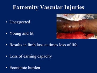
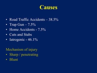
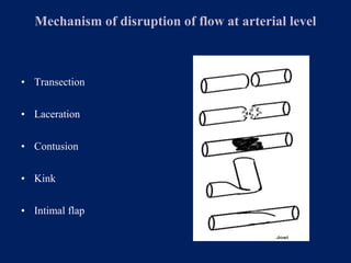
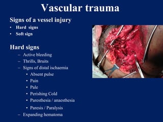
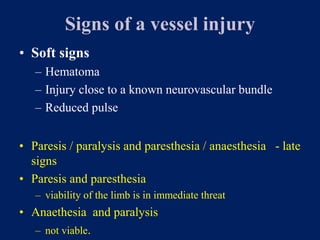
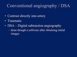
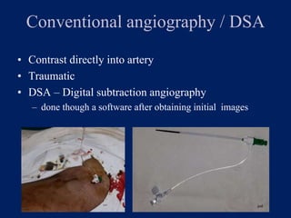
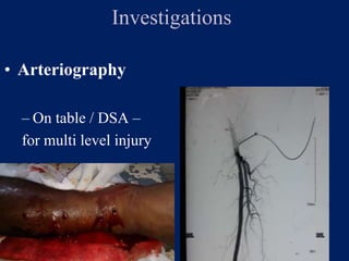
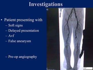
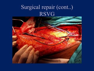
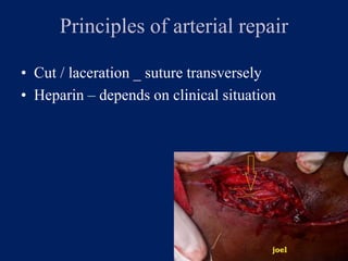
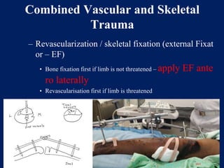
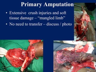
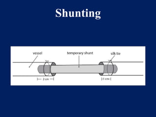
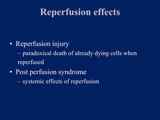
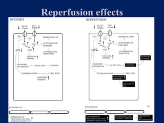
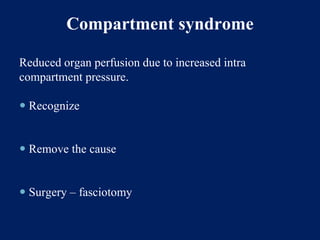
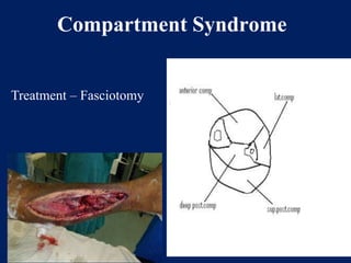
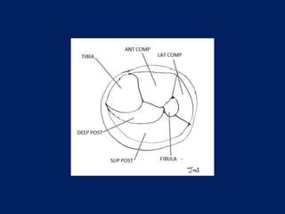
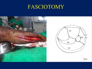
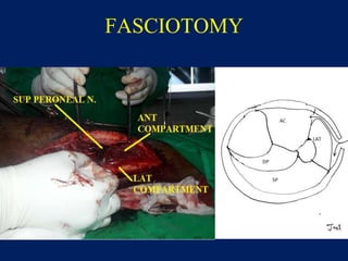
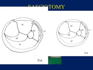
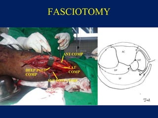
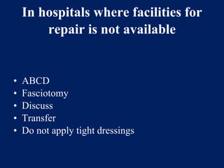
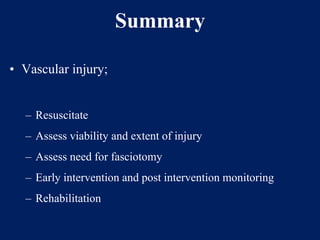
Vascular Trauma
Joel Arudchelvam
Consultant Vascular and Transplant Surgeon
Teaching Hospital Anuradhapura.
Vascular trauma /injury
• Injury to
– Arteries
– Veins
• Anatomical regions
– Extremity – limbs
– Abdomen and pelvis
– Thorax
– Head and neck
Vascular trauma /injury
• Injury to
– Arteries
– Veins
• Anatomical regions
– Extremity – limbs
– Abdomen and pelvis
– Thorax
– Head and neck
Extremity Vascular Injuries
• Unexpected
• Young and fit
• Results in limb loss at times loss of life
• Loss of earning capacity
• Economic burden
Causes
• Road Traffic Accidents – 38.5%
• Trap Gun – 7.5%
• Home Accidents - 7.5%
• Cuts and Stabs
• Iatrogenic - 46.1%
Mechanism of injury
• Sharp / penetrating
• Blunt
Mechanism of disruption of flow at arterial level
• Transection
• Laceration
• Contusion
• Kink
• Intimal flap
Vascular trauma
Signs of a vessel injury
• Hard signs
• Soft sign
Hard signs
– Active bleeding
– Thrills, Bruits
– Signs of distal ischaemia
• Absent pulse
• Pain
• Pale
• Perishing Cold
• Paresthesia / anaesthesia
• Paresis / Paralysis
– Expanding hematoma
Signs of a vessel injury
• Soft signs
– Hematoma
– Injury close to a known neurovascular bundle
– Reduced pulse
• Paresis / paralysis and paresthesia / anaesthesia - late
signs
• Paresis and paresthesia
– viability of the limb is in immediate threat
• Anaethesia and paralysis
– not viable.
Problems with diagnosing ischaemia after
trauma
• Pain
– Due to injury itself
• Pallor
– Pallor due to blood loss
• Absent pulse
– Absent due to low blood pressure. Compare with othe
limb
• Paresthesia , paresis
– Due to associated nerve, muscle injury or unresponsive
patient
Investigations
Investigations
• Hard signs
• Urgent intervention
• Soft signs
• Observe
• Investigate
Investigations
• Hand held DOPPLER
• Absent doppler flow
• Quality of signal
• ABPI
• Presence of doppler flow does not exclude vascu
lar injury
• Duplex scan (USS + DOPPLER)
• Difficult to image in trauma
• Due to
• Pain, Non cooperative patient, Dressi
ngs
• Patent distal vessels does not exclude a proximal
injury
Investigations
• Angiography
– CT angiography
– Catheter angiography
CT ANGIOGRAPHY
• Anatomy
• Site of Injury
• Soft tissue and bone
• 3D reconstruction
Conventional angiography / DSA
• Contrast directly into artery
• Traumatic
• DSA – Digital subtraction angiography
– done though a software after obtaining initial
images
Conventional angiography / DSA
• Contrast directly into artery
• Traumatic
• DSA – Digital subtraction angiography
– done though a software after obtaining initial images
Investigations
• Arteriography
– On table / DSA –
for multi level injury
Investigations
• Patient presenting with
– Soft signs
– Delayed presentation
– Avf
– False aneurysm
– Pre-op angiography
How soon we should we repair
– As soon as possible
– Effects of ischaemia
How soon we should we repair
• At ANP – 1 year
– 13 cases
– Commonest artery popliteal 53.8 %
– Mean ischaemic time – 12.67 hrs
– 4 clinically dead limb (mean time 15.75 hrs)
Surgical Repair
• Prompt transport to operating room
• General anesthesia
• Clean the entire limb
• Thigh prepared – for venous harvest
• Control of proximal and distal ends and trimming
Surgical repair (cont..)
• Balloon thrombectomy
• Systemic and distal heparinisation
• Interposition graft / Direct approxi
mation
– Unit experience – 88.2% RSVG
• Prosthesis
– lower patency
– infection
Surgical repair (cont..)
RSVG
Principles of arterial repair
• Cut / laceration _ suture transversely
• Heparin – depends on clinical situation
Combined Vascular and Skeletal
Trauma
– Revascularization / skeletal fixation (external Fixat
or – EF)
• Bone fixation first if limb is not threatened – apply EF ante
ro laterally
• Revascularisation first if limb is threatened
Primary Amputation
• Extensive crush injuries and soft
tissue damage – “mangled limb”
• No need to transfer – discuss / photo
Shunting
Reperfusion effects
• Reperfusion injury
– paradoxical death of already dying cells when
reperfused
• Post perfusion syndrome
– systemic effects of reperfusion
Reperfusion effects
Compartment syndrome
Reduced organ perfusion due to increased intra
compartment pressure.
 Recognize
 Remove the cause
 Surgery – fasciotomy
Compartment Syndrome
Treatment – Fasciotomy
FASCIOTOMY
FASCIOTOMY
FASCIOTOMY
FASCIOTOMY
Post perfusion syndrome
• ACIDOSIS
• ARDS
• AKI
• MYOCARDIA DEPRESSION
• DIC
• MYOGLOBINURIA
• HYPERKALEMIA
• ETC.
Post perfusion syndrome
• How to minimise
– hydration and maintain UOP
– keep on O2
– Mannitol
– Correct acidosis / hyperkalemia
– Inotropes
In hospitals where facilities for
repair is not available
• ABCD
• Fasciotomy
• Discuss
• Transfer
• Do not apply tight dressings
Summary
• Vascular injury;
– Resuscitate
– Assess viability and extent of injury
– Assess need for fasciotomy
– Early intervention and post intervention monitoring
– Rehabilitation
Thank You

Vasular trauma

  • 1.
    Vascular Trauma Joel Arudchelvam ConsultantVascular and Transplant Surgeon Teaching Hospital Anuradhapura.
  • 2.
    Vascular trauma /injury •Injury to – Arteries – Veins • Anatomical regions – Extremity – limbs – Abdomen and pelvis – Thorax – Head and neck
  • 3.
    Vascular trauma /injury •Injury to – Arteries – Veins • Anatomical regions – Extremity – limbs – Abdomen and pelvis – Thorax – Head and neck
  • 4.
    Extremity Vascular Injuries •Unexpected • Young and fit • Results in limb loss at times loss of life • Loss of earning capacity • Economic burden
  • 5.
    Causes • Road TrafficAccidents – 38.5% • Trap Gun – 7.5% • Home Accidents - 7.5% • Cuts and Stabs • Iatrogenic - 46.1% Mechanism of injury • Sharp / penetrating • Blunt
  • 6.
    Mechanism of disruptionof flow at arterial level • Transection • Laceration • Contusion • Kink • Intimal flap
  • 7.
    Vascular trauma Signs ofa vessel injury • Hard signs • Soft sign Hard signs – Active bleeding – Thrills, Bruits – Signs of distal ischaemia • Absent pulse • Pain • Pale • Perishing Cold • Paresthesia / anaesthesia • Paresis / Paralysis – Expanding hematoma
  • 8.
    Signs of avessel injury • Soft signs – Hematoma – Injury close to a known neurovascular bundle – Reduced pulse • Paresis / paralysis and paresthesia / anaesthesia - late signs • Paresis and paresthesia – viability of the limb is in immediate threat • Anaethesia and paralysis – not viable.
  • 9.
    Problems with diagnosingischaemia after trauma • Pain – Due to injury itself • Pallor – Pallor due to blood loss • Absent pulse – Absent due to low blood pressure. Compare with othe limb • Paresthesia , paresis – Due to associated nerve, muscle injury or unresponsive patient
  • 10.
    Investigations Investigations • Hard signs •Urgent intervention • Soft signs • Observe • Investigate
  • 11.
    Investigations • Hand heldDOPPLER • Absent doppler flow • Quality of signal • ABPI • Presence of doppler flow does not exclude vascu lar injury • Duplex scan (USS + DOPPLER) • Difficult to image in trauma • Due to • Pain, Non cooperative patient, Dressi ngs • Patent distal vessels does not exclude a proximal injury
  • 12.
    Investigations • Angiography – CTangiography – Catheter angiography
  • 13.
    CT ANGIOGRAPHY • Anatomy •Site of Injury • Soft tissue and bone • 3D reconstruction
  • 14.
    Conventional angiography /DSA • Contrast directly into artery • Traumatic • DSA – Digital subtraction angiography – done though a software after obtaining initial images
  • 15.
    Conventional angiography /DSA • Contrast directly into artery • Traumatic • DSA – Digital subtraction angiography – done though a software after obtaining initial images
  • 16.
    Investigations • Arteriography – Ontable / DSA – for multi level injury
  • 17.
    Investigations • Patient presentingwith – Soft signs – Delayed presentation – Avf – False aneurysm – Pre-op angiography
  • 18.
    How soon weshould we repair – As soon as possible – Effects of ischaemia
  • 19.
    How soon weshould we repair • At ANP – 1 year – 13 cases – Commonest artery popliteal 53.8 % – Mean ischaemic time – 12.67 hrs – 4 clinically dead limb (mean time 15.75 hrs)
  • 20.
    Surgical Repair • Prompttransport to operating room • General anesthesia • Clean the entire limb • Thigh prepared – for venous harvest • Control of proximal and distal ends and trimming
  • 21.
    Surgical repair (cont..) •Balloon thrombectomy • Systemic and distal heparinisation • Interposition graft / Direct approxi mation – Unit experience – 88.2% RSVG • Prosthesis – lower patency – infection
  • 22.
  • 23.
    Principles of arterialrepair • Cut / laceration _ suture transversely • Heparin – depends on clinical situation
  • 24.
    Combined Vascular andSkeletal Trauma – Revascularization / skeletal fixation (external Fixat or – EF) • Bone fixation first if limb is not threatened – apply EF ante ro laterally • Revascularisation first if limb is threatened
  • 25.
    Primary Amputation • Extensivecrush injuries and soft tissue damage – “mangled limb” • No need to transfer – discuss / photo
  • 26.
  • 27.
    Reperfusion effects • Reperfusioninjury – paradoxical death of already dying cells when reperfused • Post perfusion syndrome – systemic effects of reperfusion
  • 28.
  • 29.
    Compartment syndrome Reduced organperfusion due to increased intra compartment pressure.  Recognize  Remove the cause  Surgery – fasciotomy
  • 30.
  • 32.
  • 33.
  • 34.
  • 35.
  • 36.
    Post perfusion syndrome •ACIDOSIS • ARDS • AKI • MYOCARDIA DEPRESSION • DIC • MYOGLOBINURIA • HYPERKALEMIA • ETC.
  • 37.
    Post perfusion syndrome •How to minimise – hydration and maintain UOP – keep on O2 – Mannitol – Correct acidosis / hyperkalemia – Inotropes
  • 38.
    In hospitals wherefacilities for repair is not available • ABCD • Fasciotomy • Discuss • Transfer • Do not apply tight dressings
  • 39.
    Summary • Vascular injury; –Resuscitate – Assess viability and extent of injury – Assess need for fasciotomy – Early intervention and post intervention monitoring – Rehabilitation
  • 40.