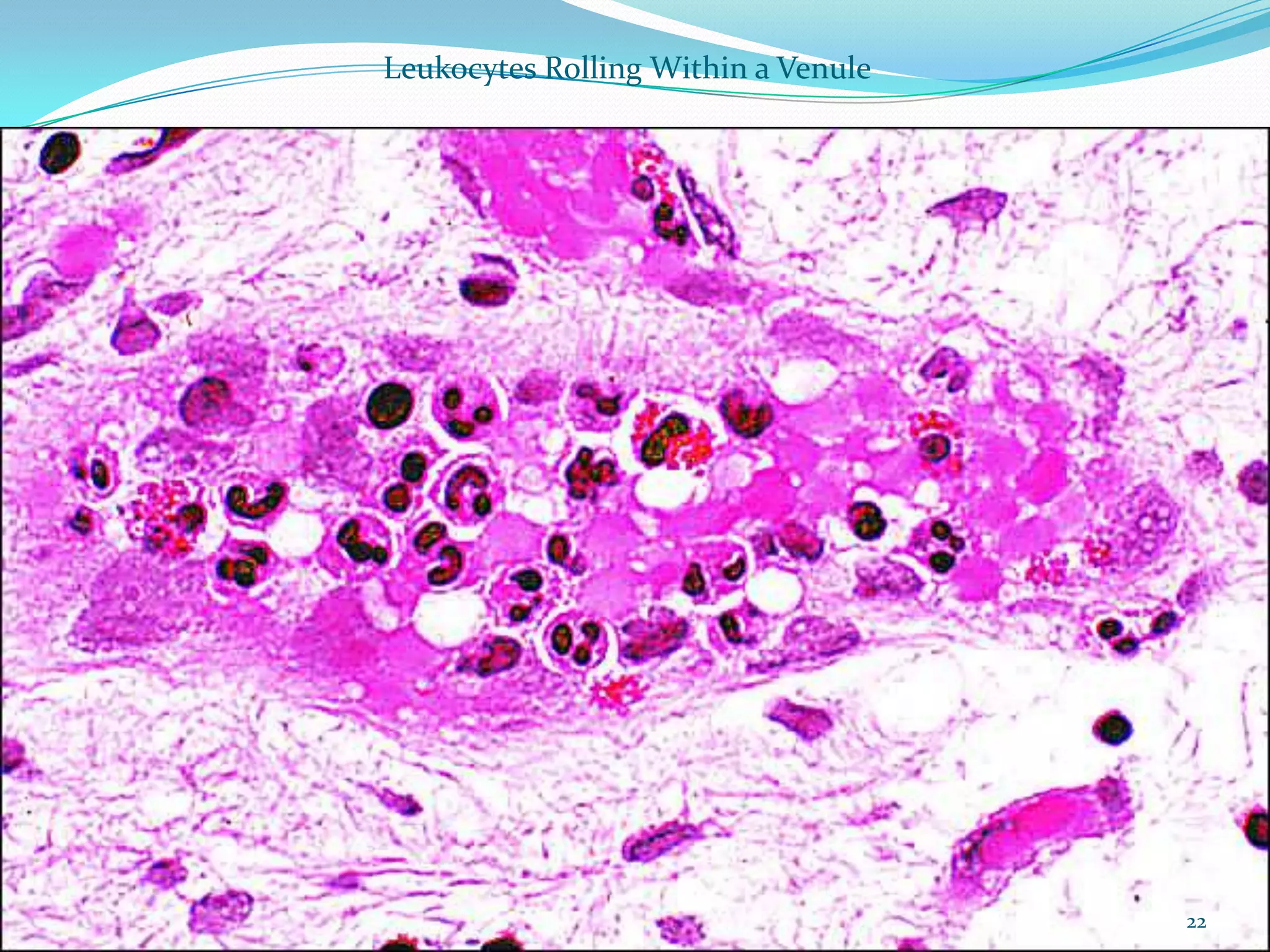
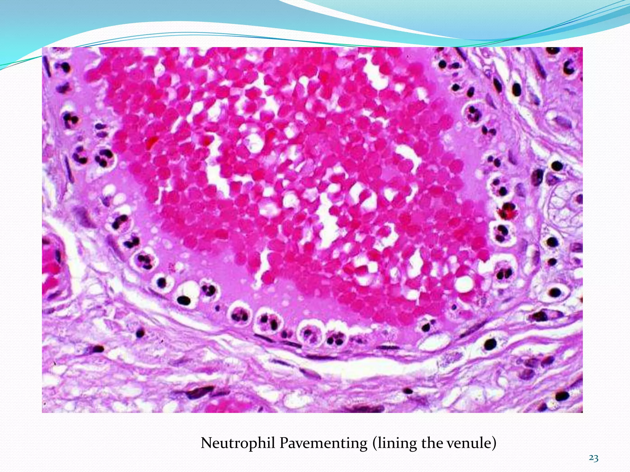
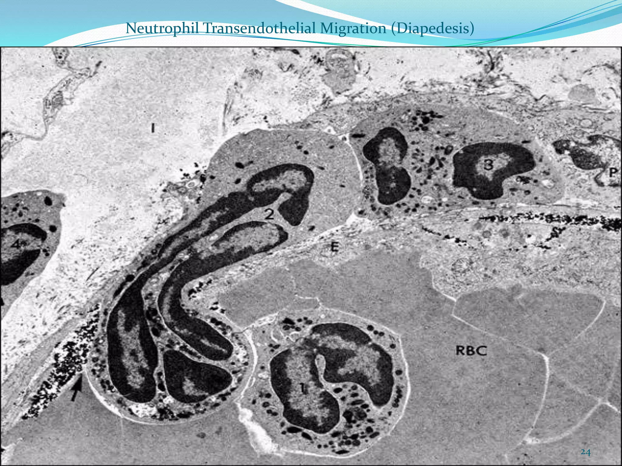
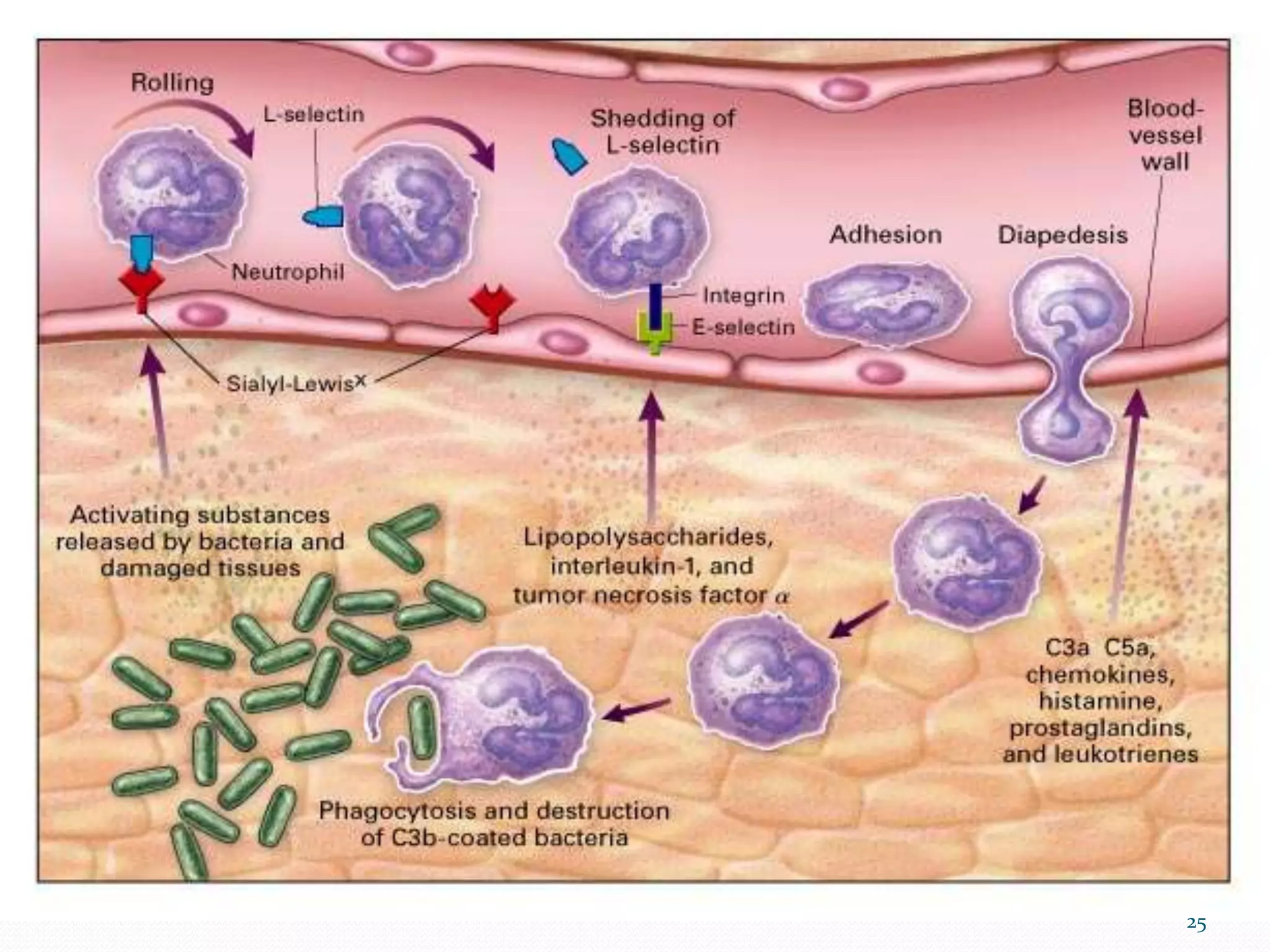
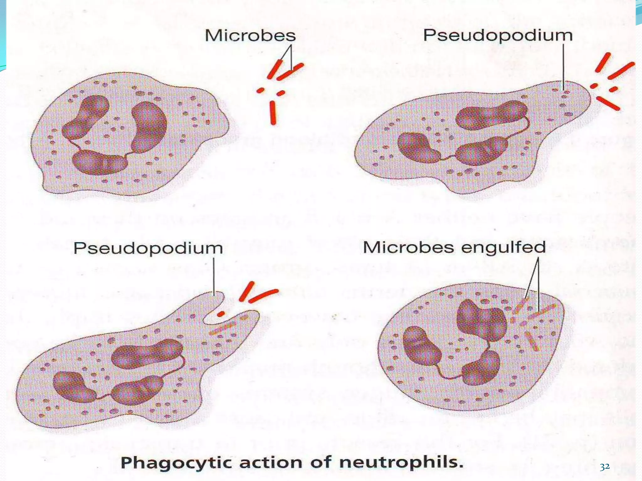
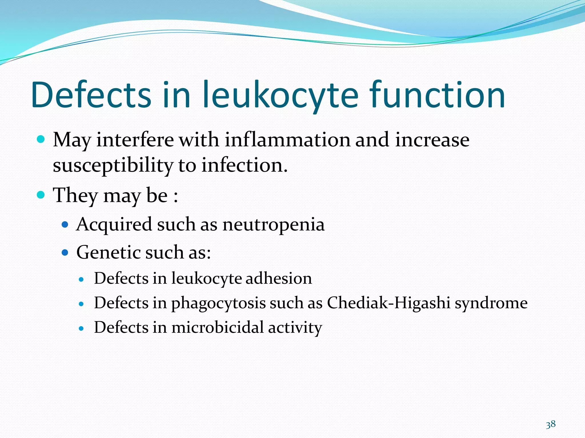
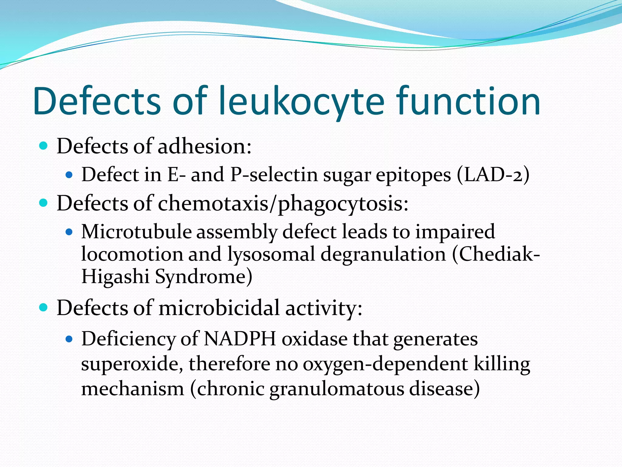
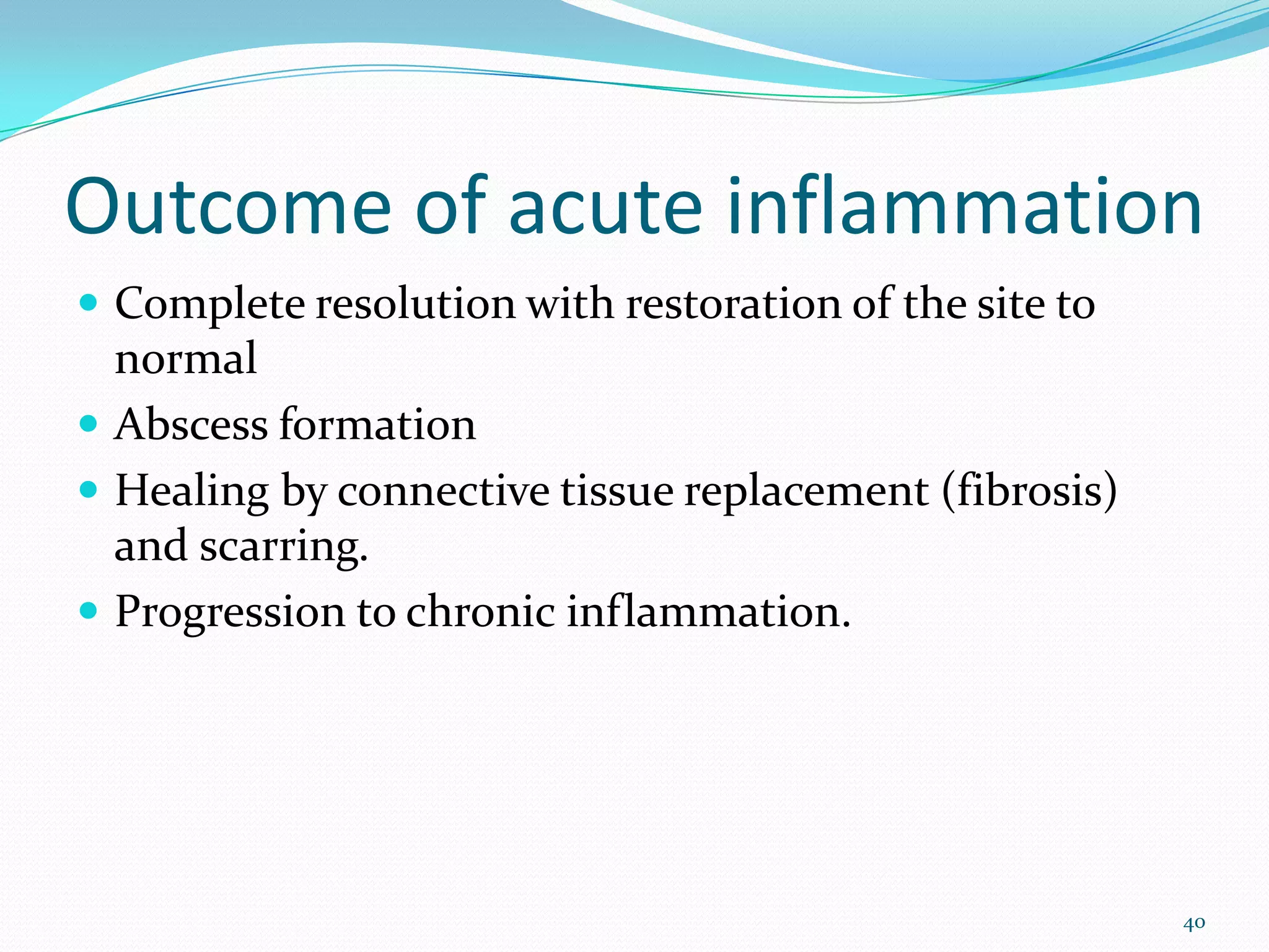
DCM2:1
PATHOLOGY LECTURE NOTES
1
DEFINITIONS
 Exudation: Escape of fluid, proteins and blood cells
from the vascular system into the interstitial tissue or
body cavities.
 Exudate: an inflammatory extra vascular fluid that
has high protein concentration and much cellular
debris.
 Edema: an excess of fluid in the interstitial tissue or
serous cavities.
2
Introduction
 Inflammation is the reaction of vascularized living
tissue to local injury.
 It is evoked by:
 Microbial infections
 Physical agents
 Chemicals
 Necrotic tissue
 Immunologic reactions
3
Introduction
 The roles of inflammation are:
 To contain and isolate injury
 To destroy invading microorganisms
 To inactivate toxins
 To prepare the tissue or organ for healing and repair
4
Acute Inflammation
 Acute inflammation is a rapid response to an injurious
agent that serves to deliver mediators of host
defense—leukocytes and plasma proteins—to the site
of injury.
 When a host encounters an injurious
agent, phagocytes that reside in all tissues try to get rid
of these agents.
5
Acute Inflammation
 At the same time, phagocytes and other host cells react
to the presence of the foreign or abnormal substance
by liberating cytokines, lipid messengers, and the
various other mediators of inflammation.
 Some of these mediators act on endothelial cells in the
vicinity and promote the efflux of plasma and the
recruitment of circulating leukocytes to the site where
the offending agent is located.
6
Acute Inflammation - continued
 As the injurious agent is eliminated and anti-
inflammatory mechanisms become active, the
process subsides and the host returns to a normal
state of health.
 If the injurious agent cannot be quickly
eliminated, the result may be chronic
inflammation.
7
Acute Inflammation
 Acute inflammation has three major components:
 alterations in vascular caliber that lead to an increase in
blood flow;
 structural changes in the microvasculature that permit
plasma proteins and leukocytes to leave the circulation;
and
 emigration of the leukocytes from the
microcirculation, their accumulation in the focus of
injury, and their activation to eliminate the offending
agent
8
Acute Inflammation
 These changes produce the classic clinical signs of
inflammation:
 Heat (Calor)
 Redness (Rubor)
 Edema (tumor)
 Pain (dolor)
 Loss of function
9
10
 Acute inflammatory reactions are triggered by a
variety of stimuli:
Infections (bacterial, viral, parasitic) and microbial
toxins
• Trauma (blunt and penetrating)
• Physical and chemical agents (thermal injury, e.g.,
burns or frostbite; irradiation; some environmental
chemicals)
• Tissue necrosis (from any cause)
• Foreign bodies (splinters, dirt, sutures)
• Immune reactions (also called hypersensitivity
reactions)
11
Changes in vascular flow and
caliber
 Initially, transient vasoconstriction of arterioles occurs
 Vasodilation follows, causing increased flow; it
accounts for the heat and redness
 Eventually slowing of the circulation occurs as a result
of increased vascular permeability, leading to stasis.
(edema)
 With slowing, leukocytic margination appears.
12
13
Vasodilation
 Brief arteriolar vasoconstriction followed by
vasodilation
 Accounts for warmth and redness
 Opens microvascular beds
 Increased intravascular pressure causes an early
transudate (protein-poor filtrate of plasma) into
interstitium (vascular permeability still not increased
yet)
Increased vascular permeability
 Leads to escape of protein rich fluid into the
intestitium.
 There is increased hydrostatic pressure, caused by
vasodilatation, decreased osmotic pressure due to
leakage of a high protein fluid, resulting in marked
outflow of fluid and edema formation.
15
Vascular leakage
 Five mechanisms known to cause vascular
leakiness
 Histamines, bradykinins, leukotrienes cause an
early, brief (15 – 30 min.) immediate transient
response in the form of endothelial cell
contraction that widens intercellular gaps of
venules (not arterioles, capillaries)
Vascular leakage
 Cytokine mediators (TNF, IL-1) induce
endothelial cell junction retraction through
cytoskeleton reorganization (4 – 6 hrs post
injury, lasting 24 hrs or more)
 Severe injuries may cause immediate direct
endothelial cell damage (necrosis, detachment)
making them leaky until they are repaired
(immediate sustained response), or may cause
delayed damage as in thermal or UV injury,
Vascular leakage
 (cont’d) or some bacterial toxins (delayed
prolonged leakage)
 Marginating and endothelial cell-adherent
leukocytes may pile-up and damage the
endothelium through activation and release of
toxic oxygen radicals and proteolytic enzymes
(leukocyte-dependent endothelial cell injury)
making the vessel leaky
Vascular leakage
 Certain mediators (VEGF) may cause increased
transcytosis via intracellular vesicles which
travel from the luminal to basement membrane
surface of the endothelial cell
 All or any combination of these events may occur
in response to a given stimulus
Cellular events: Leukocyte
extravasation and phagocytosis
 Delivers leukocytes to the site of injury.
 Sequence of events can be divided into:
 Margination, rolling and adhesion to the lumen
 Diapedesis
 Migration in the endothelial tissues towards a
chemotactic stimulus.
20
Adhesion and Transmigration
 Occurs as a result of interaction between adhesion
molecules on the leukocytes and endothelium.
 Major ligand-receptor pairs include:
 Selectins (E, P and L)
 Immunoglobulins family: ICAM and VCAM-1
 The integrins
21
Leukocytes Rolling Within a Venule
22
Neutrophil Pavementing (lining the venule)
23
Neutrophil Transendothelial Migration (Diapedesis)
24
25
Chemotaxis and leukocyte
activation
 Adherent leukocytes emigrate through
interendothelial junctions, traverse the basement
membrane and move towards the site of injury along a
gradient of chemotactic agents.
 Neutrophils emigrate first and they are followed by
monocytes and lymphocytes.
26
Chemotaxis and leukocyte
activation
 Chemotactic agents include: bacterial
products, complement fragments
(C5a), leukotrienes, and chemokines (IL-8).
 These agents bind to specific receptors on
leukocyte, resulting in signal transduction
process, which results in assembly of the contractile
elements responsible for cell movement.
27
28
Chemotaxis and leukocyte
activation
 Chemotactic agents also cause leukocyte activation
characterized by:
 Degranulation and secretion of enzymes
 Activation of an oxidative burst
 Production of arachidonic acid metabolites
 Modulation of leukocyte adhesion molecules
29
Phagocytosis
 Involves 3 steps:
 Recognition and attachment of the paricle to be
ingested. Facilitated by opsonization e.g. Fc fragment of
IgG.
 Engulfment by pseudopods, with formation of a
phagocytic vesicle, which fuses with membrane of the
lysosome to form a phagolysosome.
 Killing and degranulation of bacteria.
30
Phagocytosis
 There are 2 types of bactericidal mechanisms:
 Oxygen dependent mechanisms: this involves
production of reactive oxygen species catalysed by
enzymes such as: NADPH oxidase and
myeloperoxidase
 Oxygen independent mechanisms: these cause
increased permeability of the bacterial cell membrane.
31
32
Oxidative burst
 Reactive oxygen species formed through oxidative
burst that includes:
 Increased oxygen consumption
 Glycogenolysis
 Increased glucose oxidation
 Formation of superoxide ion
 2O2 + NADPH  2O2
-rad + NADP+ + H+
(NADPH oxidase)
 O2 + 2H+  H2O2 (dismutase)
Reactive oxygen species
 Hydrogen peroxide alone insufficient
 Myeloperoxidase oxidase (azurophilic granules)
converts hydrogen peroxide to HOCl- (in presence of
Cl- ), an oxidant/antimicrobial agent
 Therefore, PMNs can kill by halogenation, or
lipid/protein peroxidation
Degradation and Clean-up
 Reactive end-products only active within
phagolysosome
 Hydrogen peroxide broken down to water and oxygen
by catalase
 Dead microorganisms degraded by lysosomal acid
hydrolases
Leukocyte granules
 Other antimicrobials in leukocyte granules:
 Bactericidal permeability increasing protein (BPI)
 Lysozyme
 Lactoferrin
 Defensins (punch holes in membranes)
Leukocyte induced tissue injury
 Leukocytes also release some of their products into the
extracellular space. These include:
 Lysosomal enzymes
 Oxygen derived active metabolites
 Products of arachidonic acid metabolism such as
prostaglandins and leukotrienes.
 these may cause tissue damage and if persistent may
result in chronic inflammation.
37
Defects in leukocyte function
 May interfere with inflammation and increase
susceptibility to infection.
 They may be :
 Acquired such as neutropenia
 Genetic such as:
 Defects in leukocyte adhesion
 Defects in phagocytosis such as Chediak-Higashi syndrome
 Defects in microbicidal activity
38
Defects of leukocyte function
 Defects of adhesion:
 Defect in E- and P-selectin sugar epitopes (LAD-2)
 Defects of chemotaxis/phagocytosis:
 Microtubule assembly defect leads to impaired
locomotion and lysosomal degranulation (Chediak-
Higashi Syndrome)
 Defects of microbicidal activity:
 Deficiency of NADPH oxidase that generates
superoxide, therefore no oxygen-dependent killing
mechanism (chronic granulomatous disease)
Outcome of acute inflammation
 Complete resolution with restoration of the site to
normal
 Abscess formation
 Healing by connective tissue replacement (fibrosis)
and scarring.
 Progression to chronic inflammation.
40

lecture6inflammation-pptx-120913130832-phpapp01.pdf

  • 1.
  • 2.
    DEFINITIONS  Exudation: Escapeof fluid, proteins and blood cells from the vascular system into the interstitial tissue or body cavities.  Exudate: an inflammatory extra vascular fluid that has high protein concentration and much cellular debris.  Edema: an excess of fluid in the interstitial tissue or serous cavities. 2
  • 3.
    Introduction  Inflammation isthe reaction of vascularized living tissue to local injury.  It is evoked by:  Microbial infections  Physical agents  Chemicals  Necrotic tissue  Immunologic reactions 3
  • 4.
    Introduction  The rolesof inflammation are:  To contain and isolate injury  To destroy invading microorganisms  To inactivate toxins  To prepare the tissue or organ for healing and repair 4
  • 5.
    Acute Inflammation  Acuteinflammation is a rapid response to an injurious agent that serves to deliver mediators of host defense—leukocytes and plasma proteins—to the site of injury.  When a host encounters an injurious agent, phagocytes that reside in all tissues try to get rid of these agents. 5
  • 6.
    Acute Inflammation  Atthe same time, phagocytes and other host cells react to the presence of the foreign or abnormal substance by liberating cytokines, lipid messengers, and the various other mediators of inflammation.  Some of these mediators act on endothelial cells in the vicinity and promote the efflux of plasma and the recruitment of circulating leukocytes to the site where the offending agent is located. 6
  • 7.
    Acute Inflammation -continued  As the injurious agent is eliminated and anti- inflammatory mechanisms become active, the process subsides and the host returns to a normal state of health.  If the injurious agent cannot be quickly eliminated, the result may be chronic inflammation. 7
  • 8.
    Acute Inflammation  Acuteinflammation has three major components:  alterations in vascular caliber that lead to an increase in blood flow;  structural changes in the microvasculature that permit plasma proteins and leukocytes to leave the circulation; and  emigration of the leukocytes from the microcirculation, their accumulation in the focus of injury, and their activation to eliminate the offending agent 8
  • 9.
    Acute Inflammation  Thesechanges produce the classic clinical signs of inflammation:  Heat (Calor)  Redness (Rubor)  Edema (tumor)  Pain (dolor)  Loss of function 9
  • 10.
  • 11.
     Acute inflammatoryreactions are triggered by a variety of stimuli: Infections (bacterial, viral, parasitic) and microbial toxins • Trauma (blunt and penetrating) • Physical and chemical agents (thermal injury, e.g., burns or frostbite; irradiation; some environmental chemicals) • Tissue necrosis (from any cause) • Foreign bodies (splinters, dirt, sutures) • Immune reactions (also called hypersensitivity reactions) 11
  • 12.
    Changes in vascularflow and caliber  Initially, transient vasoconstriction of arterioles occurs  Vasodilation follows, causing increased flow; it accounts for the heat and redness  Eventually slowing of the circulation occurs as a result of increased vascular permeability, leading to stasis. (edema)  With slowing, leukocytic margination appears. 12
  • 13.
  • 14.
    Vasodilation  Brief arteriolarvasoconstriction followed by vasodilation  Accounts for warmth and redness  Opens microvascular beds  Increased intravascular pressure causes an early transudate (protein-poor filtrate of plasma) into interstitium (vascular permeability still not increased yet)
  • 15.
    Increased vascular permeability Leads to escape of protein rich fluid into the intestitium.  There is increased hydrostatic pressure, caused by vasodilatation, decreased osmotic pressure due to leakage of a high protein fluid, resulting in marked outflow of fluid and edema formation. 15
  • 16.
    Vascular leakage  Fivemechanisms known to cause vascular leakiness  Histamines, bradykinins, leukotrienes cause an early, brief (15 – 30 min.) immediate transient response in the form of endothelial cell contraction that widens intercellular gaps of venules (not arterioles, capillaries)
  • 17.
    Vascular leakage  Cytokinemediators (TNF, IL-1) induce endothelial cell junction retraction through cytoskeleton reorganization (4 – 6 hrs post injury, lasting 24 hrs or more)  Severe injuries may cause immediate direct endothelial cell damage (necrosis, detachment) making them leaky until they are repaired (immediate sustained response), or may cause delayed damage as in thermal or UV injury,
  • 18.
    Vascular leakage  (cont’d)or some bacterial toxins (delayed prolonged leakage)  Marginating and endothelial cell-adherent leukocytes may pile-up and damage the endothelium through activation and release of toxic oxygen radicals and proteolytic enzymes (leukocyte-dependent endothelial cell injury) making the vessel leaky
  • 19.
    Vascular leakage  Certainmediators (VEGF) may cause increased transcytosis via intracellular vesicles which travel from the luminal to basement membrane surface of the endothelial cell  All or any combination of these events may occur in response to a given stimulus
  • 20.
    Cellular events: Leukocyte extravasationand phagocytosis  Delivers leukocytes to the site of injury.  Sequence of events can be divided into:  Margination, rolling and adhesion to the lumen  Diapedesis  Migration in the endothelial tissues towards a chemotactic stimulus. 20
  • 21.
    Adhesion and Transmigration Occurs as a result of interaction between adhesion molecules on the leukocytes and endothelium.  Major ligand-receptor pairs include:  Selectins (E, P and L)  Immunoglobulins family: ICAM and VCAM-1  The integrins 21
  • 22.
  • 23.
  • 24.
  • 25.
  • 26.
    Chemotaxis and leukocyte activation Adherent leukocytes emigrate through interendothelial junctions, traverse the basement membrane and move towards the site of injury along a gradient of chemotactic agents.  Neutrophils emigrate first and they are followed by monocytes and lymphocytes. 26
  • 27.
    Chemotaxis and leukocyte activation Chemotactic agents include: bacterial products, complement fragments (C5a), leukotrienes, and chemokines (IL-8).  These agents bind to specific receptors on leukocyte, resulting in signal transduction process, which results in assembly of the contractile elements responsible for cell movement. 27
  • 28.
  • 29.
    Chemotaxis and leukocyte activation Chemotactic agents also cause leukocyte activation characterized by:  Degranulation and secretion of enzymes  Activation of an oxidative burst  Production of arachidonic acid metabolites  Modulation of leukocyte adhesion molecules 29
  • 30.
    Phagocytosis  Involves 3steps:  Recognition and attachment of the paricle to be ingested. Facilitated by opsonization e.g. Fc fragment of IgG.  Engulfment by pseudopods, with formation of a phagocytic vesicle, which fuses with membrane of the lysosome to form a phagolysosome.  Killing and degranulation of bacteria. 30
  • 31.
    Phagocytosis  There are2 types of bactericidal mechanisms:  Oxygen dependent mechanisms: this involves production of reactive oxygen species catalysed by enzymes such as: NADPH oxidase and myeloperoxidase  Oxygen independent mechanisms: these cause increased permeability of the bacterial cell membrane. 31
  • 32.
  • 33.
    Oxidative burst  Reactiveoxygen species formed through oxidative burst that includes:  Increased oxygen consumption  Glycogenolysis  Increased glucose oxidation  Formation of superoxide ion  2O2 + NADPH  2O2 -rad + NADP+ + H+ (NADPH oxidase)  O2 + 2H+  H2O2 (dismutase)
  • 34.
    Reactive oxygen species Hydrogen peroxide alone insufficient  Myeloperoxidase oxidase (azurophilic granules) converts hydrogen peroxide to HOCl- (in presence of Cl- ), an oxidant/antimicrobial agent  Therefore, PMNs can kill by halogenation, or lipid/protein peroxidation
  • 35.
    Degradation and Clean-up Reactive end-products only active within phagolysosome  Hydrogen peroxide broken down to water and oxygen by catalase  Dead microorganisms degraded by lysosomal acid hydrolases
  • 36.
    Leukocyte granules  Otherantimicrobials in leukocyte granules:  Bactericidal permeability increasing protein (BPI)  Lysozyme  Lactoferrin  Defensins (punch holes in membranes)
  • 37.
    Leukocyte induced tissueinjury  Leukocytes also release some of their products into the extracellular space. These include:  Lysosomal enzymes  Oxygen derived active metabolites  Products of arachidonic acid metabolism such as prostaglandins and leukotrienes.  these may cause tissue damage and if persistent may result in chronic inflammation. 37
  • 38.
    Defects in leukocytefunction  May interfere with inflammation and increase susceptibility to infection.  They may be :  Acquired such as neutropenia  Genetic such as:  Defects in leukocyte adhesion  Defects in phagocytosis such as Chediak-Higashi syndrome  Defects in microbicidal activity 38
  • 39.
    Defects of leukocytefunction  Defects of adhesion:  Defect in E- and P-selectin sugar epitopes (LAD-2)  Defects of chemotaxis/phagocytosis:  Microtubule assembly defect leads to impaired locomotion and lysosomal degranulation (Chediak- Higashi Syndrome)  Defects of microbicidal activity:  Deficiency of NADPH oxidase that generates superoxide, therefore no oxygen-dependent killing mechanism (chronic granulomatous disease)
  • 40.
    Outcome of acuteinflammation  Complete resolution with restoration of the site to normal  Abscess formation  Healing by connective tissue replacement (fibrosis) and scarring.  Progression to chronic inflammation. 40