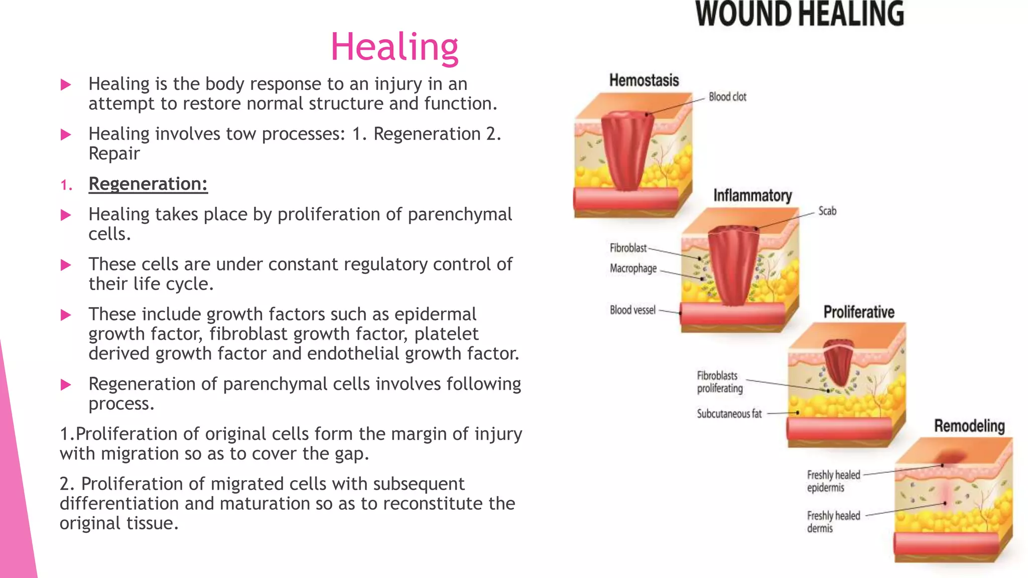
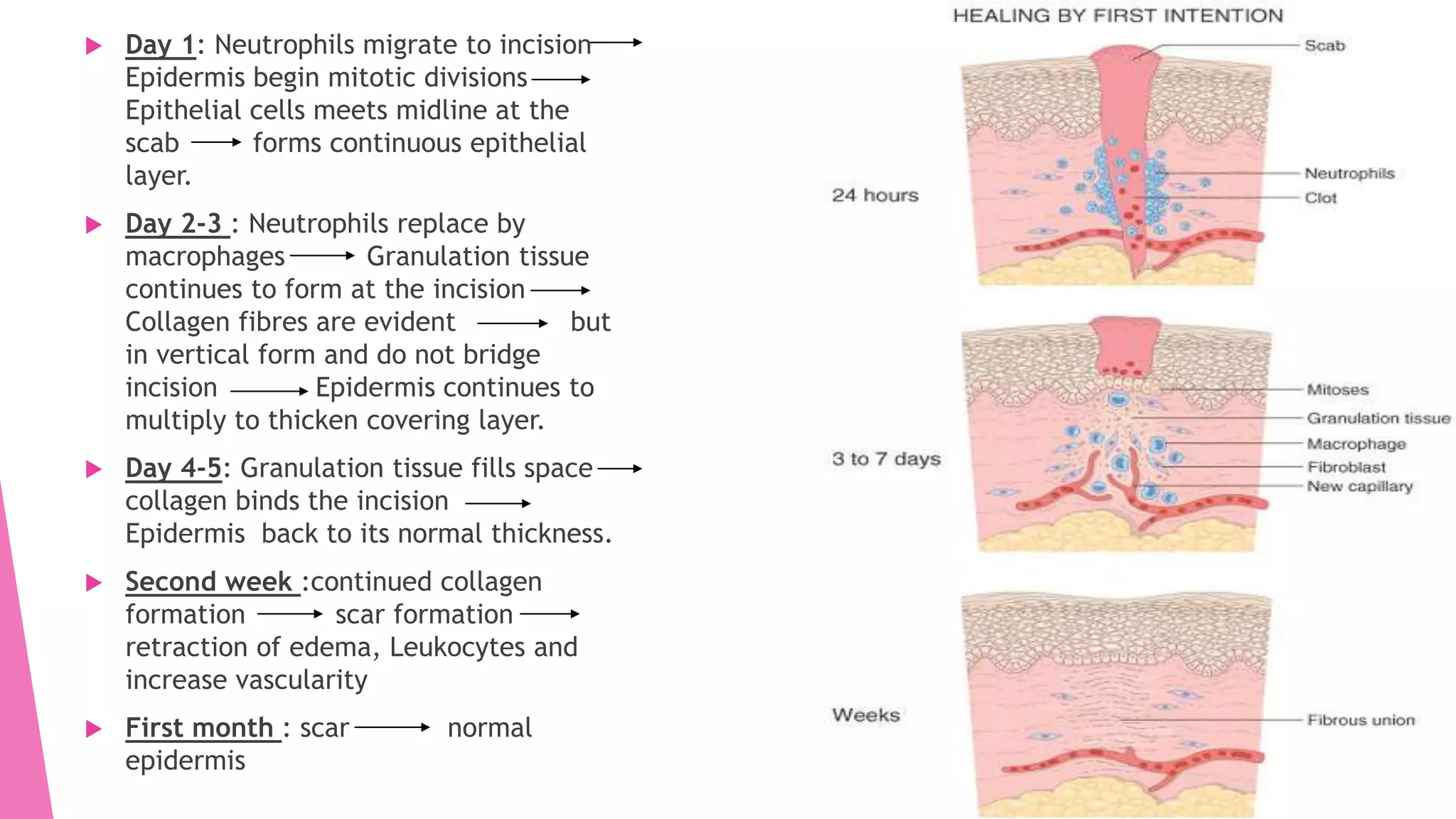
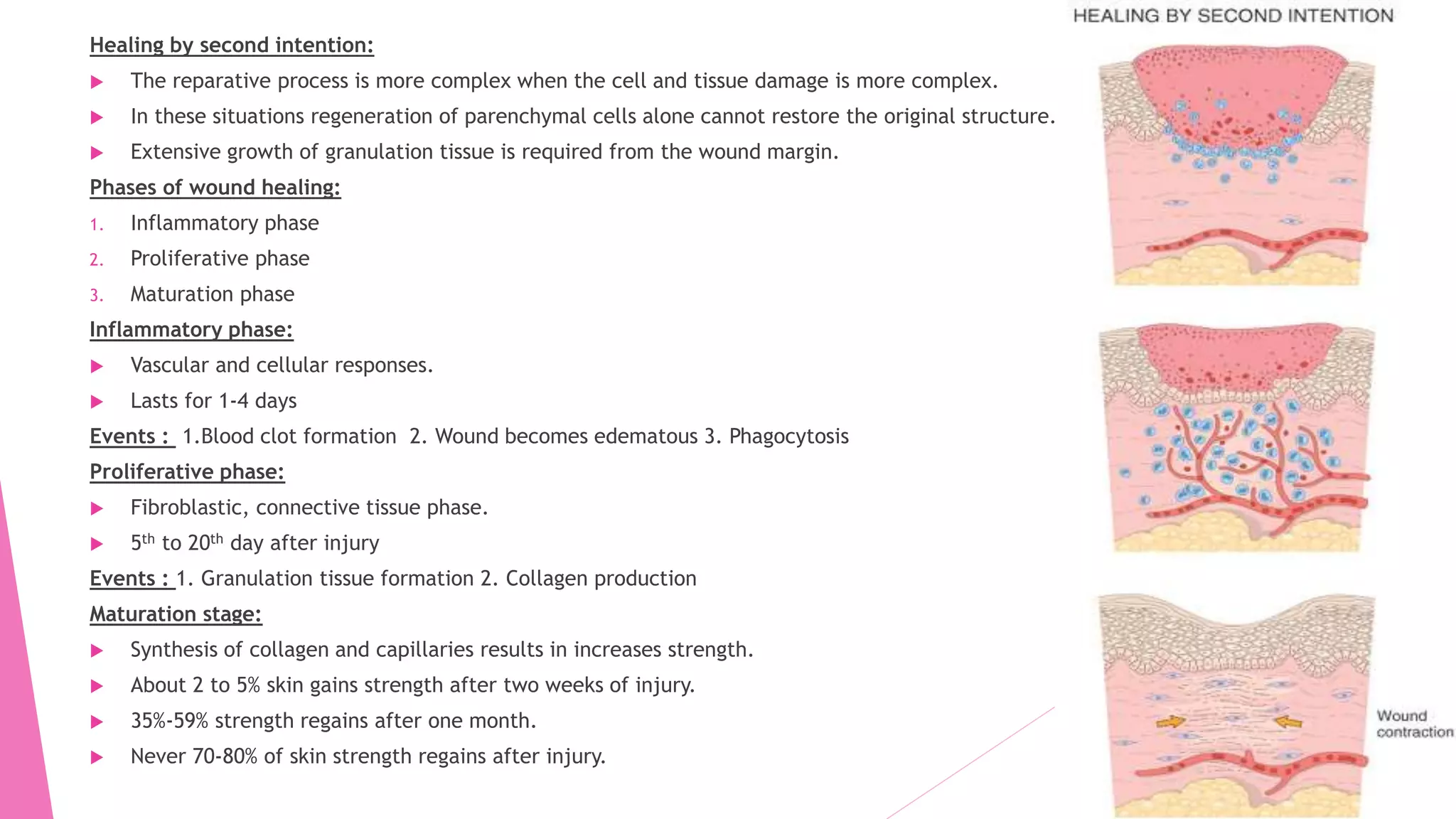
Healing and wound
contraction in
inflammation
Archana.K
Healing
 Healing is the body response to an injury in an
attempt to restore normal structure and function.
 Healing involves tow processes: 1. Regeneration 2.
Repair
1. Regeneration:
 Healing takes place by proliferation of parenchymal
cells.
 These cells are under constant regulatory control of
their life cycle.
 These include growth factors such as epidermal
growth factor, fibroblast growth factor, platelet
derived growth factor and endothelial growth factor.
 Regeneration of parenchymal cells involves following
process.
1.Proliferation of original cells form the margin of injury
with migration so as to cover the gap.
2. Proliferation of migrated cells with subsequent
differentiation and maturation so as to reconstitute the
original tissue.
 Depending upon their capacity to divide, cells of the body can be divided into three groups.
1. Labile cells:
 They multiply through out the life under normal physiologic conditions.
 Epithelial cells of epidermis, alimentary tract, respiratory tract, urinary tract, vagina, cervix,
uterine endometrium, haemopoietic cells from bone marrow.
2. Stable cells:
 These cells decrease or lose their ability to proliferate after adolescence but retain the
capacity to multiply in response to stimuli throughout adult life.
 parenchymal cells of organs like liver, pancreas, kidneys, adrenal and thyroid.
3. Permanent cells:
 These cells lose their ability to proliferate around the time of birth.
 Neuron, skeletal muscle and cardiac cells.
Repair:
 Repair is the replacement of injured tissue by fibrous tissue.
 These responses take place by participation of mesenchymal cells (consisting of connective tissue, stem
cells fibrocytes and histocytes).
 Process involves: 1. Granulation tissue formation
2. Contraction of wounds.
1. Granulation tissue formation:
 The tissue appears slightly granular and pink in colour.
 Each granule corresponds histologically proliferation of news small blood vessels by the covering of
fibroblasts and collagen.
 Three phases are observed in formation
1. Phase of inflammation:
 Trauma Blood clots Inflammatory response Exudation of plasma Neutrophils and
monocytes.
2. Phase of clearance: Enzymes released from the neutrophils and dead tissue cells and phagocytic
activity clear off the necrotic tissue, debris and red blood vessels.
3. Phase of growth of granulation tissue:
 Two processes.
1.Angiogenesis
2. Fibrogenesis
Angiogenesis:
 Formation of new blood vessels at the site of injury takes place by
proliferation of endothelial cells.
 Proliferated cells initially are solid buds with in hours turns in to lumen and
starts carrying blood.
 New blood vessels are leakier which leads to edematous appearance.
 Soon differentiate in to arterioles, capillaries, venules.
Fibrogenesis:
 The new blood vessels are amorphous ground of surface.
 Fibroblasts originate from mitotic division of fibrocytes
 They have characteristics of smooth muscle cells.
 Collagen appears by the 6th day which is formed by fibroblasts. This results in
formation of scar.
Contraction of wound:
 The wound starts contracting after 2-3 days and completes by
14th day.
 80% of wound is reduced by this time.
 Contracted wound results in rapid healing.
 Factors proposed to explain wound contraction:
1. Dehydration of wound
2. Contraction of collagen
3. Myofibroblasts and decrease in their size during healing.
Wound Healing:
 Wound healing can be accomplished in one of the following two
ways:
1. Healing by first intention
2. Healing by second intention
Healing by first intention:
 Simplest examples of wound repair is the healing of a clean,
uninfected surgical incision approximated by surgical sutures.
 Day 1: Neutrophils migrate to incision
Epidermis begin mitotic divisions
Epithelial cells meets midline at the
scab forms continuous epithelial
layer.
 Day 2-3 : Neutrophils replace by
macrophages Granulation tissue
continues to form at the incision
Collagen fibres are evident but
in vertical form and do not bridge
incision Epidermis continues to
multiply to thicken covering layer.
 Day 4-5: Granulation tissue fills space
collagen binds the incision
Epidermis back to its normal thickness.
 Second week :continued collagen
formation scar formation
retraction of edema, Leukocytes and
increase vascularity
 First month : scar normal
epidermis
Healing by second intention:
 The reparative process is more complex when the cell and tissue damage is more complex.
 In these situations regeneration of parenchymal cells alone cannot restore the original structure.
 Extensive growth of granulation tissue is required from the wound margin.
Phases of wound healing:
1. Inflammatory phase
2. Proliferative phase
3. Maturation phase
Inflammatory phase:
 Vascular and cellular responses.
 Lasts for 1-4 days
Events : 1.Blood clot formation 2. Wound becomes edematous 3. Phagocytosis
Proliferative phase:
 Fibroblastic, connective tissue phase.
 5th to 20th day after injury
Events : 1. Granulation tissue formation 2. Collagen production
Maturation stage:
 Synthesis of collagen and capillaries results in increases strength.
 About 2 to 5% skin gains strength after two weeks of injury.
 35%-59% strength regains after one month.
 Never 70-80% of skin strength regains after injury.
Thank you

Healing and wound contraction in inflammation

  • 1.
    Healing and wound contractionin inflammation Archana.K
  • 2.
    Healing  Healing isthe body response to an injury in an attempt to restore normal structure and function.  Healing involves tow processes: 1. Regeneration 2. Repair 1. Regeneration:  Healing takes place by proliferation of parenchymal cells.  These cells are under constant regulatory control of their life cycle.  These include growth factors such as epidermal growth factor, fibroblast growth factor, platelet derived growth factor and endothelial growth factor.  Regeneration of parenchymal cells involves following process. 1.Proliferation of original cells form the margin of injury with migration so as to cover the gap. 2. Proliferation of migrated cells with subsequent differentiation and maturation so as to reconstitute the original tissue.
  • 3.
     Depending upontheir capacity to divide, cells of the body can be divided into three groups. 1. Labile cells:  They multiply through out the life under normal physiologic conditions.  Epithelial cells of epidermis, alimentary tract, respiratory tract, urinary tract, vagina, cervix, uterine endometrium, haemopoietic cells from bone marrow. 2. Stable cells:  These cells decrease or lose their ability to proliferate after adolescence but retain the capacity to multiply in response to stimuli throughout adult life.  parenchymal cells of organs like liver, pancreas, kidneys, adrenal and thyroid. 3. Permanent cells:  These cells lose their ability to proliferate around the time of birth.  Neuron, skeletal muscle and cardiac cells.
  • 4.
    Repair:  Repair isthe replacement of injured tissue by fibrous tissue.  These responses take place by participation of mesenchymal cells (consisting of connective tissue, stem cells fibrocytes and histocytes).  Process involves: 1. Granulation tissue formation 2. Contraction of wounds. 1. Granulation tissue formation:  The tissue appears slightly granular and pink in colour.  Each granule corresponds histologically proliferation of news small blood vessels by the covering of fibroblasts and collagen.  Three phases are observed in formation 1. Phase of inflammation:  Trauma Blood clots Inflammatory response Exudation of plasma Neutrophils and monocytes. 2. Phase of clearance: Enzymes released from the neutrophils and dead tissue cells and phagocytic activity clear off the necrotic tissue, debris and red blood vessels.
  • 5.
    3. Phase ofgrowth of granulation tissue:  Two processes. 1.Angiogenesis 2. Fibrogenesis Angiogenesis:  Formation of new blood vessels at the site of injury takes place by proliferation of endothelial cells.  Proliferated cells initially are solid buds with in hours turns in to lumen and starts carrying blood.  New blood vessels are leakier which leads to edematous appearance.  Soon differentiate in to arterioles, capillaries, venules. Fibrogenesis:  The new blood vessels are amorphous ground of surface.  Fibroblasts originate from mitotic division of fibrocytes  They have characteristics of smooth muscle cells.  Collagen appears by the 6th day which is formed by fibroblasts. This results in formation of scar.
  • 6.
    Contraction of wound: The wound starts contracting after 2-3 days and completes by 14th day.  80% of wound is reduced by this time.  Contracted wound results in rapid healing.  Factors proposed to explain wound contraction: 1. Dehydration of wound 2. Contraction of collagen 3. Myofibroblasts and decrease in their size during healing. Wound Healing:  Wound healing can be accomplished in one of the following two ways: 1. Healing by first intention 2. Healing by second intention Healing by first intention:  Simplest examples of wound repair is the healing of a clean, uninfected surgical incision approximated by surgical sutures.
  • 7.
     Day 1:Neutrophils migrate to incision Epidermis begin mitotic divisions Epithelial cells meets midline at the scab forms continuous epithelial layer.  Day 2-3 : Neutrophils replace by macrophages Granulation tissue continues to form at the incision Collagen fibres are evident but in vertical form and do not bridge incision Epidermis continues to multiply to thicken covering layer.  Day 4-5: Granulation tissue fills space collagen binds the incision Epidermis back to its normal thickness.  Second week :continued collagen formation scar formation retraction of edema, Leukocytes and increase vascularity  First month : scar normal epidermis
  • 8.
    Healing by secondintention:  The reparative process is more complex when the cell and tissue damage is more complex.  In these situations regeneration of parenchymal cells alone cannot restore the original structure.  Extensive growth of granulation tissue is required from the wound margin. Phases of wound healing: 1. Inflammatory phase 2. Proliferative phase 3. Maturation phase Inflammatory phase:  Vascular and cellular responses.  Lasts for 1-4 days Events : 1.Blood clot formation 2. Wound becomes edematous 3. Phagocytosis Proliferative phase:  Fibroblastic, connective tissue phase.  5th to 20th day after injury Events : 1. Granulation tissue formation 2. Collagen production Maturation stage:  Synthesis of collagen and capillaries results in increases strength.  About 2 to 5% skin gains strength after two weeks of injury.  35%-59% strength regains after one month.  Never 70-80% of skin strength regains after injury.
  • 9.