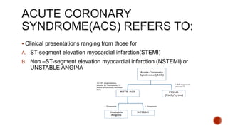
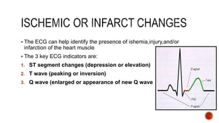
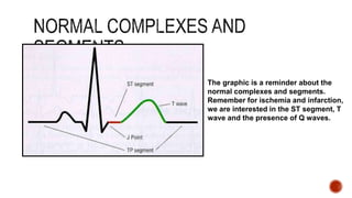
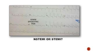
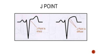
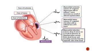
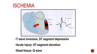
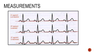
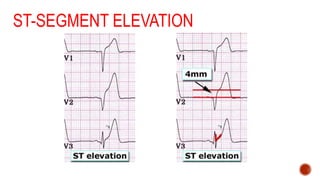
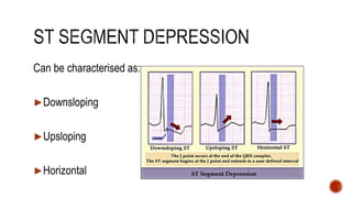
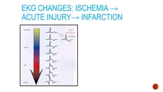
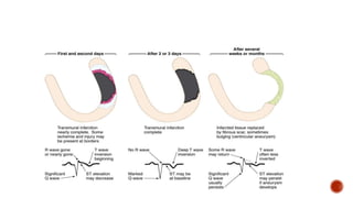
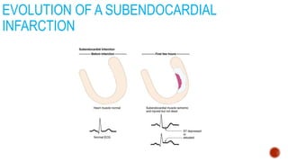
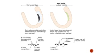
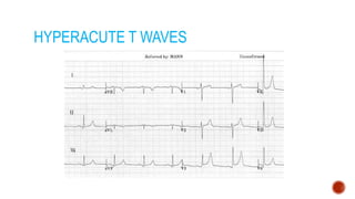
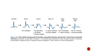
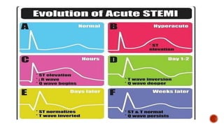
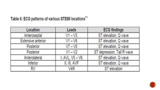
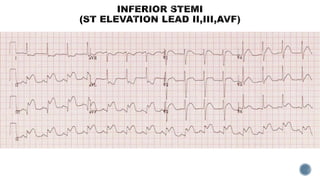
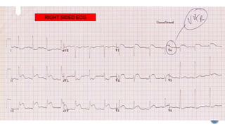
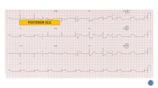
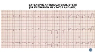
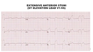
For PHT paramedics
ACS ECG
 Clinical presentations ranging from those for
A. ST-segment elevation myocardial infarction(STEMI)
B. Non –ST-segment elevation myocardial infarction (NSTEMI) or
UNSTABLE ANGINA
 The ECG can help identify the presence of ishemia,injury,and/or
infarction of the heart muscle
 The 3 key ECG indicators are:
1. ST segment changes (depression or elevation)
2. T wave (peaking or inversion)
3. Q wave (enlarged or appearance of new Q wave
The graphic is a reminder about the
normal complexes and segments.
Remember for ischemia and infarction,
we are interested in the ST segment, T
wave and the presence of Q waves.
50/M/M
EPIGESTRIC
PAIN
ISCHEMIA
•T wave inversion, ST segment depression
•Acute injury: ST segment elevation
•Dead tissue: Q wave
ST-SEGMENT ELEVATION
Can be characterised as:-
►Downsloping
►Upsloping
►Horizontal
EKG CHANGES: ISCHEMIA →
ACUTE INJURY→ INFARCTION
EVOLUTION OF A SUBENDOCARDIAL
INFARCTION
HYPERACUTE T WAVES
Q WAVES
Non Pathological Q waves
Q waves of less than 2mm are normal
Pathological Q waves
Q waves of more than 2mm
indicate full thickness myocardial
damage from an infarct
Late sign of MI (evolved)
 The presence of ST elevation in two contiguous leads in patients
with symptoms of ischaemia is the cardinal feature of STEMI.
 The cut-off points for new or presumed new ST segment elevation (in
the absence of LVH and LBBB) is the presence of ≥ 0.1 mV ST
segment elevation in all leads except leads V2-V3.
 In leads V2-V3, a cut-off point of ≥ 0.25 mV (in males < 40 years), ≥
0.2 mV (in males ≥ 40 years) and ≥0.15 mV in females is used
 The presence of a new onset or presumed new LBBB in a patient
with typical chest pain of ischaemia may indicate an infarct and
should be treated as STEMI
 In the early stages of MI, the initial ECG may be normal, equivocal or
show hyperacute T-wave changes only. In these patients if the index of
suspicion of STEMI is high, the ECG should be repeated at close
intervals of at least 15 minutes to look for progressive ST changes.
 Inferior STEMI should have an ECG recording of the right praecordial
lead (V4R) to identify concomitant right ventricular
(RV)involvement.
 In those with ST segment depression in leads V1-V3, it is advisable
to have an ECG recording of the posterior chest wall (V7-V9) to
identify a true infero-basal (formerly known as infero-posterior)STEMI.
The cut-off point for ST segment elevation in the posterior leads is ≥
0.05 mV (≥ 0.1 mV in men < 40 years).
 ST elevation in lead AVR may be a predictor of left main/3 vessel CAD
and carries an adverse prognosis
RIGHT SIDED ECG
POSTERIOR ECG
Acs ecg
Acs ecg
Acs ecg

Acs ecg

  • 1.
  • 2.
     Clinical presentationsranging from those for A. ST-segment elevation myocardial infarction(STEMI) B. Non –ST-segment elevation myocardial infarction (NSTEMI) or UNSTABLE ANGINA
  • 3.
     The ECGcan help identify the presence of ishemia,injury,and/or infarction of the heart muscle  The 3 key ECG indicators are: 1. ST segment changes (depression or elevation) 2. T wave (peaking or inversion) 3. Q wave (enlarged or appearance of new Q wave
  • 4.
    The graphic isa reminder about the normal complexes and segments. Remember for ischemia and infarction, we are interested in the ST segment, T wave and the presence of Q waves.
  • 5.
  • 8.
    ISCHEMIA •T wave inversion,ST segment depression •Acute injury: ST segment elevation •Dead tissue: Q wave
  • 10.
  • 11.
    Can be characterisedas:- ►Downsloping ►Upsloping ►Horizontal
  • 12.
    EKG CHANGES: ISCHEMIA→ ACUTE INJURY→ INFARCTION
  • 14.
    EVOLUTION OF ASUBENDOCARDIAL INFARCTION
  • 16.
  • 17.
    Q WAVES Non PathologicalQ waves Q waves of less than 2mm are normal Pathological Q waves Q waves of more than 2mm indicate full thickness myocardial damage from an infarct Late sign of MI (evolved)
  • 19.
     The presenceof ST elevation in two contiguous leads in patients with symptoms of ischaemia is the cardinal feature of STEMI.  The cut-off points for new or presumed new ST segment elevation (in the absence of LVH and LBBB) is the presence of ≥ 0.1 mV ST segment elevation in all leads except leads V2-V3.  In leads V2-V3, a cut-off point of ≥ 0.25 mV (in males < 40 years), ≥ 0.2 mV (in males ≥ 40 years) and ≥0.15 mV in females is used  The presence of a new onset or presumed new LBBB in a patient with typical chest pain of ischaemia may indicate an infarct and should be treated as STEMI
  • 20.
     In theearly stages of MI, the initial ECG may be normal, equivocal or show hyperacute T-wave changes only. In these patients if the index of suspicion of STEMI is high, the ECG should be repeated at close intervals of at least 15 minutes to look for progressive ST changes.  Inferior STEMI should have an ECG recording of the right praecordial lead (V4R) to identify concomitant right ventricular (RV)involvement.  In those with ST segment depression in leads V1-V3, it is advisable to have an ECG recording of the posterior chest wall (V7-V9) to identify a true infero-basal (formerly known as infero-posterior)STEMI. The cut-off point for ST segment elevation in the posterior leads is ≥ 0.05 mV (≥ 0.1 mV in men < 40 years).  ST elevation in lead AVR may be a predictor of left main/3 vessel CAD and carries an adverse prognosis
  • 26.
  • 27.