Download to read offline

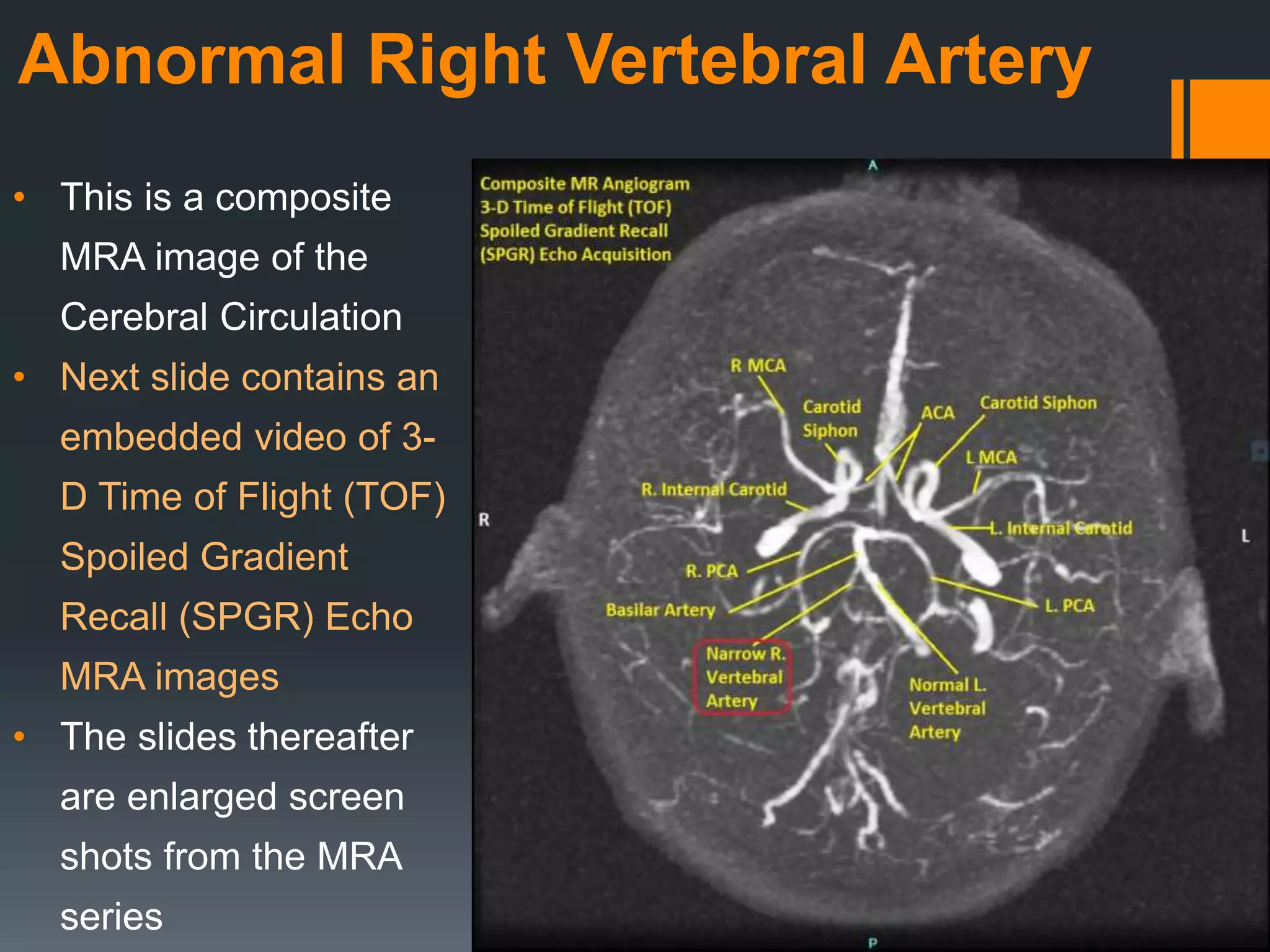


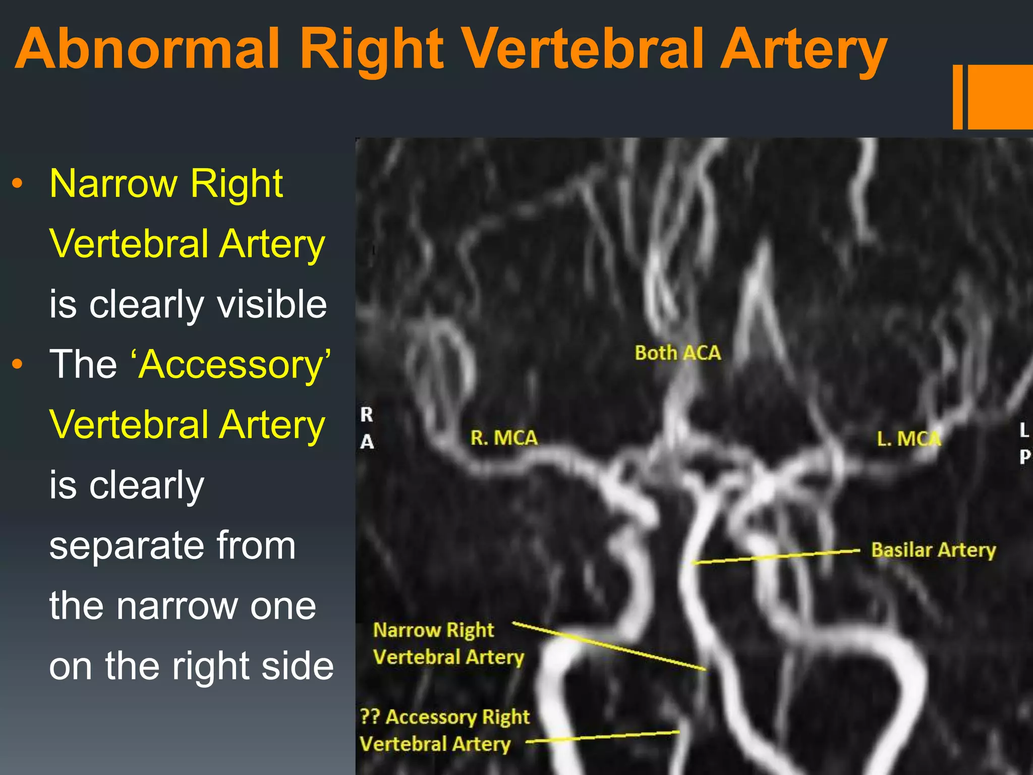


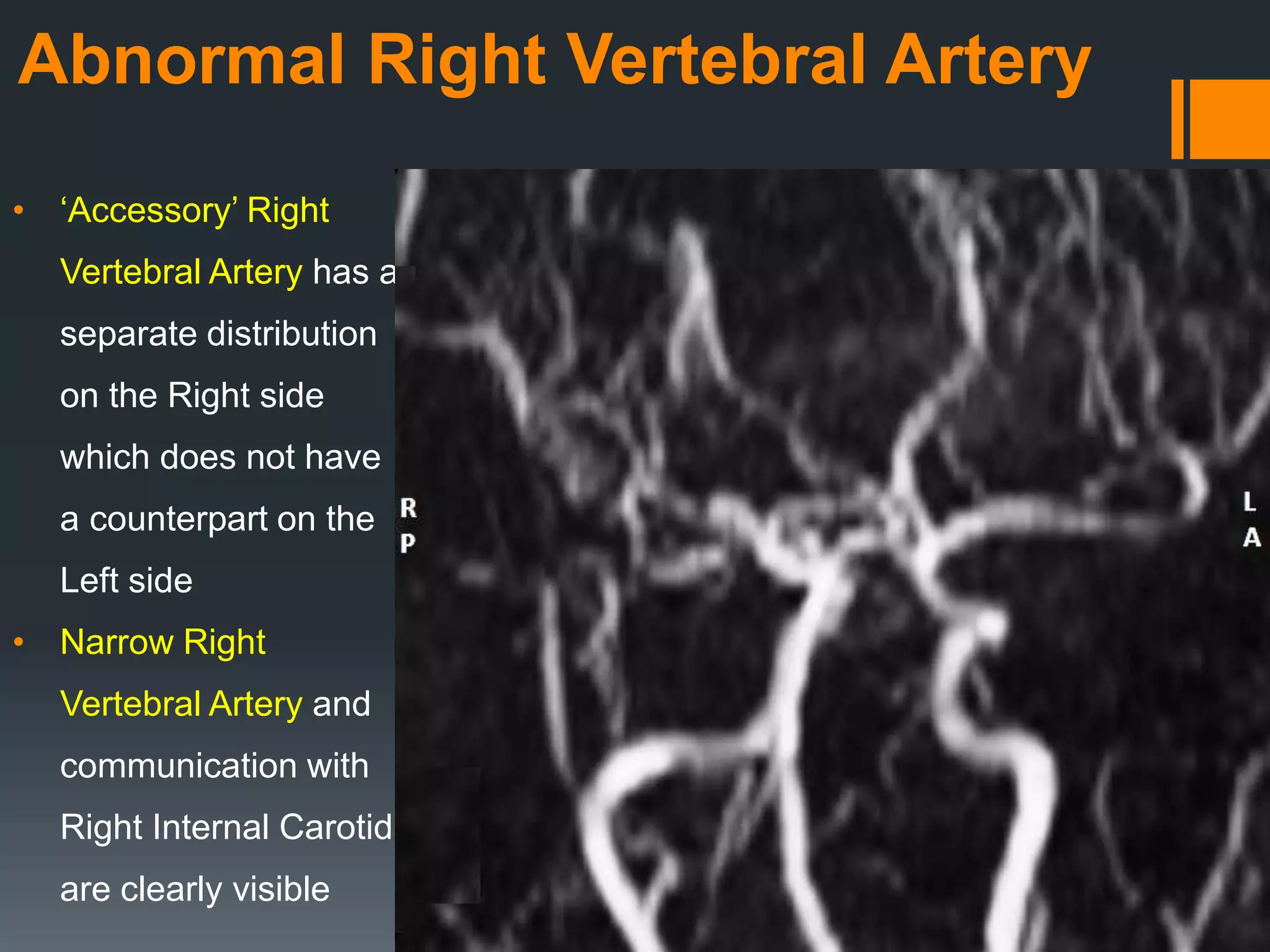


The document presents a composite MRA image illustrating abnormalities in the right vertebral artery, including a narrow artery and an accessory artery. It contains a series of time of flight (tof) MRA images that highlight the unique features of these vessels and their connections to the internal carotid artery. An embedded video further demonstrates these findings dynamically.


