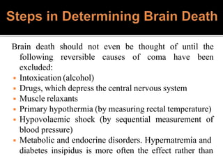
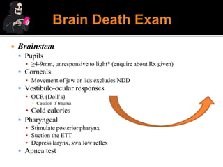
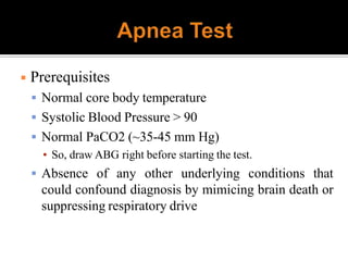
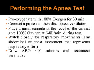
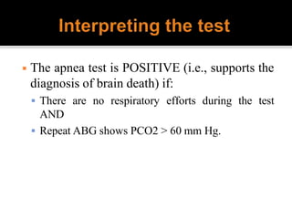
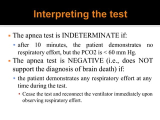
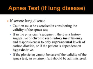
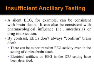
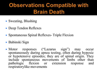
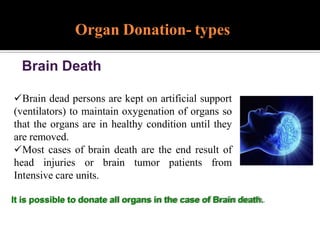
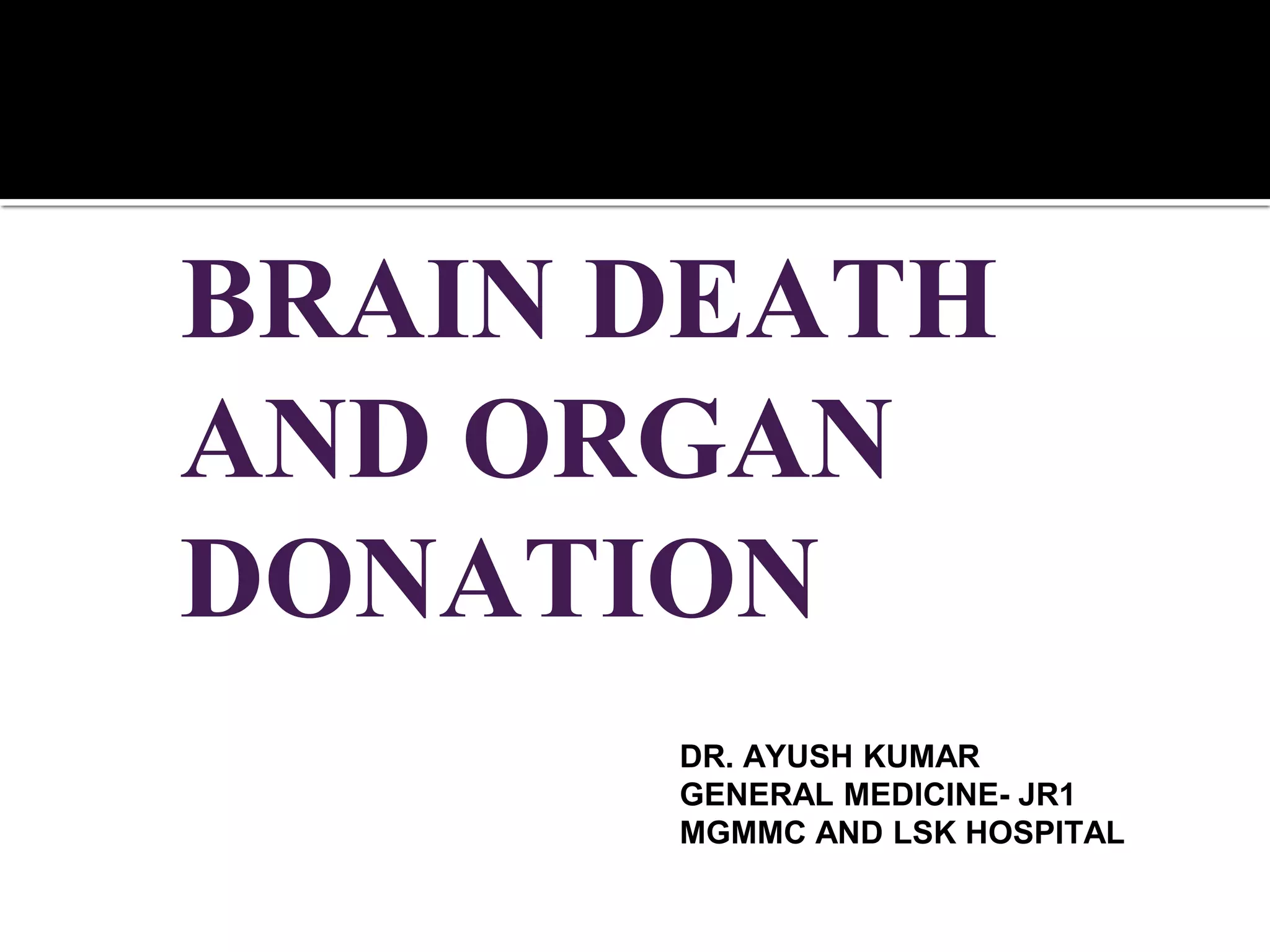
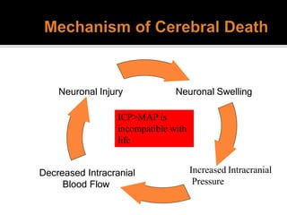
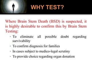
1) Brain death is defined as irreversible cessation of all functions of the entire brain, including the brainstem. Tests to determine brain death include examining brainstem reflexes and performing an apnea test.
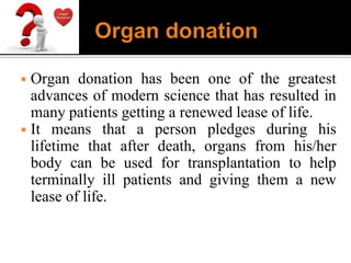
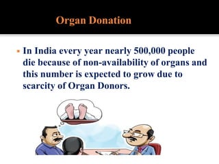
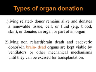
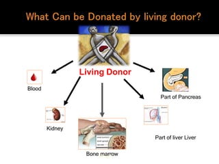
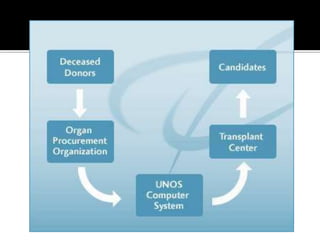
2) Organ donation provides terminally ill patients a new lease on life but organ availability is low in India. Living donors can donate renewable tissues while deceased donors are a major source of organs.
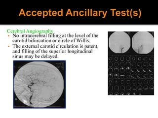
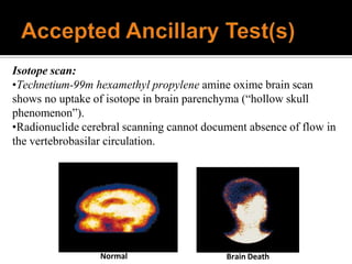
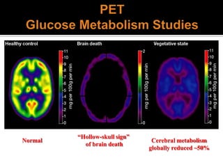
3) For organ donation to occur, all reversible causes of coma must first be excluded through testing and the diagnosis of brain death must be certified by a board of medical experts according to the law.
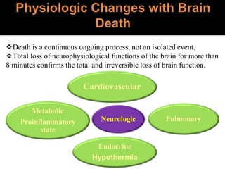
The document discusses the diagnosis of brain death through neurological criteria. It begins by describing the main parts of the brain and their functions. It then discusses the concepts, laws, and types of brain death in India. The key components of determining brain death are described, including establishing the cause of coma, achieving normal temperature and blood pressure, performing neurological examinations to check for absence of brainstem reflexes and response to stimuli, and conducting an apnea test. Potential mimics of brain death and pitfalls in the clinical evaluation are outlined. Confirmatory ancillary tests like cerebral angiography and EEG are also discussed.
This document summarizes the key updates from the 2010 American Academy of Neurology evidence-based guidelines on determining brain death. It outlines the clinical evaluation process including the neurologic assessment and apnea test to determine the irreversible cessation of brain and brainstem functions. While ancillary tests are not required, they may be used when the neurologic exam is uncertain or the apnea test cannot be done. Proper documentation of the determination is also important.
This is a presentation on brain death, its background, definition, related neurological conditions, criteria of brain death, brain stem reflexes, causes of coma, confounding factors, observation compatible with brain death, ancillary test, medical record documentation, prognosis, Management of brain death patient.
You tube link of this presentation
https://www.youtube.com/watch?v=3MzE5lHfglI&t=38s
1. Brain death is defined as the irreversible loss of function of the brain, including the brainstem. It is diagnosed through clinical examinations demonstrating coma, absence of brainstem reflexes, and apnea.
2. The diagnosis of brain death requires meeting certain prerequisites, performing a neurological exam to demonstrate no response to stimuli and no brainstem reflexes, and conducting an apnea test to show the inability to breathe independently.
3. Confirmatory tests such as an EEG, angiogram, or nuclear scan may be used but are not required for the diagnosis of brain death. Proper documentation of meeting criteria is important.
This document discusses the use of stem cells in neurosurgery. It begins with an introduction on how the brain was once thought to lack the ability for self-repair but is now known to regenerate neurons. The outline includes topics on different types of stem cells, their use in traumatic brain injury, spinal cord injury, peripheral nerve injury, brain tumors, and stroke. Methods of stem cell implantation and challenges are also covered. The document concludes by discussing the current and future prospects of neural stem cells in treatment and the need for more preclinical and clinical trials.
This document discusses brain death, its diagnosis, and organ donation. It provides definitions of brain death standards in the U.S. and U.K. Diagnosing brain death requires demonstrating irreversible loss of brain and brainstem functions through clinical exams and tests like apnea testing. Key findings that must be absent are brainstem reflexes, response to pain, and spontaneous breathing. The process of declaring brain death should be separate from discussing organ donation with family.
This document discusses decompressive craniectomy for refractory intracranial hypertension. It provides rationale and indications for decompressive craniectomy, which aims to reduce intracranial pressure by removing part of the skull. Common complications are also mentioned. Guidelines from the American Association of Neurological Surgeons are presented regarding criteria for performing decompressive craniectomy in patients with traumatic brain injury or refractory increased intracranial pressure. Outcomes of decompressive craniectomy are discussed for different patient groups.
This document discusses diffuse axonal injury (DAI) and concussion. It provides an overview of traumatic brain injury (TBI) classification, causes, mechanisms of injury, grading scales, and medicolegal importance. DAI results from acceleration/deceleration forces that cause shearing of axons throughout the brainstem, corpus callosum, and cerebral hemispheres. It is characterized by widespread damage rather than focal lesions. Concussion involves temporary dysfunction from brain impact or whipping, with risks including secondary impact syndrome. Both DAI and concussion are important forensically as they can cause death or long-term impairment without obvious anatomical signs.
An overview of Decompression hemicraniectomy in patients with large hemispheric infarctions. The presentation touches upon definition, pathophysiology, medical management, rationale for surgery, mortality, functional outcomes of DHC, and complications in a nutshell.
This document discusses various methods for monitoring the central nervous system (CNS) during and after surgery. It describes cerebral perfusion monitoring techniques like cerebral blood flow measurements, transcranial Doppler ultrasonography, near-infrared spectroscopy, and jugular bulb oximetry. Regional perfusion is also monitored using brain tissue partial oxygen tension. Cerebral metabolism is assessed using cerebral microdialysis. Cerebral function is evaluated with the bispectral index, approximate entropy, and auditory evoked potentials. The document provides details on how each technique is performed and what clinical information it provides about CNS oxygenation, blood flow, and function.
Mechanical thrombectomy with stent retrieverDr Vipul Gupta
Vipul Gupta discusses balloon assisted coiling in ruptured cerebral aneurysms and mechanical thrombectomy with stent retrievers. He summarizes several key randomized controlled trials that demonstrated the benefits of endovascular therapy using stent retrievers over standard medical therapy alone for acute ischemic stroke. The trials showed significant improvements in revascularization, clinical outcomes, and mortality. The 2015 AHA/ASA guidelines recommend endovascular therapy with stent retrievers for select patients within 6 hours of stroke onset based on the evidence from these trials. The document also reviews techniques for mechanical thrombectomy and strategies to optimize outcomes.
Nurses as the primary care providers would be the immediate health care professional to assess the patient's response and to determine whether he is improving or deteriorating. Signs of brain death can be identified and reported early by a nurse with adequate knowledge.
1. Neuroplasticity refers to the brain's ability to change and reorganize itself in response to experience or injury. It allows the brain to compensate for damage and to adjust its activity in response to new situations or information.
2. Several mechanisms underlie neuroplasticity including neuronal regeneration, synaptic plasticity, neurogenesis, gliogenesis, dendritic remodeling, and functional reorganization through processes like vicariation.
3. Neuroplasticity can be measured at the cellular level through changes in synapses and at the neural network level through reorganization of maps. Imaging techniques like MRI can also measure plastic changes in gray and white matter.
This document discusses spinal cord stimulation (SCS) and its role in pain management in New Zealand. It provides background on how SCS works and its history. It then discusses considerations for patient selection and outcomes from studies on SCS for various pain conditions. Challenges, funding issues, and requirements for establishing an SCS service in New Zealand are also examined. The document argues that more patients should be considered for SCS and that realistic funding pathways need to be established.
The document summarizes key aspects of neurophysiology, including the motor system, corticospinal system, lower motor neurons, extrapyramidal system, cerebellum, sensory system, reflex activities, and control of micturition. It describes the components and functions of these systems, as well as signs associated with lesions in different parts of the nervous system.
Stem cells show promise for treating various brain and nervous system disorders. Neural stem cells can differentiate into neurons, astrocytes, and oligodendrocytes and may be able to repair injured brain and spinal cord tissue. Preclinical and clinical trials have shown stem cells can restore function in diseases like Parkinson's and stroke. However, challenges remain in optimizing donor sources, ensuring cell viability after transplantation, and developing techniques to integrate cells and reconstruct pathways. Overcoming these challenges could realize stem cells' potential to develop new treatments.
The Fugl-Meyer Assessment of sensorimotor function (FMA) evaluates recovery in post-stroke hemiplegic patients. It measures motor impairment in five domains using a 3-point ordinal scale with a maximum score of 226. The FMA has excellent interrater and intrarater reliability as well as internal consistency. It is widely used in both clinical and research settings to quantify motor impairment recovery after stroke.
This document discusses the use of robotics in neurosurgery. It provides an overview of various robotic systems that have been used including early systems like PUMA and more recent systems like NeuroArm, ROSA, and SteadyHands. It describes different categories of robotic systems including dependent, supervisory, and shared control systems. The document also discusses benefits of robotic systems like increased accuracy and applications in telesurgery. Surgical simulation and training using virtual reality systems is also summarized.
Brain death is a clinical diagnosis based on the absence of neurologic function and irreversible coma. A complete neurologic examination is required to determine brain death, which must show no brainstem reflexes, no response to stimuli, apnea on testing, and flaccid muscle tone. Ancillary tests like EEG or angiography can assist in diagnosis but are not required. Special considerations are needed for diagnosing brain death in newborns and preterm infants due to the potential for reversible conditions and lack of brain development. Death is declared after two examinations show no changes confirming an irreversible condition.
A detailed presentation on Brain Death and Ongan transplantation.
Criteria for Brain Death are explained in detail. Legislative laws regarding the organ transplant, organ preservation are also explained.
The collection and processing of hematopoieticakshaya tomar
BASICS OF HSC COLLECTION AND PROCESSING INCLUDING ALL THE THREE SOURCES, A BRIEF ABOUT STEM CELL MOBILIZATION, STEM CELL SELECTION CRYOPRESERVATION AND DMSO
The document discusses brain death, its diagnosis and pathophysiology. It defines brain death as the complete and irreversible loss of brain function. The diagnosis involves meeting strict clinical criteria demonstrating the absence of brainstem reflexes as well as confirmatory tests like EEG. Brain death results in no prospect of survival without life support or recovery of brain function. Proper diagnosis is important for organ donation where brain death constitutes legal death.
Brain Death concepts, Its changes and life after brain death, is the body still alive?? what are the determinants of brain death and who can declare it, bio ethical dimensions of nursing care in BD
The document summarizes the process of presurgical evaluation for epilepsy patients. It discusses how modern imaging techniques like video EEG monitoring, high-resolution MRI, fMRI, PET and SPECT are used to localize the epileptogenic zone noninvasively in most patients. When noninvasive methods are insufficient, invasive EEG monitoring using subdural or depth electrodes may be used. The goal is to precisely identify the brain area responsible for seizure generation to allow its surgical resection, while avoiding damage to critical functions. A classical example where surgery is often curative is mesial temporal lobe epilepsy.
This document discusses brain death, including its historical definition, current diagnostic criteria, pathophysiology, and management of organ donors. Key points include:
- Brain death is defined as irreversible cessation of all functions of the entire brain, including the brain stem.
- Diagnosis requires two examinations at least 6 hours apart showing coma, absence of brainstem reflexes, and apnea during a standardized test. Ancillary tests like EEG can be used if clinical criteria are inconclusive.
- After brain death, pathophysiological changes occur like hypotension, diabetes insipidus, and coagulopathies due to loss of autonomic and endocrine functions regulated by the brain.
dual antiplatelet in stroke meta analysisGovind Madhaw
This journal club presentation summarizes a meta-analysis of 18 randomized controlled trials with 15,515 patients comparing dual antiplatelet therapy to mono antiplatelet therapy for secondary stroke prevention in patients with acute ischemic stroke or transient ischemic attack. The meta-analysis found dual antiplatelet therapy significantly reduced the risk of stroke recurrence and composite vascular events compared to monotherapy, without significantly increasing the risk of major bleeding. Sensitivity analyses restricting to double-blind studies showed similar results. The presentation provides details on the search methods, inclusion/exclusion criteria, data extraction, outcomes analyzed, and results of the meta-analysis.
Stem cell therapy shows promise for treating various neurological disorders. There are two main types of stem cells - embryonic stem cells which are pluripotent, and adult stem cells which are multipotent. Stem cells may promote cell replacement in damaged organs through proliferation, migration, and differentiation. Challenges include optimal cell types and doses, monitoring transplanted cells, and ensuring safety. While stem cell therapy is being studied for conditions like Alzheimer's, Parkinson's, ALS, and stroke, more research is still needed to address current obstacles in translating laboratory findings to clinical applications.
This document discusses brain death and the criteria used to diagnose it. It begins by describing different states of consciousness including coma, persistent vegetative state, and locked-in syndrome. It then defines brain death as the total and irreversible loss of brain and brainstem function. The key criteria for determining brain death are the absence of cortical function, absence of brainstem reflexes, and apnea during a specific oxygen challenge. Confirmatory tests like angiography, EEG, transcranial Doppler, and nuclear medicine scans can also support the diagnosis. Precise clinical evaluations and testing are required to distinguish brain death from other severe neurological conditions.
This document discusses brain death and the criteria used to diagnose it. It begins by describing different states of consciousness including coma, persistent vegetative state, and locked-in syndrome. It then defines brain death as the total and irreversible loss of brain and brain stem functions. The key criteria for determining brain death are the absence of cortical function, absence of brainstem reflexes, and apnea during a specific oxygen challenge. Confirmatory tests like angiography, EEG, Doppler ultrasound, and scintigraphy can also be used when clinical criteria cannot be reliably assessed.
The document discusses guidelines for determining brain death. It begins with historical context for defining death and the need for brain death criteria. Key points include:
- Brain death is the irreversible loss of all brain function, including the brain stem. It allows for legal death even if other organs are functioning.
- Tests to determine brain death include examining for the absence of brain stem reflexes and apnea testing to check for respiratory drive.
- If full criteria are met, including examinations showing no brain activity, the time of death is determined to be when respiratory criteria were met on tests.
This document discusses brain death and the criteria used to diagnose it. It begins by describing different states of altered consciousness including coma and vegetative states. It then defines brain death as the total and irreversible loss of brain and brainstem function, and notes that spinal reflexes may remain. Several prerequisites and criteria for diagnosing brain death are provided, including the absence of brainstem reflexes and apnea testing. Confirmatory tests like angiography, EEG, Doppler ultrasound and nuclear scans are also summarized. Guidelines for diagnosing brain death in children of different ages are presented.
This presentation consist information about Brain death with special emphasis to differences between Indian and Western Guidelines. Also consist information about Organ transplantation and related act.
Brain death refers to the irreversible loss of all brain function. It is diagnosed through examinations showing the absence of brainstem reflexes and apnea during an oxygen challenge. Organ donation from brain dead donors provides organs for transplantation but there is a shortage due to misperceptions about the process. The document discusses the criteria for determining brain death in adults and children, confirmatory tests, organ donation laws in India, and efforts to increase donation rates.
The document summarizes key aspects of death and brain death from a medicolegal perspective. It defines death and outlines the vital systems that cease to function at death. It discusses the presumption of death and survivorship under Indian law. The document then examines the historical definitions of death and milestones in determining brain death. It provides details on the anatomy of the normal brain and causes, mechanisms, and conditions distinct from brain death. The neurological examination for determining brain death and confirmatory testing are described. The key provisions of the Transplantation of Human Organs Act of 1994 and its amendment in 2009 are summarized.
1. Brain death is defined as the irreversible loss of all brain function, including the brainstem. The three essential findings are coma, absence of brainstem reflexes, and apnea on testing.
2. Common causes of brain death include traumatic brain injury, intracerebral hemorrhage, hypoxic-ischemic injury from cardiopulmonary arrest. A thorough exam is required to determine the cause and rule out potential confounders.
3. Evaluation of brain death involves assessing for coma, absence of brainstem reflexes on exam, and a positive apnea test showing lack of respiratory drive and rising CO2 levels. Ancillary tests like angiography or EEG can also be
Death by Neurological Criteria and Organ Donation: Bill KnightSMACC Conference
Bill Knight explains the concept of death by neurological criteria and the complexities surrounding organ donation in such situations.
Bill discusses the process of dying, the definition of death, how to approach the neurologically dead patient and how to consider organ donation.
Death is a complex topic.
Due to advancements in medical technology and processes, the definition of death is a challenging one.
Bill talks at length about the definition of death by the neurological criteria. Dying is an active process, whereas death is an event.
The acceptance of death by the neurological criteria is often challenging as Bill will highlight. Bill talks about the care of the dying or dead patient.
There is a point at which care will transition from supporting the patient to supporting the organs. This is still good care.
There is an alignment of parallel intentions – first and foremost resuscitation of patients and then failing that, proceeding to considering and actioning organ donation. This is important due to the shortage of viable donor organ worldwide.
The donation process itself is complex. Bill provides his thoughts. He insists that an intensivist be involved as this has been shown to increase the number of viable and healthy organs made available.
The timing is also important. Available evidence does not support the need for immediate procurement after brain death. Taking time to optimise perfusion and allow recovery and cardiac function is appropriate and should be done.
Bill also discusses other treatment options at the time of death such as optimising endocrine function.
Finally, Bill will provide some practical considerations when communicating with the dead patient’s family. This involves being clear on your messaging. You are supporting organs, not life.
To reinforce this point, Bill suggests not examining or talking to the patient. He also recommends using all of the available hospital support services.
Similarly, it is best to not introduce the topic of organ donation to the family yourself as the treating clinician. Utilise the Organ Procurement Organisations (or similar services) and get them involved early to speak with the family.
Join Bill Knight in his talk on the North American perspective on Organ Donation, brain death and management of the brain dead donor prior to organ donation.
For more like this, head to our podcast page. #CodaPodcast
Brain death is defined as the complete and irreversible cessation of all brain functions, including the brain stem. It can be determined by the absence of electrical activity in the brain via EEG, lack of blood flow to the brain, and the absence of all clinical brain functions upon examination. Some causes that can lead to brain death include anoxia, ischemia, intracranial hemorrhage, brain tumors, and head injuries. Proper certification of brain death requires examination by a board of doctors at least three times over a period of hours to confirm the irreversible loss of all brain functions. Recovery from brain death is not possible, as it indicates misdiagnosis, while patients in comas or vegetative states still
Brain death is defined as the complete and irreversible cessation of all brain functions, including the brain stem. It can be determined by the absence of electrical activity in the brain via EEG, lack of blood flow to the brain, and the absence of all clinical brain functions upon examination. Key steps in examining for brain death include assessing pupillary response, eye movements, motor response, and reflexes to ensure there is no function of the brain or brain stem. A board of doctors must examine the patient at least three times over six hours to certify brain death before life support is removed. While comas and vegetative states are sometimes confused with brain death, they are reversible conditions unlike the irreversible damage caused by brain death.
This document provides information about brain death. It begins with definitions of death and introduces brain death as an irreversible form of unconsciousness characterized by loss of brain function while other organs may still be functioning. The document then discusses the history of how brain death was recognized and defined. It outlines the criteria for determining brain death, including assessing brain stem reflexes and performing an apnea test. The document notes that Indian law recognizes brain death and defines a deceased person. It emphasizes the importance of establishing the cause of brain dysfunction and ruling out potential confounders before determining brain death.
This document discusses pediatric brain death. It defines brain death as the irreversible cessation of all brain functions, including the brain stem. Two examinations by two physicians are required to diagnose brain death in children, separated by 12-24 hours depending on age. The diagnostic criteria that must be fulfilled are a Glasgow Coma Scale of 3/15, apnea confirmed by an apnea test, and absent brain stem reflexes as shown by tests of cranial nerves and response to stimuli. Precautions must be taken to rule out factors that could mimic brain death.
Brain death & Potential Brain Dead Donor (PBDD)Krishn Undaviya
This document discusses brain death and the determination of brain death. It provides:
1) A definition of brain death as the irreversible loss of all functions of the brain, including the brainstem, as evidenced by coma, absence of brainstem reflexes, and apnoea.
2) Details the clinical criteria for determining brain death, including neurological examination to check for absence of brainstem reflexes and an apnoea test.
3) Outlines the general care and management of a potential organ donor who is brain dead, including maintaining body temperature and position, treating for infections, and eye and airway care.
Approach to deceased donor transplantationVishal Golay
This document discusses the process of deceased donor organ transplantation. It covers donor identification, screening, certification of brain stem death, maintenance of potential donors, and counseling of family members. Key steps include confirming brain stem death through clinical tests, screening donors based on medical criteria, obtaining consent from relatives, and providing life support to optimize organ function prior to retrieval. The overall goal is to maximize the number of transplantable organs through proper donor care and management.
Brain cut up for the general pathologistEffiong Akang
Simplified procedure for brain cut up examination for general pathologists that emphasises the importance of good clinicopathological correlation in post-mortem CNS examination. Presented at TSL workshop in Lagos on 25 November 2014
1) Organ donation rates are low, with only 3 in 1000 people dying in a way that allows organ donation, while 1 person is added to the waiting list every 10 minutes and 1 dies every 20 minutes waiting.
2) There are 3 paths to organ donation - living donation, donation after cardiac death, and donation after brain death.
3) It is important to make an early referral to the organ procurement organization whenever a patient has a severe brain injury, is intubated, end of life measures are being considered, or the family asks about donation. This allows for screening and approaching families if appropriate.
This document discusses cessation of life and determining death. It covers:
- Definitions of somatic death, molecular death, brain death, and brain stem death.
- Criteria for determining whole brain death which requires absent brain and brainstem function and apnea.
- Importance of determining the cause and time of death for organ transplantation. Different organs remain viable for varying lengths of time after circulation stops.
- Distinctions between brain death, persistent vegetative state, and brainstem injury and how each affects respiratory and cardiac function.
Adiponectin is an adipokine that is decreased in obesity and has anti-diabetic, anti-inflammatory, and cardioprotective effects. It acts through two receptors, AdipoR1 and AdipoR2, and has various actions including increasing insulin sensitivity, fatty acid oxidation, and decreasing inflammation. Low levels of adiponectin are associated with increased risk of metabolic syndrome, hypertension, chronic kidney disease, obstructive sleep apnea syndrome, diabetic retinopathy, cancer, and chronic obstructive pulmonary disease. Enhancing adiponectin expression and activity through lifestyle changes, drugs, recombinant adiponectin, or peptide mimetics may be a therapeutic approach for treating obesity and related
This document discusses renal tubular acidosis (RTA). It begins by outlining the physiology of renal acidification, including bicarbonate reabsorption in the proximal tubule and hydrogen ion secretion in the collecting duct. It then describes the four main types of RTA: proximal RTA, distal RTA, hyperkalemic distal RTA, and hyperkalemic RTA. Proximal RTA is caused by impaired bicarbonate reabsorption in the proximal tubule and results in hypokalemia. Distal RTA involves impaired hydrogen ion secretion in the collecting duct and causes severe metabolic acidosis with low bicarbonate levels and hypercalciuria. The document provides details
Monoclonal antibodies are monospecific antibodies produced by identical immune cells that are clones of a single parent cell. They bind to the same epitope or antigen. There are several types of monoclonal antibodies including murine, chimeric, humanized, and human antibodies. Monoclonal antibodies have various applications in hematology including as therapeutics for cancers, autoimmune disorders, and transplant rejection by blocking molecular targets, delivering cytotoxic compounds to tumors, or acting as signaling molecules. They can also be used diagnostically in tests like ELISA and immunohistochemistry. Common adverse effects of monoclonal antibodies include infusion reactions, immunosuppression, and increased risk of infection.
Spondyloarthritis (SpA) refers to a group of inflammatory diseases involving the spine and joints. It includes ankylosing spondylitis, psoriatic arthritis, reactive arthritis, and undifferentiated spondyloarthritis. Key features are inflammation of the spine and sacroiliac joints, peripheral arthritis, enthesitis, and extra-articular manifestations affecting the eyes, bowels, and lungs. Diagnosis involves assessing clinical features, laboratory tests like HLA-B27, and imaging of the sacroiliac joints and spine. Treatment focuses on reducing pain and inflammation with NSAIDs, TNF inhibitors, exercise and maintaining function.
Diabetic gastroparesis is a complication of diabetes that results from dysfunction of the autonomic nervous system and stomach muscles. It causes delayed emptying of food from the stomach. The presentation includes nausea, vomiting, early fullness and weight changes. Diagnosis involves testing for delayed gastric emptying. Treatment focuses on diet, glycemic control, prokinetic medications, and in severe cases, procedures like gastric stimulation. Education is important for managing the chronic nature of the condition and preventing complications like malnutrition.
The document describes various tracts of the spinal cord including their origin, pathway, termination and function. It discusses the lateral and anterior spinothalamic tracts which are involved in pain and touch sensation respectively. It also covers the dorsal column, corticospinal tract, rubrospinal tract and extrapyramidal tracts. The effects of lesions in different areas are summarized including upper and lower motor neuron lesions.
A 48-year-old female presented with complaints of chest pain, shortness of breath, and weakness. She had a history of COPD and was a chronic smoker. In the emergency room, she was semi-conscious and in respiratory distress. Despite treatment, her condition deteriorated over the next several hours with declining blood pressure and oxygen levels. Attempts to increase vasopressor medications were unsuccessful and she went into cardiac arrest. CPR was unsuccessful and she was declared dead at 8:00 AM. The cause of death was determined to be septic shock and respiratory failure due to suspected right lower lobe pneumonia exacerbating her COPD.
This document provides an overview of myelodysplastic syndrome (MDS). Key points include:
- MDS is a group of bone marrow disorders characterized by low blood cell counts, dysplastic changes in the bone marrow, and a risk of developing acute myeloid leukemia.
- It primarily affects older adults but can occur in younger patients as well. Risk factors include prior chemotherapy/radiation exposure, smoking, and certain genetic conditions.
- The disease involves malignant transformation of myeloid stem cells. Common genetic mutations impact DNA methylation and gene expression regulation.
- Patients present with anemia and related symptoms. Diagnosis involves blood and bone marrow tests showing dysplastic features. Prognosis depends on factors like blast percentage
This document provides an overview of myelodysplastic syndrome (MDS). It discusses the history, classification, pathogenesis, diagnosis and management of MDS. MDS is a heterogeneous group of clonal stem cell neoplasms characterized by cytopenia, dysplasia and risk of progression to acute myeloid leukemia. The document reviews the World Health Organization classification of MDS subtypes based on dysplastic lineages, ring sideroblasts and blast percentage. It also discusses common cytogenetic abnormalities and molecular mutations in MDS. Diagnosis involves blood counts, bone marrow morphology, histology, immunophenotyping and genetic testing. Prognostic scoring systems guide treatment and management, which may include growth factors, immunosuppress
This document discusses coronary artery disease (CAD) in young people. CAD occurring below age 45 is considered young CAD, though some studies use ages 35-55. About 85-90% of premature CAD is due to conventional risk factors like smoking, diabetes, hypertension, dyslipidemia and obesity. Smoking is the most common risk factor, present in 60-90% of young CAD patients versus 24-56% of older patients. The prevalence of diabetes, hypertension and obesity is also higher in young CAD patients compared to older patients without CAD. While dyslipidemia is important, lipid abnormalities do not differ much between young and older CAD patients. Rare causes of young CAD include cocaine use, genetic factors, and spontaneous coronary
This document discusses coronary heart disease in young adults. It finds that while most coronary disease occurs in older populations, 2-6% of acute coronary events occur in younger "premature" patients under 55 years old. Major risk factors for young adults include smoking, family history of early heart disease, male gender, and hyperlipidemia. Diagnostic tests may include electrocardiograms, stress tests, echocardiograms, CT angiograms, and calcium scoring. Aggressive risk factor modification including smoking cessation and statin therapy is important for prognosis. While short term outcomes of revascularization are good, long term mortality is still elevated compared to the general population.
This document discusses cardiac channelopathies, which are genetic disorders characterized by altered cardiac excitability without structural heart involvement. The major channelopathies discussed are Long QT syndrome (LQTS), Brugada syndrome, Catecholaminergic polymorphic ventricular tachycardia (CPVT), and Short QT syndrome. For each condition, the document outlines the genes involved, clinical manifestations, diagnostic criteria, risk stratification, and treatment recommendations including use of beta-blockers and implantable cardioverter-defibrillators.
Adhd Medication Shortage Uk - trinexpharmacy.comreignlana06
The UK is currently facing a Adhd Medication Shortage Uk, which has left many patients and their families grappling with uncertainty and frustration. ADHD, or Attention Deficit Hyperactivity Disorder, is a chronic condition that requires consistent medication to manage effectively. This shortage has highlighted the critical role these medications play in the daily lives of those affected by ADHD. Contact : +1 (747) 209 – 3649 E-mail : sales@trinexpharmacy.com
share - Lions, tigers, AI and health misinformation, oh my!.pptxTina Purnat
• Pitfalls and pivots needed to use AI effectively in public health
• Evidence-based strategies to address health misinformation effectively
• Building trust with communities online and offline
• Equipping health professionals to address questions, concerns and health misinformation
• Assessing risk and mitigating harm from adverse health narratives in communities, health workforce and health system
Osteoporosis - Definition , Evaluation and Management .pdfJim Jacob Roy
Osteoporosis is an increasing cause of morbidity among the elderly.
In this document , a brief outline of osteoporosis is given , including the risk factors of osteoporosis fractures , the indications for testing bone mineral density and the management of osteoporosis
NVBDCP.pptx Nation vector borne disease control programSapna Thakur
NVBDCP was launched in 2003-2004 . Vector-Borne Disease: Disease that results from an infection transmitted to humans and other animals by blood-feeding arthropods, such as mosquitoes, ticks, and fleas. Examples of vector-borne diseases include Dengue fever, West Nile Virus, Lyme disease, and malaria.
These lecture slides, by Dr Sidra Arshad, offer a quick overview of the physiological basis of a normal electrocardiogram.
Learning objectives:
1. Define an electrocardiogram (ECG) and electrocardiography
2. Describe how dipoles generated by the heart produce the waveforms of the ECG
3. Describe the components of a normal electrocardiogram of a typical bipolar lead (limb II)
4. Differentiate between intervals and segments
5. Enlist some common indications for obtaining an ECG
6. Describe the flow of current around the heart during the cardiac cycle
7. Discuss the placement and polarity of the leads of electrocardiograph
8. Describe the normal electrocardiograms recorded from the limb leads and explain the physiological basis of the different records that are obtained
9. Define mean electrical vector (axis) of the heart and give the normal range
10. Define the mean QRS vector
11. Describe the axes of leads (hexagonal reference system)
12. Comprehend the vectorial analysis of the normal ECG
13. Determine the mean electrical axis of the ventricular QRS and appreciate the mean axis deviation
14. Explain the concepts of current of injury, J point, and their significance
Study Resources:
1. Chapter 11, Guyton and Hall Textbook of Medical Physiology, 14th edition
2. Chapter 9, Human Physiology - From Cells to Systems, Lauralee Sherwood, 9th edition
3. Chapter 29, Ganong’s Review of Medical Physiology, 26th edition
4. Electrocardiogram, StatPearls - https://www.ncbi.nlm.nih.gov/books/NBK549803/
5. ECG in Medical Practice by ABM Abdullah, 4th edition
6. Chapter 3, Cardiology Explained, https://www.ncbi.nlm.nih.gov/books/NBK2214/
7. ECG Basics, http://www.nataliescasebook.com/tag/e-c-g-basics
Does Over-Masturbation Contribute to Chronic Prostatitis.pptxwalterHu5
In some case, your chronic prostatitis may be related to over-masturbation. Generally, natural medicine Diuretic and Anti-inflammatory Pill can help mee get a cure.
Basavarajeeyam is a Sreshta Sangraha grantha (Compiled book ), written by Neelkanta kotturu Basavaraja Virachita. It contains 25 Prakaranas, First 24 Chapters related to Rogas& 25th to Rasadravyas.
Promoting Wellbeing - Applied Social Psychology - Psychology SuperNotesPsychoTech Services
A proprietary approach developed by bringing together the best of learning theories from Psychology, design principles from the world of visualization, and pedagogical methods from over a decade of training experience, that enables you to: Learn better, faster!
2. “An individual who has sustained either
irreversible cessation of circulatory and
respiratory functions, or irreversible cessation of
all functions of the entire brain, including the
brainstem. “
Uniform Determination of Death Act (UDDA)
3. Neuronal Injury Neuronal Swelling
Decreased Intracranial
Blood Flow
Increased Intracranial
Pressure
ICP>MAP is
incompatible with
life
4. Where Brain Stem Death (BSD) is suspected, it
is highly desirable to confirm this by Brain Stem
Testing:
• To eliminate all possible doubt regarding
survivability
• To confirm diagnosis for families
• In cases subject to medico-legal scrutiny
• To provide choice regarding organ donation
6. In India, according to the Transplantation of Human Organs Act
[TOHO 1994 (Sub section 6 of Section 3)], “Brainstem death”
means the stage at which all functions of the brainstem have
permanently and irreversibly ceased and is so certified by a
“Board of Medical Experts” consisting of:
◾ The Medical Superintendent (MS) in charge of the hospital in
which “brainstem” death has occurred.
◾ A specialist, nominated by the MS in charge of the hospital, from
a panel of names approved by the Appropriate Authority.
◾ A neurologist or neurosurgeon, nominated by the MS in charge of
the hospital, from a panel of names approved by the appropriate
authority.
7. Brain death should not even be thought of until the
following reversible causes of coma have been
excluded:
◾ Intoxication (alcohol)
◾ Drugs, which depress the central nervous system
◾ Muscle relaxants
◾ Primary hypothermia (by measuring rectal temperature)
◾ Hypovolaemic shock (by sequential measurement of
blood pressure)
◾ Metabolic and endocrine disorders. Hypernatremia and
diabetes insipidus is more often the effect rather than
8. ◾ Brainstem
Pupils
▪ ≥4-9mm, unresponsive to light* (enquire about Rx given)
Corneals
▪ Movement of jaw or lids excludes NDD
Vestibulo-ocular responses
▪ OCR (Doll’s)
▪ Caution if trauma
▪ Cold calorics
Pharyngeal
▪ Stimulate posterior pharynx
▪ Suction the ETT
▪ Depress larynx, swallow reflex
Apnea test
9. ◾ Normal response = eyes always
gaze up towards roof
◾ Rapid, but steady movements and
observe for direction of gaze
Activates vestibular system
ipsilateral to head thrust
horizontal gaze
▪ Communicates with contralateral
center (CN VI)
“orchestrating” the action of the eyes
▪ Simultaneously dampens contralateral
vestibular tone, etc.
Avoided in the setting of
a patient with questionable
stability of the cervical
spine
10. ◾ 30° to the horizontal
◾ Minimum of 50cc of ice cold water into the
inner ear canal
Ensure no perforated tympanic membrane before
instilling water
Use kidney basin, prop up beside ear
◾ Start observing for eye deviation rapidly; eye
movements should be absent for 1 minute
◾ Minimum of 5 minutes before evaluating
contralateral side
11. ◾ Prerequisites
Normal core body temperature
Systolic Blood Pressure > 90
Normal PaCO2 (~35-45 mm Hg)
▪ So, draw ABG right before starting the test.
Absence of any other underlying conditions that
could confound diagnosis by mimicing brain death or
suppressing respiratory drive
12. ◾ Pre-oxygenate with 100% Oxygen for 30 min.
◾ Connect a pulse-ox, then disconnect ventilator.
◾ Place a nasal cannula at the level of the carina;
give 100% Oxygen at 6-8L/min. during test.
◾ Watch closely for respiratory movements (any
abdominal or chest movement that represents
respiratory effort)
◾ Draw ABG ~10 minutes and reconnect
ventilator.
13. ◾ The apnea test is POSITIVE (i.e., supports the
diagnosis of brain death) if:
There are no respiratory efforts during the test
AND
Repeat ABG shows PCO2 > 60 mm Hg.
14. ◾ The apnea test is INDETERMINATE if:
after 10 minutes, the patient demonstrates no
respiratory effort, but the PCO2 is < 60 mm Hg.
◾ The apnea test is NEGATIVE (i.e., does NOT
support the diagnosis of brain death) if:
the patient demonstrates any respiratory effort at any
time during the test.
▪ Cease the test and reconnect the ventilator immediately upon
observing respiratory effort.
15. ◾ If severe lung disease
Caution must be exercised in considering the
validity of the apnea test
If in the physician’s judgment, there is a history
suggestive of chronic respiratory insufficiency
and responsiveness to only supranormal levels of
carbon dioxide, or if the patient is dependent on
hypoxic drive.
If the physician cannot be sure of the validity of the
apnea test, an ancillary test should be administered.
16. Cerebral Angiography
◾ No intracerebral filling at the level of the
carotid bifurcation or circle of Willis.
◾ The external carotid circulation is patent,
and filling of the superior longitudinal
sinus may be delayed.
17. Normal Brain Death
Isotope scan:
•Technetium-99m hexamethyl propylene amine oxime brain scan
shows no uptake of isotope in brain parenchyma (“hollow skull
phenomenon”).
•Radionuclide cerebral scanning cannot document absence of flow in
the vertebrobasilar circulation.
19. ◾ A silent EEG, for example, can be consistent
with brain death. It can also be consistent with
pharmacological influence (i.e., anesthesia) or
drug intoxication.
◾ By contrast, EEGs don’t always “confirm” brain
death.
There can be minor transient EEG activity even in the
setting of clinical brain death.
Electrical artifacts on EEG in the ICU setting have
been described.
20. ◾ Children should be the same as those in adults.
◾ All these tests may be carried out twice, at an
interval of at least 6 hours according to the
internationally accepted protocol.
◾ As children are more resilient than adults, a longer
time between assessments has been advocated and
this varies according to patient’s age as follows:
◾ Term to 2 months old—48 hours
◾ Greater than 2 months to 1 year old—24 hours
◾ Greater than 1 year to less than 18 year old—12
hours
◾ Greater than 18 year old—interval optional.
21. ◾ The patient must demonstrate no response to
any stimulation.
Spontaneous movement is almost always absent.
any posturing, etc.,
function and is not
◾ Seizures,
indicates
consistent
shivering,
brainstem
with the determination of brain
death.
◾ The presence of spinal reflexes does not
exclude brain death, but if there is any doubt
then the diagnosis of brain death should be
withheld.
25. ◾ Diffuse or Multi-Focal Brain Injury
◾ Preserved Brain Stem Function
◾ Variable Interaction with Environmental
Stimuli
26. ◾ Sweating, Blushing
◾ Deep Tendon Reflexes
◾ Spontaneous Spinal Reflexes- Triple Flexion
◾ Babinski Sign
◾ Motor responses (“Lazarus sign”) may occur
spontaneously during apnea testing, often during hypoxic
or hypotensive episodes; they are of spinal origin. They
include spontaneous movements of limbs other than
pathologic flexion or extension response and
respiratorylike movements
27. ◾ Organ donation has been one of the greatest
advances of modern science that has resulted in
many patients getting a renewed lease of life.
◾ It means that a person pledges during his
lifetime that after death, organs from his/her
body can be used for transplantation to help
terminally ill patients and giving them a new
lease of life.
28. ◾ In India every year nearly 500,000 people
die because of non-availability of organs and
this number is expected to grow due to
scarcity of Organ Donors.
Organ Donation
29. Corneal blindness is very common in India.
More than 3 million cases in India.
60% are <12 years age group.
Only 1.5 lakh/year corneal donations in India.
30.
31. 1)living related- donor remains alive and donates
a renewable tissue, cell, or fluid (e.g. blood,
skin), or donates an organ or part of an organ
2)living non related(brain death and cadeveric
donor)-In brain- dead organs are kept viable by
ventilators or other mechanical mechanisms
until they can be excised for transplantation.
33. ◾ Commonly donated organs from brain death
are –kidney ,cornea ,heart, lung, liver,
pancreas, skin.
◾ Cadeveric donar-Tissues may be recovered
from donors up to 24 hours past the cessation
of heartbeat.
◾ Cadeveric donar are major source of organs
and tissues.
34. Organ Donation- types
Brain Death
Brain dead persons are kept on artificial support
(ventilators) to maintain oxygenation of organs so
that the organs are in healthy condition until they
are removed.
Most cases of brain death are the end result of
head injuries or brain tumor patients from
Intensive care units.
It is possible to donate all organs in the case of Brain death.
35. Cardiac Death
Due to the lack of circulation of blood the vital
organs quickly become unusable for
transplantation.
However, if the person is on a ventilator and if it
is medically clear that the person cannot survive,
then the family can consider Organ donation for
certain vital organs.
Such donations typically take place in the operating room.
Organ Donation- types
36. ◾ Almost everywhere organ donation is voluntary-
Two voluntary systems include –
1.Opt In - Where the donor gives consent
2.Opt Out - Where anyone who has not refused is
considered as a donor
In India we have the Opt in system, while many
western countries practice the opt out system
37. changing its name from ‘Transplantation
Human Organs Act’ to ‘Transplantation
◾ It is proposed to amend the THO Act by
of
of
Human Organs & Tissues Act’
◾ Law will broaden the definition of ‘near relative’
to include grandparents, grandchildren, uncles
and aunts.
◾ Also, not-so-close relatives who have stayed
with the patient can donate organs, provided
there is no commercial dealing.
38. ◾ Hospital Organ Donation Registry
(HODR)coordinates the process of cadaver organ
donation
◾ During lifetime, a person can pledge for organ
donation by filling up a donor form in the
presence of two witnesses, one of who shall be a
near relative and send the same to HODR
◾ The organ donor form could be obtained from
HODR either personally or through mail
39.
40. Hepatitis B or Hepatitis C may be acceptable for
HBV/C recipients
IV drug abuse or practicing homosexual
Untreated bacterial, fungal or viral infection (treated
infection may be considered)
Malignancies other than primary brain tumours and
nonmelanoma skin cancers
41. ◾ 10-20% donors are lost to cardiovascular collapse
as patient evolves to brain death
Volume Depletion in BD
◾ Causes multifactorial
Underlying medical condition – blood loss, etc
Prior management – osmotic therapy for ICP
Neuro-hormonal cascade
Capillary Leak
Diabetes Insipidus
◾ 50% of potential BD donors are volume responsive