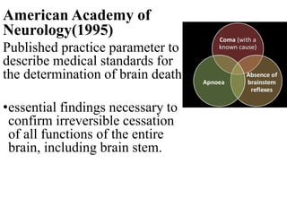
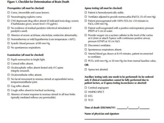
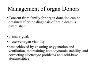
Moderator: Prof. Dr R. N. Khadgaray
Presenter: Dr Kundan Kishor Ghimire
UCMS-TH
BRAIN DEATH
Objectives
•Will be able to know when to, how to, and
why to declare a patient as brain dead.
Historical Perspective
•Historically death defined as ā€œpermanent cessation of
circulatory and respiratory function.ā€
•However, in 1968 a committee at Harvard Medical School
decided to redefine death by using ā€œirreversible comaā€ as
a new criterion.
• advancements in CPR techniques and widespread
mechanical ventilators use, number of patients could be
kept temporarily ā€˜alive.
• Consequently, Harvard criteria was developed and formed
the basis for all brain death guidelines.
BRAIN DEATH
Uniform Determination of Death
Act(UDDA) 1981
•An individual who has sustained either
•Irreversible cessation of circulatory and
respiratory functions, or
•Irreversible cessation of all functions of the entire
brain, including the brain stem
American Academy of
Neurology(1995)
Published practice parameter to
describe medical standards for
the determination of brain death
•essential findings necessary to
confirm irreversible cessation
of all functions of the entire
brain, including brain stem.
Pathophysiology of brain
death
cerebral hypoxia, traumatic head
injury, subarachnoid or intracerebral
hemorrhage and bacterial meningitis.
primary brainstem pathology
or conditions causing cerebral
edema. E.g DKA, HE, Water
intoxication, eclampsia,
Aspirin overdose, Malignant
hypertension.
irreversible rise
in ICP
increased ICP compresses the
entire brain including the
brainstem
total brain infract
•Rise in ICP āž”ļø massive release of catecholamine stores
āž”ļø increased systemic arterial blood pressure, sometimes
associated with reflex bradycardia, bradyarrhythmia, or
both.
•Decreased cardiac output, pulmonary edema, and cardiac
ischemia may be encountered.
•Once total brain infarction has occurred āž”ļø catecholamines
depletion āž”ļø loss of sympathetic output āž”ļø peripheral
vasodilation and systemic hypotension.
•Other common problems after brain death include diabetes
insipidus, hypothermia, metabolic acidosis and cardiac
arrest.
Physiological change after brain
death
Neurological Changes:-
• temperature regulation center in hypothalamus is impaired.
• patient becomes poikilothermic and hypothermic.
Cardiovascular Changes:-
• Raised ICP may cause rise in catecholamines, leading to
'sympathetic storm', responsible for the increased blood pressure,
cardiac dysrhythmias, ECG abnormalities, myocardial damage
and renal impairment.
Pulmonary Changes:-
• During the sympathetic storm, rapid rise in left atrial pressure
exceeds pulmonary artery pressure may result in capillary
disruption, protein-rich pulmonary edema and interstitial
hemorrhage.
Endocrine Changes:-
• Neurogenic diabetes insipidus.
• Electrolyte disturbances: hypernatremia, hypokalemia,
hypocalcaemia, hypophosphatemia and hypomagnesemia occur
rapidly without treatment.
•Interrupt hypothalamic-pituitary axis leading to
serum hormone depletion.
• Hypothyroidism & adrenal insufficiency
•Fall in insulin levels
Leads to systemic hyperglycemia with severe
osmotic diuresis and profound hypovolemia.
Hematological:-
•Release of fibrinolytic agents and plasminogen activators
into the circulation can cause coagulation defects, DIC.
Incidence of pathophysiological changes following
brain death:
•- Hypotension 81%
•- Diabetes Insipidus 65%
•- DIC 28%
•- Cardiac arrhythmias 25%
•- Pulmonary edema 18%
•- Metabolic acidosis 11%
DIAGNOSTIC CRITERIA
•Two examinations, separated by at least 6
hours
•Done by 2 or 3 physicians independent of the
transplant team
•At least one physician should be a specialist
in neurology/neurosurgery/neuroanesthesia
•Prerequisites
•Inclusion criteria
•Exclusion criteria
•Demonstration of clinical signs of brain death
•Ancillary tests for brain death
PREREQUISITES:
Inclusion criteria
•patient must be totally unresponsive and ventilator
dependent.
•Irreversible cause of brain death of known etiology:
evidence of brain injury-clinical, radiological.
PREREQUISITES:
Exclusion criteria
•No drug intoxication or poisoning
•Recent administration or continued presence of
neuromuscular blockers.
•Guillain-BarrĆ© or the locked-in syndrome
PREREQUISITES:
Exclusion criteria
•Metabolic or endocrine factors:
• Hypothermia(<32ĀŗC)
• Hypoglycemia/ Hyperglycemia/ ketoacidosis
• Hepatic failure/ Reye’s syndrome
• Uremia
• Hyponatremia
• Hypercalcemia
• Panhypopituitarism
• Myxedema
• Adrenal cortical failure
Demonstration of clinical signs of brain death
1. Coma or unresponsiveness
•GCS of 3
•No cerebral motor response in all extremities or
facial muscles to painful nail-bed pressure or
supraorbital ridge pressure.
2. Absence of brainstem reflexes
A. Pupils
a) No response to bright light
b) Size: midposition (4 mm) to dilated (9 mm)
B. Facial sensation and facial motor response
a) No corneal reflex to touch with a cotton swab
b) No jaw reflex
c) No grimacing to deep pressure on nail bed,
supraorbital ridge, or temporomandibular joint
C . Pharyngeal(gag) and tracheal(cough) reflexes
a) No response after stimulation of the posterior
pharynx with tongue blade
b) No cough response to tracheal suctioning
D. Ocular movement
a) No oculocephalic reflex (testing only when no
fracture or instability of the cervical spine is
apparent)
b) No occulo-vestibular reflex
Oculocephalic reflex
Oculo-Vestibular Reflex
ā€œ Caloric Testingā€
3. Apnea test:
considered as most important test.
• main components
• Prevent hypoxemia
• Ensure an adequate PaCO2
• Observe that spontaneous respiratory effort is
absent
Prerequisites
•Normothermia (Core temperature>35ĀŗC or
97ĀŗF).
•Normotension (SBP >=100 mm Hg or MAP>60
mm of Hg)
•Euvolemia.
• Eucapnia(PaCO2 35-45 mm Hg)
• no prior evidence of CO2 retention
Procedure
•Adjust vasopressors to a systolic blood pressure 100
mm Hg
•Pre-oxygenate with 100% oxygen for at least 10
minutes to PaO2 of 200 mm Hg.
•Reduce ventilation frequency to 10 breath to eucapnia
•Reduce PEEP to 5 cm H2O.
•If oxygen saturation remains 95%, obtain baseline
ABG
•Disconnect the patient from the ventilator.
•Deliver 100% O2
•4-6 L/m via a insufflation catheter inserted at the
level of carina.
•6 L/m by T piece attached to the ETT tube
•Look closely for the respiratory effort for 8-10 mins.
•If no respiratory drive observed after 8 min draw
blood for blood gas analysis and reconnect ventilator
Apnea test- Positive
•respiratory movements- absent
•and arterial PaCO2 is ≄ 60 mm Hg or 20 mm Hg
increase in PaCO2 over a baseline
Apnea test- Indeterminate
•Respiratory movement-absent
•PaCO2<60 mm Hg.
Apnea test- Negative
•respiratory movements-present.
Criteria to abort test
•Presence of respiratory movements
•systolic blood pressure becomes < 90 mm Hg.
•significant oxygen desaturation.
•New cardiac arrhythmias.
Factors that can interfere the clinical
diagnosis of brain death
•Severe facial or cervical spine trauma
•Preexisting pupillary abnormalities
•Toxic levels of any sedative drugs, aminoglycosides,
TCA, anticholinergics, antiepileptic drugs,
chemotherapeutic agents, or neuromuscular blocking
agents
•Sleep apnea or severe pulmonary disease resulting in
chronic retention of CO2
Ancillary Testing
Recommended when
•cause of coma is not known
•Apnea testing inconclusive or aborted
•confounding clinical conditions limit the
clinical examination.
•Skull or cervical injuries
•Cardiovascular instability
•To reassure family member and medical staff
Choice of test
•Ideal ancillary test should meet all of the
following criteria:
• no false positives results
• sufficient on its own to establish is or is not present.
• not susceptible to ā€confoundersā€ such as drug effect
or metabolic disturbances.
•standardized in technology, technique and
classification of results
•available, safe and readily applied in all medical
centers with ICUs,
Ancillary test for brain death
•Electroencephalography
•Cerebral Angiography
•Nuclear brain scanning
•Transcranial doppler ultrasonography
•CT Scan
•MRI/ MRI-angiography
•SSEP
Spinal reflex
•Movements originating from the spinal cord or
peripheral nerve.
•Common(33-75%), triggered by tactile stimuli or
spontaneously.
•Examples include:
• Undulating toe flexion response- planter tactile
stimulation
• Triple flexion response with flexion at the hip, knee and
ankle.
• Pronator extensor reflex- head turning.
• Facial myokymia, repetitive twitching of facial muscles.
• Lazarus sign
• truncal movements including asymmetrical opisthotonic
posturing of the trunk and preservation of superficial and
deep abdominal reflexes.
Brain death in child
•Most commonly occurs as a result of trauma and
anoxic encephalopathy.
•Anatomic neurodevelopment continues by 2 years of
age or beyond the first dacade of life.
•Presence of open fontanelles and open sutures makes
the skull an expandable chamber.
•ICP may not exceed MAP and cerebral blood flow
continues.
History: determination of the proximate cause of
coma to ensure absence of remediable or reversible
conditions.
Physical examination criteria:
1. coma and apnea
2. absence of brainstem function
• Midposition or fully dilated pupils
• Absence of spontaneous eye movements.
• Absence of movement of bulbar musculature and
corneal, gag, cough, sucking and rooting reflexes.
• Absence of respiratory movements with standardized
testing for apnea.
3. patient must not be hypothermic or hypotensive for
age.
4. Flaccid tone and absence of spontaneous or induced
movements, excluding spinal cord events.
5. Examination results should remain consistent with
brain death throughout the observation and testing
period.
•Exclusion of preterm infants younger than 37 weeks
of gestational age.
•Observational period:
•24 hours: neonates (37 weeks of gestation to term
infants 30 days of age)
•12 hours: Infants and children (>30 days to 18 years)
Management of organ Donors
•Consent from family for organ donation can be
obtained after the diagnosis of brain death is
established.
•primary goal:
•preserve organ viability.
•best achieved by ensuring oxygenation and
ventilation, maintaining homodynamic stability, and
correcting electrolyte problems and acid-base
abnormalities.
ā€¢ā€œRules of 100’sā€
•- Maintain SBP > 100 mmHg
•- HR < 100 BPM
•- UOP > 100 ml/hr
•- PaO2 > 100 mmHg
•Methylprednisolone 15mg/kg bolus
•Triiodothyronine (T3) 4mcg bolus, 3mcg/hr.
•Arginine vasopressin 1 unit bolus, 0.5-4 units/hr
•Insulin infusion 1 unit/hr minimum
•Donor organ preservation ( cold ischemic time):
• Heart & Lungs 3-4 hrs
• Pancreas 6 hrs
• Liver 8 hrs
• Kidneys 36 hrs
Conditions Distinct From Brain Death
•Deep coma
•Persistent Vegetative State
•Minimally Responsive State
•Locked in syndrome
Deep coma
•Non-responsive to external stimuli
•Dysfunctional cerebrum
• Brain stem intact spontaneous breathing and
heartbeat
Vegetative state
•Appears to be wakeful with cycles of eyes
closure and opening.
•Normal Sleep-Wake Cycles
•No Response to Environmental Stimuli
•Can ventilate themselves
•Lack of higher brain function to control
emotions, consciousness and cognition.
•Hypoxic brain injury.
Minimally Responsive State
•Unlike vegetative state, patients with MCS have
partial preservation of conscious awareness.
•Diffuse or Multi-Focal Brain Injury
Locked in syndrome
• Lesion to the brainstem, most frequently an ischemic
pontine lesion.
•complete disruption of the motor pathways
• face, trunk and limb movements, including
breathing, swallowing and phonation.
• Consciousness and cortical functions are preserved.
References
• Miller’s Anesthesia, 8th Edition
• ICU book.
• American Academy of Neurology guidelines for brain death
determination.
• Textbook of Neuroanaesthesia and Critical Care.
Thank you

Brain death

  • 1.
    Moderator: Prof. DrR. N. Khadgaray Presenter: Dr Kundan Kishor Ghimire UCMS-TH BRAIN DEATH
  • 2.
    Objectives •Will be ableto know when to, how to, and why to declare a patient as brain dead.
  • 3.
    Historical Perspective •Historically deathdefined as ā€œpermanent cessation of circulatory and respiratory function.ā€ •However, in 1968 a committee at Harvard Medical School decided to redefine death by using ā€œirreversible comaā€ as a new criterion.
  • 4.
    • advancements inCPR techniques and widespread mechanical ventilators use, number of patients could be kept temporarily ā€˜alive. • Consequently, Harvard criteria was developed and formed the basis for all brain death guidelines.
  • 5.
    BRAIN DEATH Uniform Determinationof Death Act(UDDA) 1981 •An individual who has sustained either •Irreversible cessation of circulatory and respiratory functions, or •Irreversible cessation of all functions of the entire brain, including the brain stem
  • 6.
    American Academy of Neurology(1995) Publishedpractice parameter to describe medical standards for the determination of brain death •essential findings necessary to confirm irreversible cessation of all functions of the entire brain, including brain stem.
  • 7.
  • 8.
    cerebral hypoxia, traumatichead injury, subarachnoid or intracerebral hemorrhage and bacterial meningitis. primary brainstem pathology or conditions causing cerebral edema. E.g DKA, HE, Water intoxication, eclampsia, Aspirin overdose, Malignant hypertension. irreversible rise in ICP increased ICP compresses the entire brain including the brainstem total brain infract
  • 9.
    •Rise in ICPāž”ļø massive release of catecholamine stores āž”ļø increased systemic arterial blood pressure, sometimes associated with reflex bradycardia, bradyarrhythmia, or both. •Decreased cardiac output, pulmonary edema, and cardiac ischemia may be encountered.
  • 10.
    •Once total braininfarction has occurred āž”ļø catecholamines depletion āž”ļø loss of sympathetic output āž”ļø peripheral vasodilation and systemic hypotension. •Other common problems after brain death include diabetes insipidus, hypothermia, metabolic acidosis and cardiac arrest.
  • 11.
    Physiological change afterbrain death Neurological Changes:- • temperature regulation center in hypothalamus is impaired. • patient becomes poikilothermic and hypothermic. Cardiovascular Changes:- • Raised ICP may cause rise in catecholamines, leading to 'sympathetic storm', responsible for the increased blood pressure, cardiac dysrhythmias, ECG abnormalities, myocardial damage and renal impairment.
  • 12.
    Pulmonary Changes:- • Duringthe sympathetic storm, rapid rise in left atrial pressure exceeds pulmonary artery pressure may result in capillary disruption, protein-rich pulmonary edema and interstitial hemorrhage. Endocrine Changes:- • Neurogenic diabetes insipidus. • Electrolyte disturbances: hypernatremia, hypokalemia, hypocalcaemia, hypophosphatemia and hypomagnesemia occur rapidly without treatment.
  • 13.
    •Interrupt hypothalamic-pituitary axisleading to serum hormone depletion. • Hypothyroidism & adrenal insufficiency •Fall in insulin levels Leads to systemic hyperglycemia with severe osmotic diuresis and profound hypovolemia.
  • 14.
    Hematological:- •Release of fibrinolyticagents and plasminogen activators into the circulation can cause coagulation defects, DIC.
  • 15.
    Incidence of pathophysiologicalchanges following brain death: •- Hypotension 81% •- Diabetes Insipidus 65% •- DIC 28% •- Cardiac arrhythmias 25% •- Pulmonary edema 18% •- Metabolic acidosis 11%
  • 16.
    DIAGNOSTIC CRITERIA •Two examinations,separated by at least 6 hours •Done by 2 or 3 physicians independent of the transplant team •At least one physician should be a specialist in neurology/neurosurgery/neuroanesthesia
  • 17.
    •Prerequisites •Inclusion criteria •Exclusion criteria •Demonstrationof clinical signs of brain death •Ancillary tests for brain death
  • 18.
    PREREQUISITES: Inclusion criteria •patient mustbe totally unresponsive and ventilator dependent. •Irreversible cause of brain death of known etiology: evidence of brain injury-clinical, radiological.
  • 19.
    PREREQUISITES: Exclusion criteria •No drugintoxication or poisoning •Recent administration or continued presence of neuromuscular blockers. •Guillain-BarrĆ© or the locked-in syndrome
  • 20.
    PREREQUISITES: Exclusion criteria •Metabolic orendocrine factors: • Hypothermia(<32ĀŗC) • Hypoglycemia/ Hyperglycemia/ ketoacidosis • Hepatic failure/ Reye’s syndrome • Uremia • Hyponatremia • Hypercalcemia • Panhypopituitarism • Myxedema • Adrenal cortical failure
  • 21.
    Demonstration of clinicalsigns of brain death 1. Coma or unresponsiveness •GCS of 3 •No cerebral motor response in all extremities or facial muscles to painful nail-bed pressure or supraorbital ridge pressure.
  • 22.
    2. Absence ofbrainstem reflexes A. Pupils a) No response to bright light b) Size: midposition (4 mm) to dilated (9 mm) B. Facial sensation and facial motor response a) No corneal reflex to touch with a cotton swab b) No jaw reflex c) No grimacing to deep pressure on nail bed, supraorbital ridge, or temporomandibular joint
  • 23.
    C . Pharyngeal(gag)and tracheal(cough) reflexes a) No response after stimulation of the posterior pharynx with tongue blade b) No cough response to tracheal suctioning D. Ocular movement a) No oculocephalic reflex (testing only when no fracture or instability of the cervical spine is apparent) b) No occulo-vestibular reflex
  • 24.
  • 25.
  • 26.
    3. Apnea test: consideredas most important test. • main components • Prevent hypoxemia • Ensure an adequate PaCO2 • Observe that spontaneous respiratory effort is absent
  • 27.
    Prerequisites •Normothermia (Core temperature>35ĀŗCor 97ĀŗF). •Normotension (SBP >=100 mm Hg or MAP>60 mm of Hg) •Euvolemia. • Eucapnia(PaCO2 35-45 mm Hg) • no prior evidence of CO2 retention
  • 28.
    Procedure •Adjust vasopressors toa systolic blood pressure 100 mm Hg •Pre-oxygenate with 100% oxygen for at least 10 minutes to PaO2 of 200 mm Hg. •Reduce ventilation frequency to 10 breath to eucapnia •Reduce PEEP to 5 cm H2O. •If oxygen saturation remains 95%, obtain baseline ABG
  • 29.
    •Disconnect the patientfrom the ventilator. •Deliver 100% O2 •4-6 L/m via a insufflation catheter inserted at the level of carina. •6 L/m by T piece attached to the ETT tube •Look closely for the respiratory effort for 8-10 mins. •If no respiratory drive observed after 8 min draw blood for blood gas analysis and reconnect ventilator
  • 30.
    Apnea test- Positive •respiratorymovements- absent •and arterial PaCO2 is ≄ 60 mm Hg or 20 mm Hg increase in PaCO2 over a baseline Apnea test- Indeterminate •Respiratory movement-absent •PaCO2<60 mm Hg. Apnea test- Negative •respiratory movements-present.
  • 31.
    Criteria to aborttest •Presence of respiratory movements •systolic blood pressure becomes < 90 mm Hg. •significant oxygen desaturation. •New cardiac arrhythmias.
  • 32.
    Factors that caninterfere the clinical diagnosis of brain death •Severe facial or cervical spine trauma •Preexisting pupillary abnormalities •Toxic levels of any sedative drugs, aminoglycosides, TCA, anticholinergics, antiepileptic drugs, chemotherapeutic agents, or neuromuscular blocking agents •Sleep apnea or severe pulmonary disease resulting in chronic retention of CO2
  • 33.
    Ancillary Testing Recommended when •causeof coma is not known •Apnea testing inconclusive or aborted •confounding clinical conditions limit the clinical examination. •Skull or cervical injuries •Cardiovascular instability •To reassure family member and medical staff
  • 34.
    Choice of test •Idealancillary test should meet all of the following criteria: • no false positives results • sufficient on its own to establish is or is not present. • not susceptible to ā€confoundersā€ such as drug effect or metabolic disturbances. •standardized in technology, technique and classification of results •available, safe and readily applied in all medical centers with ICUs,
  • 35.
    Ancillary test forbrain death •Electroencephalography •Cerebral Angiography •Nuclear brain scanning •Transcranial doppler ultrasonography •CT Scan •MRI/ MRI-angiography •SSEP
  • 37.
    Spinal reflex •Movements originatingfrom the spinal cord or peripheral nerve. •Common(33-75%), triggered by tactile stimuli or spontaneously. •Examples include: • Undulating toe flexion response- planter tactile stimulation • Triple flexion response with flexion at the hip, knee and ankle. • Pronator extensor reflex- head turning. • Facial myokymia, repetitive twitching of facial muscles. • Lazarus sign • truncal movements including asymmetrical opisthotonic posturing of the trunk and preservation of superficial and deep abdominal reflexes.
  • 38.
    Brain death inchild •Most commonly occurs as a result of trauma and anoxic encephalopathy. •Anatomic neurodevelopment continues by 2 years of age or beyond the first dacade of life. •Presence of open fontanelles and open sutures makes the skull an expandable chamber. •ICP may not exceed MAP and cerebral blood flow continues.
  • 39.
    History: determination ofthe proximate cause of coma to ensure absence of remediable or reversible conditions. Physical examination criteria: 1. coma and apnea 2. absence of brainstem function • Midposition or fully dilated pupils • Absence of spontaneous eye movements. • Absence of movement of bulbar musculature and corneal, gag, cough, sucking and rooting reflexes. • Absence of respiratory movements with standardized testing for apnea.
  • 40.
    3. patient mustnot be hypothermic or hypotensive for age. 4. Flaccid tone and absence of spontaneous or induced movements, excluding spinal cord events. 5. Examination results should remain consistent with brain death throughout the observation and testing period.
  • 41.
    •Exclusion of preterminfants younger than 37 weeks of gestational age. •Observational period: •24 hours: neonates (37 weeks of gestation to term infants 30 days of age) •12 hours: Infants and children (>30 days to 18 years)
  • 42.
    Management of organDonors •Consent from family for organ donation can be obtained after the diagnosis of brain death is established. •primary goal: •preserve organ viability. •best achieved by ensuring oxygenation and ventilation, maintaining homodynamic stability, and correcting electrolyte problems and acid-base abnormalities.
  • 43.
    ā€¢ā€œRules of 100’sā€ •-Maintain SBP > 100 mmHg •- HR < 100 BPM •- UOP > 100 ml/hr •- PaO2 > 100 mmHg
  • 44.
    •Methylprednisolone 15mg/kg bolus •Triiodothyronine(T3) 4mcg bolus, 3mcg/hr. •Arginine vasopressin 1 unit bolus, 0.5-4 units/hr •Insulin infusion 1 unit/hr minimum
  • 45.
    •Donor organ preservation( cold ischemic time): • Heart & Lungs 3-4 hrs • Pancreas 6 hrs • Liver 8 hrs • Kidneys 36 hrs
  • 46.
    Conditions Distinct FromBrain Death •Deep coma •Persistent Vegetative State •Minimally Responsive State •Locked in syndrome
  • 47.
    Deep coma •Non-responsive toexternal stimuli •Dysfunctional cerebrum • Brain stem intact spontaneous breathing and heartbeat
  • 48.
    Vegetative state •Appears tobe wakeful with cycles of eyes closure and opening. •Normal Sleep-Wake Cycles •No Response to Environmental Stimuli •Can ventilate themselves •Lack of higher brain function to control emotions, consciousness and cognition. •Hypoxic brain injury.
  • 49.
    Minimally Responsive State •Unlikevegetative state, patients with MCS have partial preservation of conscious awareness. •Diffuse or Multi-Focal Brain Injury
  • 50.
    Locked in syndrome •Lesion to the brainstem, most frequently an ischemic pontine lesion. •complete disruption of the motor pathways • face, trunk and limb movements, including breathing, swallowing and phonation. • Consciousness and cortical functions are preserved.
  • 51.
    References • Miller’s Anesthesia,8th Edition • ICU book. • American Academy of Neurology guidelines for brain death determination. • Textbook of Neuroanaesthesia and Critical Care.
  • 52.

Editor's Notes

  • #2Ā Midbrain- cranial nerve III, pupillary function, eye movement Pons- cranial nerve IV,V,VI- conjugate eye movement, corneal reflex, medulla- CN IX,X- pharyngeal(gag) reflex, tracheal (cough) reflex. respiration
  • #3Ā Brain stem testing done- to eliminate all possible doubt regarding survivability, to confirm dx for families, provide choice regarding organ donation.
  • #6Ā The act did not define the accepted medical standards.
  • #9Ā Brain edema focal initially-then spreads- increase ICP- ICP>arterial BP-cerebral circulation ceases-aseptic necrosis of brain-liquified mass-respirator brain
  • #10Ā Decreased cardiac output, pulmonary edema, and cardiac ischemia may be encountered while brain death is evolving.
  • #12Ā Neural connection between temperature-regulating centre and peripheral body tissue is lost and the pt becomes poikilothermic.
  • #13Ā Hypothalamic pituitary and endocrine- preserved to a certain degree for a certain period after onset of brain death. Thyroid hormones and vasopressin levels are markedly reduced after brain death.
  • #15Ā Release from damaged brain tissue
  • #18Ā Steps in determining brain death- clinical evaluation, neurological assessment, ancillary test and documentation
  • #19Ā Cause of coma- severe traumatic brain injury, subarachnoid or intracerebral hemorrhage, extensive ischemic strokes, cerebral edema secondary to fulminant hepatic failure and hypoxic ischemic brain injury.
  • #20Ā CNS depressants- anesthetic agents, BDZs,barbiturates, opiates, and tricyclic antidepressant. Conditions mimicking coma- GBS, locked in syndrome and severe hypothyroidism.
  • #21Ā Medical conditions confounding clinical assessment- no severe electrolyte, acid base, endocrine or circulatory(shock) disturbance. Hypothermia- depresses CNS function and can misdx brain death, also delays PaCO2 increase necessary for apnea testing. Thyroid dysfunction and Addisonian crisis can affect muscle function or result in coma. Na:115-160mmol/l, glucose 3-20mmol/l, Mg/PO4: 0.5-3mmol/l, K:1-2mmol/l, O2:>10kPa, CO2<6kpa, MAP>60mm Hg Clinically can be tested by knee/ ankle jerk. Or use PNS
  • #22Ā Brain originating motor response, including response to pain stimulus above the neck or other brain originating movements- seizures, decrebrate or decorticate. Lazarus sign can occur spontaneously during apnea testing and are considered to have spinal origin during hypoxic or hypotensive episodes.
  • #23Ā Pupil shape- round, oval or irregular. Pupil- CNs-II and III, corneal reflex- V and VII, no motor response- CNs V and VII.
  • #24Ā Cough or gag- CNs IX and X, Oculo-vestibular-CNs III, VI and VIII, Pharyngeal(gag) reflex, tracheal(cough) reflex Movement of the eye should be absent during 1 min of observation. Both sides are tsted, with an interval of several minutes.
  • #25Ā Mechanical/gravitational forces stimulate vestibular responses. On brisk rotatation of head from side to side. Intact lower brainstem with impaired cerebral hemisphere- eye will deviate away from the direction of rotation and maintain an forward field of view. Lower brainstem damaged- eye will follow direction of head rotation.
  • #26Ā Thermal energy stimulates vestibular responses. After injection of 50 ml of cold saline, intact brainstem- both eye will deviate slowly towards the irrigated ear, and horizontal nystagmus in opposite ear. Conjugate eye movement is lost when lower brainstem is damaged.
  • #27Ā PaCO2 >60mm Hg produces maximum stimulation of brainstem Prevent hypoxemia- so preoxygenate with 100% FiO2 for 10 mins to increase PaO2>200 mm Hg
  • #28Ā Hypotension may result in compromised cerebral perfusion leading to loss of electroencephalographic activity causing a SPURIOUS dx.
  • #29Ā Pre-oxygenate with 100% O2 for at least 10 mins. To PaO2 of 200 mm Hg
  • #30Ā Respirations- abdominal or chest excursions and may include a brief gasp.
  • #31Ā If the test is inconclusive but the patient is hemodynamically stable during procedure, it may be repeated for a longer period of time (10-15 minutes) after the patient is again adequately pre-oxygenated.
  • #32Ā Abort the test if pt becomes unstable at any point during apnea test. SpO2 <85% for longer than 30 s.
  • #34Ā Ancillary- an alternative test to one that otherwise, for any reason cannot be conducted. In adults, ancillary tests are not needed for the clinical diagnosis of brain death and cannot replace a neurologic examination.
  • #35Ā No false positive- when the test confirms brain death there should be none that recover or have the potential to recover. Sufficient-wheather there is total and irreversible destruction of brainstem or the entire brain
  • #36Ā Unfortunately no currently available test meets all of these criteria.
  • #38Ā Undulating- alternating flexion-extension of toes with passive displacement of foot Lazarus sign- bilateral arm flexion, shoulder adduction, hand raising to chest/neck, triggered by head flexion and sternal stimulation. Opisthotonic posture- backward arching of head, neck and spine Associated with sweating, flushing, tachycardia
  • #39Ā Preterm- brainstem reflexes may not be completely developed.
  • #48Ā Sleep like state from which patient cannot be aroused
  • #49Ā No response to environmental stimuli- not aware of surroundings
  • #51Ā Embolic occlusion of basilar artery.