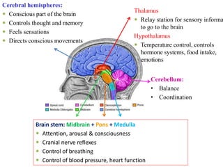
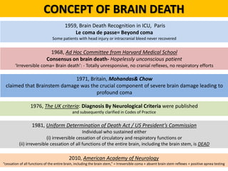
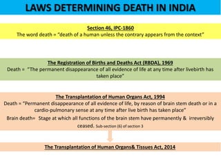
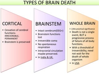
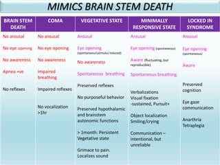
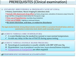
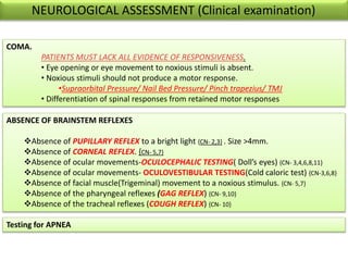
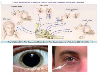
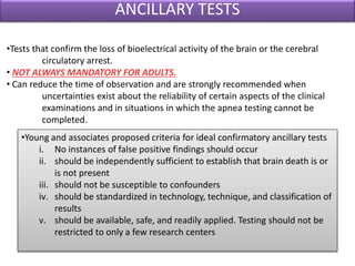
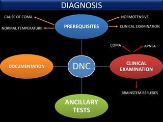
The document discusses the diagnosis of brain death through neurological criteria. It begins by describing the main parts of the brain and their functions. It then discusses the concepts, laws, and types of brain death in India. The key components of determining brain death are described, including establishing the cause of coma, achieving normal temperature and blood pressure, performing neurological examinations to check for absence of brainstem reflexes and response to stimuli, and conducting an apnea test. Potential mimics of brain death and pitfalls in the clinical evaluation are outlined. Confirmatory ancillary tests like cerebral angiography and EEG are also discussed.
Brain death and care for cadaveric organ donarKrishna R
The document discusses the history and criteria for determining brain death. It begins by outlining the evolution of the concept of brain death from the 1960s onwards. Key events included distinguishing brain death from cardiac death with the advent of life support technologies. The document then discusses the anatomy and functions of the brainstem and how brainstem death criteria focus on irreversible loss of brainstem function. It provides details on the tests and criteria used in India to determine brain death, including loss of consciousness, absence of brainstem reflexes, apnea testing, and rule out of confounding factors. Spinal cord reflexes that can occur after brain death are also addressed.
Brain Death and Preparation for Organ DonationRanjith Thampi
This document discusses brain death, including definitions, causes, mechanisms, diagnostic criteria and confirmatory tests. It provides details on:
- Loss of brainstem and cortical function constituting brain death
- Common causes like stroke, trauma, hypoxia
- Mechanism of increased intracranial pressure leading to circulatory arrest
- Clinical criteria including apnea testing over multiple examinations
- Confirmatory tests like EEG, evoked potentials, angiography and imaging to demonstrate lack of cerebral blood flow
This is a presentation on brain death, its background, definition, related neurological conditions, criteria of brain death, brain stem reflexes, causes of coma, confounding factors, observation compatible with brain death, ancillary test, medical record documentation, prognosis, Management of brain death patient.
You tube link of this presentation
https://www.youtube.com/watch?v=3MzE5lHfglI&t=38s
Brain death occurs when there is irreversible cessation of all functions of the entire brain, including the brain stem. It can be caused by conditions that lead to loss of oxygen to the brain like cerebral anoxia, hemorrhage or trauma. Diagnosing brain death involves assessing for the absence of brain stem reflexes and response to stimuli, as well as a positive apnea test where the patient fails to breathe independently with high carbon dioxide levels. Ancillary tests like EEG, angiography or PET scans may be used to confirm the diagnosis when clinical assessment is limited.
Hypertonic Saline Versus Mannitol for Increased Intracranial Pressure ManagementAde Wijaya
Mannitol has traditionally been used as the first-line osmotic agent for decreasing intracranial pressure in various conditions such as traumatic brain injury, hemorrhage, and cerebral infarction. However, mannitol can cause renal failure and hypovolemia as adverse effects. Hypertonic saline formulations may reduce ICP without volume contraction and less nephrotoxicity risk. A recent meta-analysis found that hypertonic saline is more effective than mannitol for treating elevated ICP.
Nurses as the primary care providers would be the immediate health care professional to assess the patient's response and to determine whether he is improving or deteriorating. Signs of brain death can be identified and reported early by a nurse with adequate knowledge.
This document discusses cerebral blood flow and its regulation. It begins with an introduction to the components inside the skull and the Monro-Kellie doctrine. It then covers the anatomy of brain circulation discovered by Willis in 1664, including the anterior and posterior circulations and collateral pathways. Regulation of cerebral blood flow is achieved through hemodynamic autoregulation, metabolic and chemical mediators, neural control, and circulatory peptides. Clinical measurement techniques include laser Doppler flowmetry, transcranial Doppler, and imaging modalities like CT, MRI, PET, and SPECT. Factors like age, hypertension, and failure of autoregulation can impact cerebral blood flow and its regulation.
1. Brain death is defined as the irreversible loss of function of the brain, including the brainstem. It is diagnosed through clinical examinations demonstrating coma, absence of brainstem reflexes, and apnea.
2. The diagnosis of brain death requires meeting certain prerequisites, performing a neurological exam to demonstrate no response to stimuli and no brainstem reflexes, and conducting an apnea test to show the inability to breathe independently.
3. Confirmatory tests such as an EEG, angiogram, or nuclear scan may be used but are not required for the diagnosis of brain death. Proper documentation of meeting criteria is important.
This document discusses brain death, including its definition, criteria, and controversies. It begins by explaining how advances in medicine led to the development of neurological criteria for determining death. It then covers the anatomical and physiological basis of brain death, focusing on damage to the brainstem and reticular formation. The document outlines the clinical criteria for diagnosing brain death, including the absence of brainstem reflexes and apnea testing. It notes complicating factors and observation periods. The history and acceptance of brain death criteria internationally is reviewed. Some bioethical controversies are also presented, such as debates around the definition of death and higher brain formulations.
Brain death current concepts and legal issues in indiaNeurologyKota
This document discusses the history and criteria for determining brain death. It begins by outlining the historical definitions of death from the 1960s onwards, which evolved from cardiopulmonary criteria to brain-based criteria with the development of life-support technologies. The document then examines the anatomical basis of brainstem death and causes that can lead to it. It provides details on the clinical evaluation process for determining brain death in both adults and children based on guidelines from the US and UK. The document also discusses legal aspects of brain death certification in India according to the Transplantation of Human Organs Act.
1. The document discusses the history and techniques of intracranial pressure (ICP) monitoring. It describes historical figures who contributed to the understanding of ICP and various monitoring methods that have been developed over time.
2. The current gold standard for ICP monitoring is an external ventricular drain, though fiberoptic and strain gauge monitors provide alternatives. Newer methods like optic nerve sheath ultrasound provide noninvasive options.
3. Careful analysis of ICP waveforms can provide insights into intracranial compliance and dynamics that help guide management of conditions with elevated ICP like traumatic brain injury.
This document discusses brain death and the criteria used to diagnose it. It begins by describing different states of consciousness including coma, persistent vegetative state, and locked-in syndrome. It then defines brain death as the total and irreversible loss of brain and brainstem function. The key criteria for determining brain death are the absence of cortical function, absence of brainstem reflexes, and apnea during a specific oxygen challenge. Confirmatory tests like angiography, EEG, transcranial Doppler, and nuclear medicine scans can also support the diagnosis. Precise clinical evaluations and testing are required to distinguish brain death from other severe neurological conditions.
This document discusses targeted temperature management (TTM), previously known as therapeutic hypothermia. It provides background on the mechanisms, history, recommendations and methods for TTM. Key points include that inducing mild hypothermia (32-36°C) for 24 hours after cardiac arrest can reduce neurological injury and improve outcomes. Several methods are described for cooling patients, including surface cooling with blankets/pads and internal cooling via intravenous fluids or catheters. Guidelines recommend TTM for comatose cardiac arrest patients with return of spontaneous circulation.
Intracranial pressure - waveforms and monitoringjoemdas
The document discusses intracranial pressure (ICP) waveforms and monitoring. It defines the components of the intracranial vault and describes the normal ICP waveform consisting of P1, P2, and P3 waves representing arterial pulsation, intracranial compliance, and venous pulsation, respectively. It also discusses Lundberg waves including A waves resulting from increased cerebrovascular volume due to vasodilation, B waves related to respiratory fluctuations in PaCO2, and C waves corresponding to Traube-Hering-Meyer fluctuations. The gold standard for ICP monitoring is external ventricular drainage connected to an external strain gauge, which allows CSF drainage but carries risks of infection and hemorrhage. Int
This document discusses brain death, including its prerequisites, neurological exam criteria, ancillary tests, mimics, and prognosis. It notes that brain death is diagnosed through meeting specific criteria including deep coma, absence of brainstem reflexes on exam, and apnea test showing no respiratory effort and rising carbon dioxide levels. Ancillary tests like EEG, evoked potentials, and blood flow studies can help in certain cases. True brain death is considered irreversible, though very young patients may in rare cases survive longer with life support.
Fluid responsiveness in critically ill patientsUbaidur Rahaman
This document discusses fluid responsiveness in critically ill patients. It begins by defining fluid responsiveness as an increase in cardiac index after fluid infusion. It then describes three scenarios involving fluid resuscitation: patients with acute blood or fluid loss requiring immediate resuscitation, patients with suspected septic shock where early goal-directed therapy with fluids is important, and ICU patients who have already received fluids and their fluid responsiveness needs to be assessed. The document discusses various methods of assessing preload and preload dependence, including filling pressures, volumes, respiratory variations in inferior vena cava diameter, inspiratory drops in right atrial pressure, and predicting responsiveness through respiratory variations in parameters related to cardiac index. It emphasizes that preload alone
This presentation consist information about Brain death with special emphasis to differences between Indian and Western Guidelines. Also consist information about Organ transplantation and related act.
This document discusses brain death and organ donation. It begins by outlining the history of defining brain death from 1959 to present. It then explains how brain death is determined, including establishing the cause of coma, performing a clinical examination to demonstrate signs of brain death like coma, brainstem areflexia and apnea, and confirming with ancillary tests if needed. The document provides details on specific tests like the apnea test and discusses special considerations for determining brain death in children. It also outlines conditions that must be excluded before declaring brain death. Finally, it discusses the importance of organ donation and the types of donations possible.
1. Awake craniotomy is a surgical procedure performed with the patient awake to allow mapping of brain functions while removing a brain tumor.
2. During surgery, a neurosurgeon performs cortical mapping to identify vital brain areas that should not be disturbed while removing the tumor.
3. Awake craniotomy provides benefits over surgery under general anesthesia such as higher rates of total tumor resection, fewer permanent neurological deficits, and shorter hospital stays. However, it requires careful patient selection and management of anesthesia to balance pain and cooperation.
A 44-year-old woman collapsed at work and received bystander CPR and defibrillation. She was intubated by EMS and taken to the ED with a pulse but exhibiting seizure activity and a minimally responsive neurological exam. The document then discusses therapeutic hypothermia for cardiac arrest survivors including how to induce and maintain cooling, potential complications, and prognostic factors.
An anatomic coma involves mechanical destruction of the brainstem or cortex from events like hemorrhagic stroke or car accidents. A metabolic coma results from global disruption of metabolic processes due to electrolyte imbalances. Determining brain death requires two independent examinations by qualified physicians demonstrating the absence of brainstem and cortical function and apnea on testing. Organ donation follows a process of determining medical suitability and optimizing donor organ management to potentially save multiple lives through transplantation.
ICU management of traumatic brain injury FemiOpadotun
This document provides information on the management of traumatic brain injury (TBI) in the intensive care unit (ICU). It discusses the epidemiology, pathophysiology, clinical features, and management of TBI. Key points include that TBI is a leading cause of death and disability, with falls, motor vehicle accidents, and assaults being common causes. Primary brain injury results directly from trauma, while secondary brain injury involves downstream effects that can exacerbate damage. Clinical assessment involves the Glasgow Coma Scale and monitoring for signs of increased intracranial pressure. Aggressive ICU management is aimed at preventing secondary injury.
This document discusses altered mental status and provides information on understanding consciousness, conducting an examination of a patient with impaired consciousness, generating a differential diagnosis, and considering various etiologies that can cause altered mental status including focal brain lesions, diffuse brain injuries, infections, toxicities, and metabolic derangements. Three clinical cases are presented and specific conditions such as hypoxia, hypoglycemia, and hepatic encephalopathy are discussed in further detail.
This document discusses brain death, including its historical definition, current diagnostic criteria, pathophysiology, and management of organ donors. Key points include:
- Brain death is defined as irreversible cessation of all functions of the entire brain, including the brain stem.
- Diagnosis requires two examinations at least 6 hours apart showing coma, absence of brainstem reflexes, and apnea during a standardized test. Ancillary tests like EEG can be used if clinical criteria are inconclusive.
- After brain death, pathophysiological changes occur like hypotension, diabetes insipidus, and coagulopathies due to loss of autonomic and endocrine functions regulated by the brain.
The document discusses principles for managing traumatic brain injury, including maintaining optimal cerebral blood flow, limiting intracranial hypertension, and preventing secondary brain insults. It covers monitoring of intracranial pressure, cerebral blood flow, oxygenation and other factors. The goal of treatment is to keep ICP below 20 mmHg and CPP between 50-70 mmHg through various medical, surgical and pharmacologic interventions. More aggressive therapies include hypothermia, decompressive craniectomy or barbiturate coma for refractory intracranial hypertension.
Management of Traumatic Brain Injury in ICUDr.Tarek Sabry
This document discusses the management of traumatic brain injury (TBI) patients in the intensive care unit (ICU). It outlines the key aspects of care including general monitoring, intracranial pressure (ICP) monitoring, analgesia and sedation, mechanical ventilation, hemodynamic support, maintaining normothermia and cerebral perfusion pressure, and preventing secondary insults. The goal of management is to stabilize the patient, prevent intracranial hypertension, and maintain adequate cerebral blood flow and oxygenation through various treatment strategies and intensive care measures.
1) Brain death is defined as irreversible cessation of all functions of the entire brain, including the brainstem. Tests to determine brain death include examining brainstem reflexes and performing an apnea test.
2) Organ donation provides terminally ill patients a new lease on life but organ availability is low in India. Living donors can donate renewable tissues while deceased donors are a major source of organs.
3) For organ donation to occur, all reversible causes of coma must first be excluded through testing and the diagnosis of brain death must be certified by a board of medical experts according to the law.
This document provides information about brain death. It begins with definitions of death and introduces brain death as an irreversible form of unconsciousness characterized by loss of brain function while other organs may still be functioning. The document then discusses the history of how brain death was recognized and defined. It outlines the criteria for determining brain death, including assessing brain stem reflexes and performing an apnea test. The document notes that Indian law recognizes brain death and defines a deceased person. It emphasizes the importance of establishing the cause of brain dysfunction and ruling out potential confounders before determining brain death.
The document discusses guidelines for determining brain death. It begins with historical context for defining death and the need for brain death criteria. Key points include:
- Brain death is the irreversible loss of all brain function, including the brain stem. It allows for legal death even if other organs are functioning.
- Tests to determine brain death include examining for the absence of brain stem reflexes and apnea testing to check for respiratory drive.
- If full criteria are met, including examinations showing no brain activity, the time of death is determined to be when respiratory criteria were met on tests.
1. The document discusses the approach to evaluating and managing a comatose patient. It defines consciousness and the components of arousal and content of consciousness.
2. Evaluation of a comatose patient involves obtaining a detailed history, performing a physical exam including neurological assessment of pupil size and reactivity, eye and motor movements, and determining the level of coma.
3. Management begins with addressing airway, breathing, and circulation (ABCs), treating potentially life-threatening metabolic disorders, evaluating for increased intracranial pressure, and providing supportive care measures.
A detailed presentation on Brain Death and Ongan transplantation.
Criteria for Brain Death are explained in detail. Legislative laws regarding the organ transplant, organ preservation are also explained.
The document discusses changes that occur after death, including cooling of the body, hypostasis (livor mortis), rigor mortis, and decomposition. It provides details on the timing and processes of each change and factors that can influence them. Multiple criteria for determining brain death are also outlined from different medical organizations between 1966-1985. The document recommends brain death be recognized as death and that two specialists should diagnose it.
This document provides an overview of coma, including its anatomical and physiological bases, definition, causes, evaluation, and management. Coma requires dysfunction of the pontine reticular activating system and/or bilateral cerebral hemispheres. Common causes include drug overdose, metabolic derangements, head trauma, anoxia, and stroke. Evaluation involves assessing ABCs, looking for signs of increased intracranial pressure, and performing tests to identify potential causes. Management priorities are supporting ABCs, treating potentially reversible causes like hypoglycemia, and controlling intracranial pressure if elevated.
The document discusses the pathophysiology of brain death in pediatric patients. It begins with a brief history of the concept of brain death and outlines the key findings required for a diagnosis, including coma, absence of brainstem reflexes, and apnea. The document then describes the specific brainstem reflexes and other tests used in the clinical examination, including precautions that must be taken. It emphasizes that determination of brain death requires two examinations by different physicians, separated by an observation period, as well as an apnea test. The role of ancillary tests is also outlined.
1) Brain death is the irreversible loss of all brain and brainstem functions and is diagnosed clinically through examination of coma, absent brainstem reflexes, and apnea on testing.
2) The role of the intensivist is to determine if the patient meets criteria for brain death through clinical examination and ancillary testing, and to prepare potential organ donors.
3) Brain death results from severe brain injury or lack of oxygen that causes raised intracranial pressure, cessation of cerebral blood flow, and ultimately complete necrosis of brain tissue.
The document discusses brain death, its diagnosis and pathophysiology. It defines brain death as the complete and irreversible loss of brain function. The diagnosis involves meeting strict clinical criteria demonstrating the absence of brainstem reflexes as well as confirmatory tests like EEG. Brain death results in no prospect of survival without life support or recovery of brain function. Proper diagnosis is important for organ donation where brain death constitutes legal death.
The document summarizes key aspects of death and brain death from a medicolegal perspective. It defines death and outlines the vital systems that cease to function at death. It discusses the presumption of death and survivorship under Indian law. The document then examines the historical definitions of death and milestones in determining brain death. It provides details on the anatomy of the normal brain and causes, mechanisms, and conditions distinct from brain death. The neurological examination for determining brain death and confirmatory testing are described. The key provisions of the Transplantation of Human Organs Act of 1994 and its amendment in 2009 are summarized.
Brain Death concepts, Its changes and life after brain death, is the body still alive?? what are the determinants of brain death and who can declare it, bio ethical dimensions of nursing care in BD
1. Brain death is defined as the irreversible loss of all brain function, including the brainstem. The three essential findings are coma, absence of brainstem reflexes, and apnea on testing.
2. Common causes of brain death include traumatic brain injury, intracerebral hemorrhage, hypoxic-ischemic injury from cardiopulmonary arrest. A thorough exam is required to determine the cause and rule out potential confounders.
3. Evaluation of brain death involves assessing for coma, absence of brainstem reflexes on exam, and a positive apnea test showing lack of respiratory drive and rising CO2 levels. Ancillary tests like angiography or EEG can also be
Dr. S. K. Nawaz Ahmed's document discusses death and its causes. It defines death as the irreversible loss of brain function, including brainstem function. It describes the stages of death as somatic/clinical death followed by molecular/cellular death. It discusses the modes of death including asphyxia, coma, and syncope based on which organ system fails first. The document also discusses brainstem death criteria, vegetative and permanent vegetative states, and India's Transplantation of Human Organs and Tissues Act regarding organ donation.
Brain cut up for the general pathologistEffiong Akang
Simplified procedure for brain cut up examination for general pathologists that emphasises the importance of good clinicopathological correlation in post-mortem CNS examination. Presented at TSL workshop in Lagos on 25 November 2014
The document discusses care of unconscious patients. It defines various levels of consciousness including alertness, lethargy, stupor, semi-coma, and coma. It also defines vegetative state and brain death. Causes of unconsciousness can be brain-derived like brain injury or hemorrhage, or systemic like low blood sugar, cardiac issues, infections, or intoxication. Common causes interrupt energy delivery, alter neurophysiology, cause osmolar abnormalities, or result in hypoxia or hypercapnia. Myxedema coma, a rare complication of hypothyroidism, is also discussed. Clinical manifestations of decreased consciousness range from behavioral changes to pupillary and motor responses depending on the level of alert
Approach to deceased donor transplantationVishal Golay
This document discusses the process of deceased donor organ transplantation. It covers donor identification, screening, certification of brain stem death, maintenance of potential donors, and counseling of family members. Key steps include confirming brain stem death through clinical tests, screening donors based on medical criteria, obtaining consent from relatives, and providing life support to optimize organ function prior to retrieval. The overall goal is to maximize the number of transplantable organs through proper donor care and management.
Epilepsy is a chronic neurological disorder characterized by recurrent seizures. Seizures occur when there is a sudden, uncontrolled electrical disturbance in the brain. The document discusses the types of seizures including generalized tonic-clonic, absence, myoclonic, complex partial, and simple partial seizures. It also outlines the diagnostic studies, medical management with anti-seizure medications, and surgical options that include resection of the seizure focus area or disconnecting communication between brain hemispheres. Vagus nerve stimulation is another treatment approach where a device implanted in the chest delivers electrical pulses to the vagus nerve in the neck to help prevent seizures.
This document discusses brain death and the criteria used to diagnose it. It begins by describing different states of consciousness including coma, persistent vegetative state, and locked-in syndrome. It then defines brain death as the total and irreversible loss of brain and brain stem functions. The key criteria for determining brain death are the absence of cortical function, absence of brainstem reflexes, and apnea during a specific oxygen challenge. Confirmatory tests like angiography, EEG, Doppler ultrasound, and scintigraphy can also be used when clinical criteria cannot be reliably assessed.
The document discusses various causes of upper airway obstruction including congenital lesions in infants, inflammatory and traumatic issues in children, and complications that can arise from prolonged intubation. It also outlines the steps for performing an adult tracheostomy, including making an incision, separating tissues and strap muscles, opening the trachea, inserting a tracheostomy tube, and potential complications. Different tracheostomy tubes and a percutaneous dilational tracheostomy procedure are also mentioned.
This document discusses the history and development of double lumen tubes (DLTs) for lung separation during surgery. It describes some of the key innovators in DLT design from the 1930s to 1960s, including Gale and Waters, Magill, Carlens, Bryce-Smith, and Robertshaw. Their designs improved aspects like cuff placement, tube shape and size, and ease of insertion. The document also reviews anatomical considerations for optimal placement of right and left-sided DLTs, as well as alternatives that can be used if standard DLT placement is not possible.
This document discusses various topics related to child development and welfare. It begins by defining behavior and outlining different types of behavioral development in children. It then discusses behavioral problems, juvenile delinquency, theories of delinquency, and measures to prevent delinquency. Next, it covers topics such as children in difficult circumstances, the girl child and gender bias, child abuse, its types and effects. Other sections discuss battered baby syndrome, street children, child marriage, child placement options, and legislation related to children.
The document provides a historical overview of malaria. Some key points:
- Malaria symptoms have been described as early as 2700 BC in China and 400 BC by Hippocratic writings.
- In 1880, Charles Louis Alphonse Laveran discovered the malaria parasite in blood samples.
- In 1897, Ronald Ross demonstrated that malaria can be transmitted from infected patients to mosquitoes.
- In 1898, it was shown that the human malaria parasites Plasmodium falciparum, P. vivax, and P. malariae complete their life cycle within mosquitoes.
- Major developments in the 20th century included the 1955 WHO malaria eradication program using DDT and
The document discusses various methods for testing the efficacy of disinfectants, including:
1. Koch's method, which tests the ability of a disinfectant to kill Bacillus anthraces spores.
2. Rideal Walker and Chick-Martin tests, which determine the phenol coefficient of a disinfectant by comparing its bactericidal effects to phenol under clean and dirty conditions.
3. In-use and capacity use dilution tests assess the ability of a disinfectant to kill microbes when diluted in conditions mimicking actual use over time in the presence of organic matter.
4. No single test can reliably determine a disinfectant's efficacy
The document discusses the family life cycle and the role of family in health and disease. It describes the family life cycle as having phases such as formation, extension, and contraction. Family provides socialization, personality formation, and care for dependents. Stress on the family can impact health, and family support influences conditions like heart disease recovery rates. The family is a key context for health behaviors and support during illness.
The document discusses the spectrum of health and diseases and the determinants of health. It states that health lies on a continuum from positive health to death. The determinants of health include both uncontrollable factors like genetics and gender as well as controllable factors like diet, smoking, and exercise. Biological determinants discussed include body weight, genetics, hormones, blood pressure, cholesterol, and glucose regulation. Behavioral determinants include physical activity and social relationships. Social determinants discussed are socioeconomic status, housing, work, and unemployment.
The document discusses various methods for water purification on both large and small scales. It describes the multi-stage process of water treatment including storage, filtration through slow sand filters, and disinfection through chlorination or other methods. Key aspects of slow sand filtration are explained such as the vital biological layer and filtration rates. Advantages of slow sand filtration include high quality water output and simple design and operation.
There are several methods for measuring mortality in a population. Crude death rates measure the number of deaths per 1000 people in a given population over a period of time. Specific death rates measure mortality for particular causes or groups. Standardized rates allow for comparisons between populations by adjusting for differences in age structure. Common metrics include crude death rates, specific death rates, case fatality rates, proportional mortality rates, and survival rates.
Communicating effectively and consistently with students can help them feel at ease during their learning experience and provide the instructor with a communication trail to track the course's progress. This workshop will take you through constructing an engaging course container to facilitate effective communication.
Strategies for Effective Upskilling is a presentation by Chinwendu Peace in a Your Skill Boost Masterclass organisation by the Excellence Foundation for South Sudan on 08th and 09th June 2024 from 1 PM to 3 PM on each day.
Beyond Degrees - Empowering the Workforce in the Context of Skills-First.pptxEduSkills OECD
Iván Bornacelly, Policy Analyst at the OECD Centre for Skills, OECD, presents at the webinar 'Tackling job market gaps with a skills-first approach' on 12 June 2024
বাংলাদেশের অর্থনৈতিক সমীক্ষা ২০২৪ [Bangladesh Economic Review 2024 Bangla.pdf] কম্পিউটার , ট্যাব ও স্মার্ট ফোন ভার্সন সহ সম্পূর্ণ বাংলা ই-বুক বা pdf বই " সুচিপত্র ...বুকমার্ক মেনু 🔖 ও হাইপার লিংক মেনু 📝👆 যুক্ত ..
আমাদের সবার জন্য খুব খুব গুরুত্বপূর্ণ একটি বই ..বিসিএস, ব্যাংক, ইউনিভার্সিটি ভর্তি ও যে কোন প্রতিযোগিতা মূলক পরীক্ষার জন্য এর খুব ইম্পরট্যান্ট একটি বিষয় ...তাছাড়া বাংলাদেশের সাম্প্রতিক যে কোন ডাটা বা তথ্য এই বইতে পাবেন ...
তাই একজন নাগরিক হিসাবে এই তথ্য গুলো আপনার জানা প্রয়োজন ...।
বিসিএস ও ব্যাংক এর লিখিত পরীক্ষা ...+এছাড়া মাধ্যমিক ও উচ্চমাধ্যমিকের স্টুডেন্টদের জন্য অনেক কাজে আসবে ...
Temple of Asclepius in Thrace. Excavation resultsKrassimira Luka
The temple and the sanctuary around were dedicated to Asklepios Zmidrenus. This name has been known since 1875 when an inscription dedicated to him was discovered in Rome. The inscription is dated in 227 AD and was left by soldiers originating from the city of Philippopolis (modern Plovdiv).
Gender and Mental Health - Counselling and Family Therapy Applications and In...PsychoTech Services
A proprietary approach developed by bringing together the best of learning theories from Psychology, design principles from the world of visualization, and pedagogical methods from over a decade of training experience, that enables you to: Learn better, faster!
This document provides an overview of wound healing, its functions, stages, mechanisms, factors affecting it, and complications.
A wound is a break in the integrity of the skin or tissues, which may be associated with disruption of the structure and function.
Healing is the body’s response to injury in an attempt to restore normal structure and functions.
Healing can occur in two ways: Regeneration and Repair
There are 4 phases of wound healing: hemostasis, inflammation, proliferation, and remodeling. This document also describes the mechanism of wound healing. Factors that affect healing include infection, uncontrolled diabetes, poor nutrition, age, anemia, the presence of foreign bodies, etc.
Complications of wound healing like infection, hyperpigmentation of scar, contractures, and keloid formation.
2. Cerebral hemispheres:
Conscious part of the brain
Controls thought and memory
Feels sensations
Directs conscious movements
Thalamus
Relay station for sensory informa
to go to the brain
Hypothalamus
Temperature control, controls
hormone systems, food intake,
emotions
Cerebellum:
• Balance
• Coordination
Brain stem: Midbrain + Pons + Medulla
Attention, arousal & consciousness
Cranial nerve reflexes
Control of breathing
Control of blood pressure, heart function
6. 1959, Brain Death Recognition in ICU, Paris
Le coma de passe= Beyond coma
Some patients with head injury or intracranial bleed never recovered
1968, Ad Hoc Committee from Harvard Medical School
Consensus on brain death- Hopelessly unconscious patient
‘Irreversible coma= Brain death’: - Totally unresponsive, no cranial reflexes, no respiratory efforts
1981, Uniform Determination of Death Act / US President’s Commission
Individual who sustained either
(i) irreversible cessation of circulatory and respiratory functions or
(ii) irreversible cessation of all functions of the entire brain, including the brain stem, is DEAD
2010, American Academy of Neurology
“cessation of all functions of the entire brain, including the brain stem,” = Irreversible coma + absent brain stem reflexes + positive apnea testing
1976, The UK criteria: Diagnosis By Neurological Criteria were published
and subsequently clarified in Codes of Practice
CONCEPT OF BRAIN DEATH
1971, Britain, Mohandas& Chow
claimed that Brainstem damage was the crucial component of severe brain damage leading to
profound coma
7. LAWS DETERMINING DEATH IN INDIA
The Registration of Births and Deaths Act (RBDA), 1969
Death = “The permanent disappearance of all evidence of life at any time after livebirth has
taken place”
Section 46, IPC-1860
The word death = “death of a human unless the contrary appears from the context”
The Transplantation of Human Organs Act, 1994
Death = “Permanent disappearance of all evidence of life, by reason of brain stem death or in a
cardio-pulmonary sense at any time after live birth has taken place”
Brain death= Stage at which all functions of the brain stem have permanently & irreversibly
ceased. Sub-section (6) of section 3
The Transplantation of Human Organs& Tissues Act, 2014
8.
9.
10. TYPES OF BRAIN DEATH
CORTICAL
• Cessation of cerebral
functions-
IRREVERSIBLE,
PERMANENT
• Brainstem is preserved
BRAINSTEM
• Intact cerebrum(EEG+)
• Brainstem functions
lost
• Irreversible coma
• No spontaneous
respiration
• Intracranial circulation
maybe preserved.
• In India & UK.
WHOLE BRAIN
• Little practical significance
• Death is not a single
event, BUT a
progressive process
of failure of all body
functions.
• With a threshold of
irreversibility, need
not wait for the
death of whole
organism
• In USA
11. MIMICS BRAIN STEM DEATH
BRAIN STEM
DEATH
COMA VEGETATIVE STATE MINIMALLY
RESPONSIVE STATE
LOCKED IN
SYNDROME
No arousal
No eye opening
No awareness
Apnea +ve
No reflexes
No arousal
No eye opening
No awareness
Impaired
breathing
Impaired reflexes
No vocalization
>1hr
Arousal
Eye opening
(spontaneousstimulus induced)
No awareness
Spontaneous breathing
Preserved reflexes
No purposeful behavior
Preserved hypothalamic
and brainstem
autonomic functions
> 1month: Persistent
Vegetative state
Grimace to pain.
Localizes sound
Arousal
Eye opening (spontaneous)
Aware (fluctuating, but
reproducible)
Spontaneous breathing
Verbalizations
Visual fixation
-sustained, Pursuit+
Object localization
Smiling/crying
Communication –
intentional, but
unreliable
Arousal
Eye opening
(spontaneous)
Aware
Preserved
cognition
Eye gaze
communication
Anarthria
Tetraplegia
12. • Time of death
• Fill up the check list
• Date and signature
• Contact organ
procurement organization
• Cerebral angiography
• EEG
• Nuclear scan
• TCD
• CTA
• MRI/MRA
• Coma
• Absence of brainstem
reflexes
• Apnea
• Establish irreversible and
proximate cause of coma
• Achieve normal core temperature
• Achieve normal systolic blood
pressure
• Perform one neurological
examination.
PREREQUISITES
NEUROLOGIC
ASSESSMENT
DOCUMENTATION
ANCILLARY
TESTS
THE DETERMINATION OF BRAIN DEATH
13. 1) ESTABLISH IRREVERSIBLE & PROXIMATE CAUSE OF COMA:
History, Examination, Neuro-imaging & Laboratory tests
Exclude CNS-depressant drug effect by history, drug screen, calculation of clearance using 5 times the
drug’s half-life, or drug plasma levels below the therapeutic range
Prior use of hypothermia may delay drug metabolism
Prior use of NMBs: ongoing or continued infusion
No severe electrolyte, acid-base, or endocrine disturbance
The legal alcohol limit for driving (blood alcohol content 0.08%) is a practical threshold below which an
examination to determine brain death could reasonably proceed
2) ACHIEVE NORMAL CORE TEMPERATURE:
A warm blanket may be needed for normal or near-normal temperature.
Avoids any delay in the rise of PaCO2 during apnea testing.
3) ACHIEVE NORMAL SYSTOLIC BLOOD PRESSURE:
Neurological examination is usually reliable with SBP ≥100 mm Hg
Hypotension: Loss of peripheral vascular tone, hypovolemia(Diabetes Insipidus)
Vasopressors or Vasopressin are often required
4) PERFORM ONE NEUROLGICAL EXAMINATION
PREREQUISITES (Clinical examination)
14. NEUROLOGICAL ASSESSMENT (Clinical examination)
COMA.
PATIENTS MUST LACK ALL EVIDENCE OF RESPONSIVENESS.
• Eye opening or eye movement to noxious stimuli is absent.
• Noxious stimuli should not produce a motor response.
•Supraorbital Pressure/ Nail Bed Pressure/ Pinch trapezius/ TMJ
• Differentiation of spinal responses from retained motor responses
ABSENCE OF BRAINSTEM REFLEXES
Absence of PUPILLARY REFLEX to a bright light {CN- 2,3} . Size >4mm.
Absence of CORNEAL REFLEX. {CN- 5,7}
Absence of ocular movements-OCULOCEPHALIC TESTING( Doll’s eyes) {CN- 3,4,6,8,11}
Absence of ocular movements- OCULOVESTIBULAR TESTING(Cold caloric test) {CN-3,6,8}
Absence of facial muscle(Trigeminal) movement to a noxious stimulus. {CN- 5,7}
Absence of the pharyngeal reflexes (GAG REFLEX) {CN- 9,10}
Absence of the tracheal reflexes (COUGH REFLEX) {CN- 10}
Testing for APNEA
15.
16.
17. MANDATORY POINTS TO BE NOTED:
Consultant Physician and/or Neurologist/Neurosurgeon should conduct
a thorough and methodological clinical examination of the neurological
status & functioning of the patient
Examination should remain consistent with prescribed brain stem death
guidelines throughout the observation and testing period
Two tests are necessary, separated by no less than 6 hours in adults
Once a time interval has been set for the second test, it must be
carried out within that interval, either by the examining physician or by
suitable designated medical practitioner
In children,
1 week to 2 months : 48hours
2 months to 1 year: 24 hours
>1 year: 12 hours
18. APNEA TESTING PREREQUISITES:
Normotension
Normothermia
Euvolemia
Eucapnia
Absence of hypoxia
No prior evidence of
CO2 retention
Adjust Vasopressors to a SBP ≥100 mm Hg.
Preoxygenate: 10 minutes. 100% O2, PaO2 >200 mm Hg.
Eucapnia: Ventilation@Rate-10 bpm, PEEP- 5cm H2O
Baseline ABG, If SpO2 remains >95%
Disconnect the patient from the ventilator.
Keep Oxygenating( Insufflation catheter through ETT, close to
the carina, 100% O2 @ 6L/min
Watch for respiratory movements for 8–10min
(Brief gasp, abdominal or chest excursions)
ABORT:
SBP : <90 mm Hg
SpO2 <85% for > 30sec
Respiratory drive +
Arrhythmias
Retry with T-piece, CPAP 10cm
H2O, 100% O2 @ 12 L/min
Repeat ABG, If no respiratory drive is observed for 8min
Apnea test POSITIVE
•No respiratory
movements
•PaCO2 ≥60 mm Hg
•∆PaCO2 > 20mm Hg
INCONCLUSIVE Apnea Test
Repeat after 10-15min after
adequate preoxygenation
19. PITFALLS IN CLINICAL EVALUATION
•Interfere with the clinical diagnosis of brain death.
•Confirmatory tests are recommended for the diagnosis..
Severe facial trauma
Pre existing pupillary abnormalities
Sleep apnea
Toxic levels of sedatives
Amino glycosides
Tricyclic antidepressants
Anticholinergics
Neuromuscular blockers
Chemotherapeutic agents
Antiepileptics
Severe pulmonary diseases
Lazarus sign
Sweating
Blushing
Tachycardia
Normal BP without supports
Absence of diabetes insipidus
Occasional deep tendon reflexes
Superficial abdominal reflexes
Triple flexion response
Babinski response
20. The most common movements:
• Finger jerks
• Undulating toe flexion sign (initial plantar flexion of great toe followed by sequential brief plantar
flexion of the 2nd, 3rd, 4th & 5th toes)
• Triple flexion response (flexion of thigh, leg and dorsiflexion of foot)
• Lazarus sign
• Pronation-extension reflex
• Facial myokymia (bag of worms appearance= continuous twitching of small bands/strips of muscles
giving an undulating/rippling appearance to overlying skin)
21. LAZARUS SIGN
Spontaneous body movements may be observed during the apnea test, while the body is
being prepared for transport, at the time of a skin incision for the retrieval of organs, or in
synchrony with the respiration produced by mechanical ventilation.
BIZARRE, SEEMINGLY PURPOSEFUL movements of the upper extremities, in which the arms
flexed quickly to the chest from the patient’s side, the shoulders adducted, and in some
patients, the hands crossed or opposed just below the chin.
Remember that a patient may move in response to noxious stimulation and that full curarization
is required for surgical procedures involving brain death.
22.
23. STEPS TO BE FOLLOWED WHEN DIAGNOSIS OF BRAIN DEATH IS BEING CONSIDERED
TIME OF DEATH = TIME OF SECOND POSITIVE APNEA TEST
TESTING
INVESTIGATIONS
INFORM TRANSPLANT COORDINATOR
• 1st test done by consultant of ICU unit
• 2nd test done by two consultants (one from the nominated panel)
• Second series of tests done 6 hours after the first
Available / at least sent to the laboratory (by the end of 1st series of tests)
HIV/HCV/ HbsAg
Blood Grouping & Typing
LFTs, WBC, TC/DC
PT, PTT, Platelets
• ONLY After 1st positive apnea test
• ABORT if transplant team examines/enquires/
talks to the candidate’s relatives before
informing them
• Interact with the family & Keep the ICU informed at all times
• Informs other personnel of the transplant team
• Obtain all drugs & fluids necessary, after the first positive apnea test
• Complete all legal formalities except Form-8
24. Neurological examination &
Apnea testing
Inconsistent Incomplete/ Unreliable Consistent
BRAINSTEMDEATHDETERMINATIONPROCESS
FLOWCHART
COMPLETECHECKLIST
INITIATEORGANPROCUREMENTPROCESS
Confirmatory tests
Negative/
Not consistent
Positive/
Consistent
Repeat clinical exam
CANNOT BE DONE
RELIABLE & complete
clinical exam DONE
Repeat confirmatory
tests within 24hrs
NO FURTHER TESTING
25. ANCILLARY TESTS
•Tests that confirm the loss of bioelectrical activity of the brain or the cerebral
circulatory arrest.
• NOT ALWAYS MANDATORY FOR ADULTS.
• Can reduce the time of observation and are strongly recommended when
uncertainties exist about the reliability of certain aspects of the clinical
examinations and in situations in which the apnea testing cannot be
completed.
•Young and associates proposed criteria for ideal confirmatory ancillary tests
i. No instances of false positive findings should occur
ii. should be independently sufficient to establish that brain death is or
is not present
iii. should not be susceptible to confounders
iv. should be standardized in technology, technique, and classification of
results
v. should be available, safe, and readily applied. Testing should not be
restricted to only a few research centers
26. ANCILLARY TESTS (….contd)
CEREBRAL ANGIOGRAPHY (S4VA) , that demonstrates an absent cerebral circulation
remains the GOLD-STANDARD supplementary test for the diagnosis of brain-death
• The contrast medium should be injected in the aortic arch under high pressure and
reach both anterior and posterior circulations.
• No intra-cerebral filling should be detected at the level of entry of the carotid or
vertebral artery to the skull.
• The external carotid circulation should be patent.
• The filling of the superior longitudinal sinus may be delayed.
27. ANCILLARY TESTS (….contd)
ELECTROENCEPHALOGRAM:
• Loss of bioelectric brain activity for at least 30 minutes of recording, is a reliable test.
• EEG may also not show electrical activity in barbiturate coma.
• Devices in the ICU may cause artifacts, leading to spurious results.
• Affected by hypothermia, drug administration and metabolic disturbances.
• A minimum of 8 scalp electrodes should be used.
• Inter-electrode impedance should be between 100 and 10,000 Ω .
• The integrity of the entire recording system should be tested.
• The distance between electrodes should be at least 10 cm.
• The sensitivity should be increased to at least 2 V for 30 minutes.
• The high-frequency filter setting should not be set below 30 Hz & the low-frequency setting should not be above 1 Hz.
• Electroencephalography should demonstrate a lack of reactivity to intense somato-
sensory or audiovisual stimuli.
28. ANCILLARY TESTS (….contd)
TRANSCRANIAL DOPPLER ULTRASONOGRAPHY:
Subject to technical problems
Noninvasive & found to be highly sensitive to determine absent cerebral perfusion
• The abnormalities should include either reverberating flow or small systolic peaks in
early systole.
• A finding of a complete absence of flow may not be reliable owing to inadequate trans-
temporal windows for insonation.
• There should be bilateral insonation and anterior and posterior insonation.
• The probe should be placed at the temporal bone, above the zygomatic arch and the
vertebro-basilar arteries, through the sub-occipital trans-cranial window.
• Insonation through the orbital window can be considered to obtain a reliable signal.
• TCD may be less reliable in patients with a prior craniotomy.
29. ANCILLARY TESTS (….contd)
CEREBRAL SCINTIGRAPHY (technetium Tc 99m hexametazime(HMPAO), have been widely
performed. There is no uptake of isotope in the brain (“hollow skull phenomenon”) in brain-
dead patients
• The isotope should be injected within 30 minutes after its reconstitution.
• Anterior and both lateral planar image counts (500,000) of the head should be obtained at
several time points: immediately, between 30 and 60 minutes later, and at 2 hours.
• A correct IV injection may be confirmed with additional images of the liver demonstrating
uptake (optional).
• No radionuclide localization in the middle cerebral artery, anterior cerebral artery, or basilar
artery territories of the cerebral hemispheres (hollow skull phenomenon).
• No tracer in superior sagittal sinus (minimal tracer can come from the scalp).
30. •Most of these tests have been validated against the gold standard of bedside diagnosis and
are not 100% specific or sensitive.
•May have a place in patients in whom the results of specific components of clinical testing
cannot be reliably evaluated
•Currently do not form part of the mandatory diagnostic requirements in most
countries, including India.
Multimodal Evoked Potentials. (SSEP/BERA)
Computed tomographic Angiography(CTA) – 7 point, 10 point scoring and Perfusion(CTP)
Positron Emission Tomography(PET)
Magnetic resonance Angiography(MRA) and Perfusion(MRP)
ANCILLARY TESTS (….contd)
31. NEONATES,INFANTS&CHILDREN PREREQUISITES:
a) Correct hypothermia, hypotension, metabolic disturbances
b) Discontinue sedatives, analgesics, NMBs, anticonvulsants (based on t ½)
i. Supra therapeutic/ High therapeutic range: No diagnosis based on
Neurological examination alone.
ii. Mid and low therapeutic range: unlikely to affect diagnosis
a) Defer neurological examination for 24-48hrs: Immediately after CPR, severe cute brain
injuries
NUMBER OF EXAMINATIONS, EXAMINERS:
2 examinations separated by an observation period (1st = determines if criteria are
met for BD, 2nd =confirms that the child fulfilled BD criteria)
Examinations by 2 different attending physicians.
Apnea test by Physician in charge of the ventilator
OBSERVATION PERIODS:
A. Neonates(37wk gestation to 30days age): 24hrs
B. Infants(>30days) and Children(<18yrs): 12hrs
APNEA TESTING:
PaCO2 >60mm Hg or ∆PaCO2 >20mm Hg.
Desaturation <85%, or inability to reach PaCO2 60mm Hg: Perform Ancillary tests.
ANCILLARY TESTS:
Never a substitute for clinical examination
If tests are unreliable, waiting period= 24hrs. Supportive care must be continued
during this period.
34. WHOCERTIFIES?“THEPANEL” RMP in charge of hospital
Head of the Institute, RMO, ARMO, Duty RMO
No clearance from Appropriate authority
RMP (Physician/ Surgeon/ Intensivist)
Clearance from Appropriate authority
Neurologist or Neuro-surgeon
Intensivists & Anaesthesiologists
Clearance from Appropriate authority
RMP treating the aforesaid patient
No clearance from Appropriate authority4
2
3
1
1st & 2nd medical examinations of Form-8 of THO rules: Category 2&3 doctors
No category 3 doctor? Request from any panel member from another hospital
Appropriate authority = Director of Medical & Rural Health Services
35. Need for Declaration of Brain Death
Once an unequivocal diagnosis of brain death has been made,
Most medical and legal authorities agree that continuing treatment is
not in the interest of the patient or is ethically permissible.
This is not related to withdrawing support to allow a patient to die, but
rather to ceasing a futile intervention in a patient who is already dead.
Acceptance of this approach would
Reduce human distress
Lead to the rational use of the limited ICU facilities
Increase the availability of organs.
Solid organs can be donated only after confirmation of brain death.
Unless medical personnel provide family members with information that
all cognitive and life support functions have irreversibly stopped, the
family may harbor false hopes for the loved one's recovery.
36. Ethical Issues in Declaring Brain Death
•The DDR (Dead Donor Rule) is the formalization of the widely held belief that it
is wrong to kill one person to save the life of another, leading to the conclusion
that people should already be dead before vital organs are removed, an act that
would certainly kill them.
•The DDR is neither a law nor a regulation — it is a description of an ethical
norm: an organ donor must be dead before vital organs are removed.
37. OFFENSES & PENALITIES
CHAPTER VI, THOA,1994
PUNISHMENT FOR REMOVAL OF HUMAN ORGANS WITHOUT AUTHORITY:
Any person who
Renders his service to or at any hospital
Conducts, associates with, or helps in any manner for removal of any organ
5 years Imprisonment + up to Rs.10,000
If he/she is a RMP
Reported to the appropriate authority, state medical council
Cancel registration for 2 years for the first offence
Permanent cancellation for subsequent offence.
38. OFFENSES & PENALITIES
CHAPTER VI, THOA,1994
PUNISHMENT FOR REMOVAL OF HUMAN ORGANS WITHOUT AUTHORITY:
Any person who
Makes or receives payment for the supply of, or offers to supply any human organ
Seeks to find a person who is willing to supply any organ for payment
Offers to supply any organ for payment
Initiates or negotiates any arrangement for payment
Takes part in management/control of a body of persons(society/firm/company)
Advertisements – publishes/ distributes/ causes to be published/distributed
Inviting persons to supply organs for payment
Offering to supply organs for payment
Term not less than 2 years (up to 7 years) +
Fine not less than Rs.10000 (up to Rs.20,000)
42. Demand for organs- much higher than organs which are being available
With demand, the responsibility to caring for potential organ donors is also rising
Appropriate multidisciplinary team approach for successful organ transplantation
WHY…..???
For graft survival after donation, all available organs in the donor need to be
maintained at their normal physiological condition until the time of organ retrieval
Early identification of a potential donor is critical in starting early medical
management for successful organ transplantation
43. WHO CERTIFIES…??
A panel of four doctors:
(I) Hospital administrator
(II) Resident doctor of the hospital
(III) Neurologist/Neurosurgeon/Physician, Intensivist, and Anesthetist
(IV) Treating doctor.
In the recent 2014 rules apart from neurologist or neurosurgeons,
anesthetists, critical care specialists, intensivists, physicians, or
surgeons have been included to facilitate brain death declaration
In MLC cases, Role of Forensic medicine..????
Police clearance is MUST. (Superintendent of Police or Deputy Inspector General)
44. POTENTIAL ORGAN DONOR
• An individual who has suffered a fatal injury to the brain (with impending or
actual brain death), yet has intact cardiovascular function.
•Cause of death:
• Severe traumatic head injury
•Others: Primary Brain tumors, CVAs, drug overdose.
i. Meet age and Brain dead criteria. {Newborn to 65years. Ideally 10-50 years}
ii. Infection free. Antibiotic coverage and negative cultures are necessary
iii. STD screening. HIV, HTLV, Syphilis, Viral hepatitis
iv. No history of carcinoma, except primary brain or low-grade skin caner
v. Free of severe systemic disease
vi. Relatively normal organ functions
vii. Hemo-dynamically salvageable
viii.At least one organ that has a reasonable likelihood of functioning well
(post transplantation)
46. ABSOLUTE CONTRAINDICATIONS
•Active visceral or hematologic neoplasm
•Clinical signs that indicate organ is unlikely to function well
•Transmissible infections that will adversely affect recipient
•HIV
•Active HBV
•Encephalitis of unknown cause
•CJD
•Disseminated TB
Organ donation exclusion criteria related to HIV risk:
•Drugs( iv, im, sc) for nonmedical reasons in past 5 years
•Persons with hemophilia, who received human derived clotting factors
•Persons engaged in sex in exchange for money, drugs or with another man in the past 5
years
•Persons exposed to known/suspected HIV infected blood in past 12 months
•Inmates of correctional systems.
If the risk to the recipient of not performing the transplant is deemed to be greater than the
risk of transmitting disease, then it is carried out & the recipient is informed the potential risk
48. PATHOPHYSIOLOGY OF BRAIN DEATH
Severe damage to neuronal tissue
Intra-cranial edema
Raised ICP
Reduces cerebral perfusion
Reduces cerebral blood flow
Trans-tentoral herniation
Coning of the foramen magnum
Crushing of the brain stem
Permanent dysfunction
VISCIOUS
Late neuronal injury
Hypoxic ischemic
brain injury
MUNRO-KELLY
DOCTRINE
1. VASOGENIC EDEMA(↑ vascular permeability)
2. CYTOGENIC EDEMA (altered cellular osmoregulation)
CUSHING’S
REFLEX
49. Death is an ONGOING PROCESS, rather than isolated event.
Total loss of neuro-physiological functions for >8 minutes confirms
total &irreversible loss of brain function.
Complete cardiovascular and autonomic uncoupling occurs.
Hypernatremia and diabetes insipidus are more often the effect
rather than the cause of brain death.
Even with maximal support, cardio-respiratory deterioration
leading to somatic death will occur within days.
Damage to the RF may lead to loss of cognition, persistent
unconsciousness and coma.
51. CARDIOVASCULAR
• Physiological, histological, biochemical and electrocardiographic evidence of "damage" to
the heart at the time of brain death.
• Induced by the vascular regulatory injury and the diffuse metabolic injury.
• The autonomic storm results from a sudden increase in ICP and cerebral ischemia (Cushing's
reflex), a last-ditch effort by the brain to maintain its perfusion.
• The magnitude of the rise in catecholamines and the sequelae thereof depend on the rate
of rise in intracranial pressurel5
54. RESPIRATORY
•LUNGS- most often assumed to be unsuitable for transplantation.
•Only 10-20% of lungs are eligible for transplantation
• 2 factors: Neurogenic pulmonary edema (NPE) and inflammatory acute lung injury
Catecholamine
storm
Systemic VC
↑Afterload
↑ LA & LV pressures
↑ Pulmonary blood volume
↑ Pulmonary artery pressure
Massive pulmonary capillary pressure
Pulmonary edema
↑ Hydrostatic pressure
Endothelial damage
Capillary leakage
Respiratory arrhythmias
Other factors:
Pro-inflammatory mediators
Chest wall trauma
Atelectasis
Aspiration
Long term Mechanical ventilation
55. TEMPERATURE
Loss of thermoregulatory control
Exposure to cold ambient temperature
Massive infusion of cold IvF & Blood
Loss of protective mechanisms ( Vasoconstriction, Shivering)
↓ BMR
Preventive Hypothermia is attempted rather than treating hypothermia.
Once hypothermia sets in, it is difficult to warm the patient.
Hypothermia
Induces Arrhythmias
Alters coagulation cascade
Interferes with O2 delivery to tissues
Affects cardiac function
POIKILOTHERMIC
56. RENAL
Biomarkers for renal tubular injury increases as early as 30 minutes after onset .
Both pro-inflammatory and pro-coagulant effects are contributing factors
Pre-transplant workup (of Brain-Dead Donors) showed more T-Lymphocytes and
Macrophages than the live donors.
The Na/K ATPase mechanism requires ATP produced from mitochondria to
maintain Na/K ratio. ( ionic gradient)
• Levels of ATP in human cadaver kidneys correlate well with post-transplantation
function
Aggravating factors
Marked vasoconstriction (Autonomic Storm)
Hypovolemia
Diabetes Insipidus
Marked histological changes associated with
renal dysfunction have been described in the
kidneys of brain dead organ donors maintained
for prolonged periods on Vasopressin and
adrenaline
57. HEMATOLOGICAL
Necrotic brain
tissue
Thromboplastin
Plasminogen activators
DIC
Passage of cerebral tissue into the circulation may also initiate the clotting cascade,
with a consumptive coagulopathy ensuing
Brain Death → Catecholamines →Stress Response → Tendency to coagulate↑
Anemia and coagulopathy are common
Deranged coagulation profile (PT & aPTT)
Thrombocytopenia
↑Total Cell counts
58. ENDOCRINE
HYPOTHALAMO-PITUTORY-NEUROENDOCRINE AXIS
• Posterior pituitary function is clinically lost in 80% BDDs
•Anterior pituitary: relatively preserved because of pituitary blood flow
•Normal ACTH, GH, TSH
•SICK-EUTHYROID SYNDROME
•Diabetes Insipidus
•Hyperglycemia
•↓ insulin secretion
•↑ insulin resistance
•Hyperosmolarity.
Untreated Hyperglycemia: pancreatic cell damage & alters Renal Transplant outcome.
59.
60. CARDIOVASCULAR SUPPORT
Maintain tissue perfusion ( adequate Cardiac output& Tissue perfusion pressure)
Correct intravascular fluid depletion : aggressive fluid resuscitation.
Invasive BP, CVP. (if unstable, Pulmonary artery catheter to monitor Q, PAOP, SVR)
Choice of fluid? { Hb, Albumin, Electrolytes, coagulation profile, ongoing losses }
Vasopressor(s) support. Prefer agents with both Ionotropic and Vasopressor properties
DOPAMINE (2-5 mcg/kg/min)- improves perfusion of transplantable abdominal organs
DOBUTAMINE/ NORADRENALINE
Arrhythmias found to occur in 27-56% in any form.
Diuretics, Diabetes Insipidus, Acidosis: further worsen arrhythmias
Correct the coexisting conditions first & then arrhythmias can be managed.
MAP >60mm Hg
CVP up to 15mm HgULTIMATE GOAL= NORMOTENSIVE, EUVOLEMIC STATE
61. VENTILATORY SUPPORT
Lung protective mechanisms.
TV(6-9ml/kg) with optimal PEEP to maintain PaO2 > 70-80mm Hg
Optimal PEEP may reduce high FiO2 requirements. ( FiO2 to maintain SpO2 >90%)
Avoid PEEP > 15cm H2O
HOB 30-40 degrees
Tracheal cuff pressure 25cm H2O
Maintain normocarbia. (if not possible, moderate hypercarbia is permitted)
Recruitment maneuvers initially, repeat after apnea testing and tracheal suction.
If hyperventilation is employed, discontinue after diagnosis of Brain Death
Avoid administration of excess IVF. (Consider Diuretics, if marked fluid overload)
ABG every 4 hours, After 30min of change in settings
Paw< 40 m H2O
Pplat <35 cm H2O
FiO2- [ SpO2>92% and PaO2 > 70-80mm Hg]
Rate- To maintain normocarbia and pH
Auto PEEP < 5 cm H2O
TV- 6-9ml/kg
62. FLUIDS & ELECTROLYTES
s. Na Fluid
> 150 mEq/L 0.2% NS
<133 mEq/L 0.9% NS
< 128mEq/L 3% NS
s. K Correction
>5.8 mEq/L 50cc 5%D, 15U Insulin, 50mEq NaHCO3
<3.4 mEq/L 20mEq KCl, 2 doses over 1 hour
<3.1 mEq/L 20mEq KCl, 3 doses over 1 hour
< 2.9 mEq/L 20mEq KCl, 4 doses over 1 hour
↓ Mg <1.5mg/dl: 4gm MgSO4 over 2 hours
↓ P <2.2mg/dl: 30 mmol Potassium/ Sodium phosphate over 2 hours and repeat
↓ Ionized Ca <2.1 mEq/L: 10cc of 10% Ca.Gluconate, slow iv and repeat if needed
64. BLOOD & COAGULATION
Coagulopathy:
PLC< 65,000/CC: 1 Platelet pack transfusion (5-6 units of platelets)
PT>15sec, aPTT >38s seconds: 4 units of FFP, repeat after 30min& 1 hour
Fibrinogen <100mg/dl, 6 units of cryoprecipitate. Repeat after 1 and 2 hours.
Anemia:
Reserve 2-4 units of PRBC.
Maintain Hct 28-30% with transfusion
Insert NGT, if UGI bleed is suspected