The document discusses the anatomy of the thorax, including:
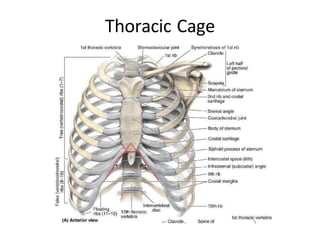
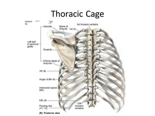
1) The thorax is the region between the neck and abdomen containing the thoracic cage or wall which includes the sternum, 12 pairs of ribs, and 12 thoracic vertebrae, enclosing the thoracic cavity.
2) The thoracic cage protects the abdominal viscera and contains various joints including costovertebral, costotransverse, and costochondral joints.
3) The thoracic wall has intrinsic muscles between the ribs and extrinsic muscles on the exterior surface.


![Joint between manubrium and body of
sternum
Manubriosternal joint :
Secondary cartilagenous ( Symphysis)
[Synostosis in elderly]
Joint in between body of sternum & xiphoid
process
Xiphisternal joint :
Primary cartilagenous (Synchondrosis)
Intervertebral Joints
Sternoclavicular Joint
Joints of thoracic cage](https://image.slidesharecdn.com/thoracicwall5-2-15-240108010119-cb2faa55/85/thoracic_wall_5-2-15-pptx-13-320.jpg)


