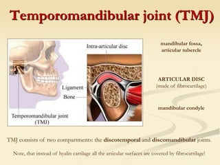
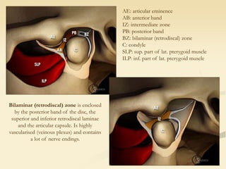
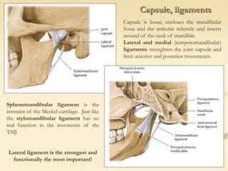
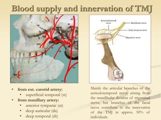
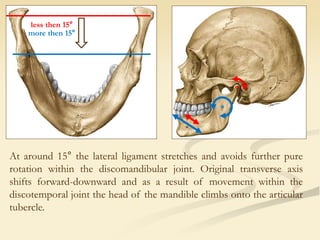
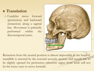
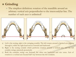
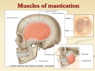
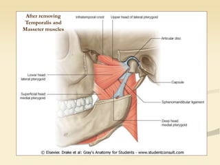
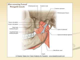
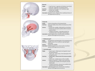
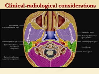
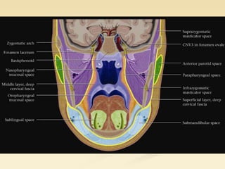
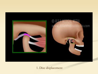
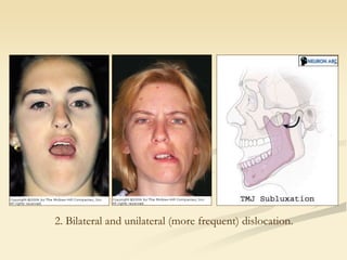
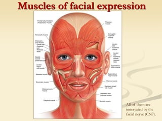
Temporomandibular joint
Muscles and mechanism of mastication
Muscles of facial expression
Mark Kozsurek, M.D., Ph.D.
mark@kozsurek.hu
EM I., 03/12/2018
Probably you’ve already seen in the dissection room...
Or will see soon...
coronoid process
condylar process
head or condyle of the mandible
pterygoid fovea
masseteric tuberosity
pterygoid tuberosity
mandibular fossa
articular eminence
petrotympanic fissure
(of Glaser)
articular eminence
mandibular fossa
petrotympanic fissure
(of Glaser)
Temporomandibular joint (TMJ)
mandibular fossa,
articular tubercle
mandibular condyle
ARTICULAR DISC
(made of fibrocartilage)
TMJ consists of two compartments: the discotemporal and discomandibular joints.
Note, that instead of hyalin cartilage all the articular surfaces are covered by fibrocartilage!
superior retrodiscal lamina
(elastic fibres)
inferior retrodiscal lamina
(collagen fibres)
• articular disc is thicker anteriorly and
posteriorly
• anterior border of the disc is in contact
with the upper insertion of the superior
belly of the lat. pterygoid muscle
• position of the disc is determined by the
superior part of lat. pterygoid muscle and
the elastic fibres of sup. retrodiscal lamina
AE: articular eminence
AB: anterior band
IZ: intermediate zone
PB: posterior band
BZ: bilaminar (retrodiscal) zone
C: condyle
SLP: sup. part of lat. pterygoid muscle
ILP: inf. part of lat. pterygoid muscle
Bilaminar (retrodiscal) zone is enclosed
by the posterior band of the disc, the
superior and inferior retrodiscal laminae
and the articular capsule. Is highly
vascularised (veinous plexus) and contains
a lot of nerve endings.
Capsule, ligaments
Capsule is loose, encloses the mandibular
fossa and the articular tubercle and inserts
around of the neck of mandible.
Lateral and medial (temporomandibular)
ligaments strenghten the joint capsule and
limit anterior and posterior movements.
Sphenomandibular ligament is the
remnant of the Meckel-cartilage. Just like
the stylomandibular ligament has no
real function in the movments of the
TMJ.
Lateral ligament is the strongest and
functionally the most important!
Blood supply and innervation of TMJ
• from ext. carotid artery:
• superficial temporal (st)
• from maxillary artery:
• anterior tympanic (at)
• deep auricular (da)
• deep temporal (dt)
Mainly the articular branches of the
auriculotemporal nerve arising from
the mandibular division of trigeminal
nerve, but branches of the facial
nerve contribute to the innervation
of the TMJ in approx. 50% of
individuals.
Biomechanics of mastication
 Open-close movement
(synonyms: abduction-adduction or elevation-depression)
 1st phase: rotation in the discomandibular joint around a
bicondylar axis, e.g. during talk;
 2nd phase: gliding in the discotemporal joint around a
transverse axis connecting the two mandibular foramina.
New axis
of rotation
At around 15° the lateral ligament stretches and avoids further pure
rotation within the discomandibular joint. Original transverse axis
shifts forward-downward and as a result of movement within the
discotemporal joint the head of the mandible climbs onto the articular
tubercle.
less then 15°
more then 15°
 Translation
 Condyles move forward
(protrusion) and backward
(retraction) along a sagittal
line. Movement is primarily
performed within the
discotemporal joints.
Retraction from the neutral position is almost impossible as the head of
mandible is arrested by the external acoustic meatus, and mouth has to
be slightly opened for protrusion otherwise upper front teeth will not
let the lower ones to move forward.
 Grinding
 The simpliest definition: rotation of the mandible around an
arbitrary vertical axis perpendicular to the intercondylar line. The
number of such axes is unlimited!
a) Left is the resting, right is the swinging condyle. The left rotates around a vertical axis passing
through it, while the right head moves forward and backward.
b) Right is the resting condyle which performs rotation around a vertical axis, and the left
condyle swings anteriorly-posteriorly.
c) Both the condyles swing: one forward, the other one backward and vice versa. Axis of
rotation is a vertical axis located half way between the two condyles.
Muscles of mastication
After removing
Temporalis and
Masseter muscles
After removing Lateral
Pterygoid muscle
Clinical-radiological considerations
1. Disc displacement
2. Bilateral and unilateral (more frequent) dislocation.
Muscles of facial expression
All of them are
innervated by the
facial nerve (CN7).
Thank you for your attention!

tmj_facialexpr_em1_kom2018.ppt

  • 1.
    Temporomandibular joint Muscles andmechanism of mastication Muscles of facial expression Mark Kozsurek, M.D., Ph.D. mark@kozsurek.hu EM I., 03/12/2018
  • 2.
    Probably you’ve alreadyseen in the dissection room... Or will see soon... coronoid process condylar process head or condyle of the mandible pterygoid fovea masseteric tuberosity pterygoid tuberosity
  • 3.
    mandibular fossa articular eminence petrotympanicfissure (of Glaser) articular eminence mandibular fossa petrotympanic fissure (of Glaser)
  • 4.
    Temporomandibular joint (TMJ) mandibularfossa, articular tubercle mandibular condyle ARTICULAR DISC (made of fibrocartilage) TMJ consists of two compartments: the discotemporal and discomandibular joints. Note, that instead of hyalin cartilage all the articular surfaces are covered by fibrocartilage!
  • 5.
    superior retrodiscal lamina (elasticfibres) inferior retrodiscal lamina (collagen fibres) • articular disc is thicker anteriorly and posteriorly • anterior border of the disc is in contact with the upper insertion of the superior belly of the lat. pterygoid muscle • position of the disc is determined by the superior part of lat. pterygoid muscle and the elastic fibres of sup. retrodiscal lamina
  • 6.
    AE: articular eminence AB:anterior band IZ: intermediate zone PB: posterior band BZ: bilaminar (retrodiscal) zone C: condyle SLP: sup. part of lat. pterygoid muscle ILP: inf. part of lat. pterygoid muscle Bilaminar (retrodiscal) zone is enclosed by the posterior band of the disc, the superior and inferior retrodiscal laminae and the articular capsule. Is highly vascularised (veinous plexus) and contains a lot of nerve endings.
  • 7.
    Capsule, ligaments Capsule isloose, encloses the mandibular fossa and the articular tubercle and inserts around of the neck of mandible. Lateral and medial (temporomandibular) ligaments strenghten the joint capsule and limit anterior and posterior movements. Sphenomandibular ligament is the remnant of the Meckel-cartilage. Just like the stylomandibular ligament has no real function in the movments of the TMJ. Lateral ligament is the strongest and functionally the most important!
  • 8.
    Blood supply andinnervation of TMJ • from ext. carotid artery: • superficial temporal (st) • from maxillary artery: • anterior tympanic (at) • deep auricular (da) • deep temporal (dt) Mainly the articular branches of the auriculotemporal nerve arising from the mandibular division of trigeminal nerve, but branches of the facial nerve contribute to the innervation of the TMJ in approx. 50% of individuals.
  • 9.
    Biomechanics of mastication Open-close movement (synonyms: abduction-adduction or elevation-depression)  1st phase: rotation in the discomandibular joint around a bicondylar axis, e.g. during talk;  2nd phase: gliding in the discotemporal joint around a transverse axis connecting the two mandibular foramina. New axis of rotation
  • 10.
    At around 15°the lateral ligament stretches and avoids further pure rotation within the discomandibular joint. Original transverse axis shifts forward-downward and as a result of movement within the discotemporal joint the head of the mandible climbs onto the articular tubercle. less then 15° more then 15°
  • 12.
     Translation  Condylesmove forward (protrusion) and backward (retraction) along a sagittal line. Movement is primarily performed within the discotemporal joints. Retraction from the neutral position is almost impossible as the head of mandible is arrested by the external acoustic meatus, and mouth has to be slightly opened for protrusion otherwise upper front teeth will not let the lower ones to move forward.
  • 13.
     Grinding  Thesimpliest definition: rotation of the mandible around an arbitrary vertical axis perpendicular to the intercondylar line. The number of such axes is unlimited! a) Left is the resting, right is the swinging condyle. The left rotates around a vertical axis passing through it, while the right head moves forward and backward. b) Right is the resting condyle which performs rotation around a vertical axis, and the left condyle swings anteriorly-posteriorly. c) Both the condyles swing: one forward, the other one backward and vice versa. Axis of rotation is a vertical axis located half way between the two condyles.
  • 14.
  • 15.
  • 16.
  • 18.
  • 20.
  • 21.
    2. Bilateral andunilateral (more frequent) dislocation.
  • 23.
    Muscles of facialexpression All of them are innervated by the facial nerve (CN7).
  • 24.
    Thank you foryour attention!