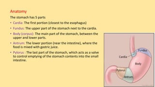
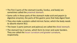
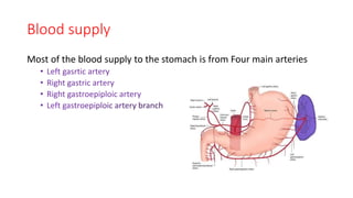
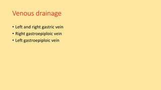
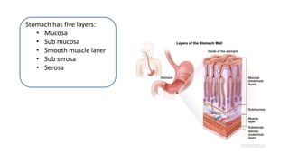
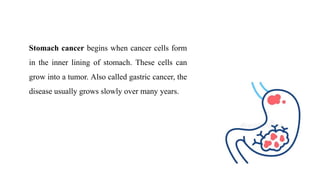
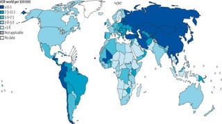
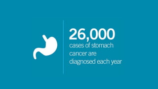
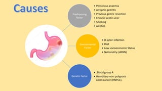
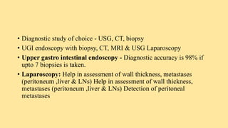
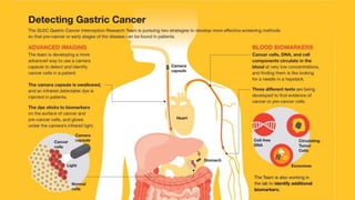
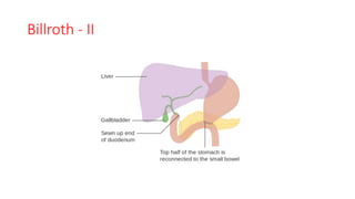
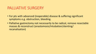
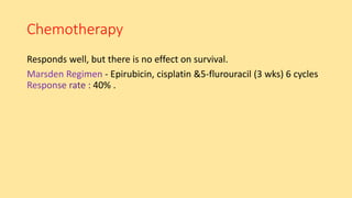
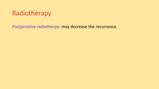
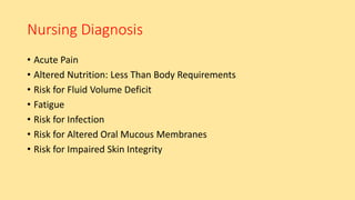
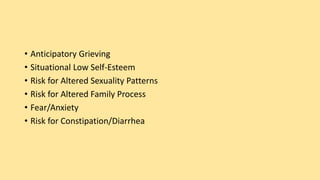
Presented by:
Ms. Elizabeth M.Sc (N)
Asst. Professor,
Dept of MSN,
NNC, GNSU
Anatomy
The stomach has 5 parts
• Cardia: The first portion (closest to the esophagus)
• Fundus: The upper part of the stomach next to the cardia.
• Body (corpus): The main part of the stomach, between the
upper and lower parts.
• Antrum: The lower portion (near the intestine), where the
food is mixed with gastric juice.
• Pylorus : The last part of the stomach, which acts as a valve
to control emptying of the stomach contents into the small
intestine.
• The first 3 parts of the stomach (cardia, fundus, and body) are
sometimes called the
• Some cells in these parts of the stomach make acid and pepsin (a
digestive enzyme), the parts of the gastric juice that help digest food.
• They also make a protein called intrinsic factor, which the body needs
to absorb vitamin B12.
• The lower 2 parts (antrum and pylorus) are called the
• The stomach has 2 curves, which form its inner and outer borders.
They are called the ,
respectively.
Blood supply
Most of the blood supply to the stomach is from Four main arteries
• Left gasrtic artery
• Right gastric artery
• Right gastroepiploic artery
Venous drainage
• Left and right gastric vein
• Right gastroepiploic vein
• Left gastroepiploic vein
Lymphatic drainage
It has into four zones:
• Superior gastric
• Suprapyloric
• Pancreaticolienal
• Inferior gastric/subpyloric
Stomach has five layers:
• Mucosa
• Sub mucosa
• Smooth muscle layer
• Sub serosa
• Serosa
Stomach cancer begins when cancer cells form
in the inner lining of stomach. These cells can
grow into a tumor. Also called gastric cancer, the
disease usually grows slowly over many years.
Predisposing
factor
• Pernicious anaemia
• Atrophic gastritis
• Previous gastric resection
• Chronic peptic ulcer
• Smoking
• Alcohol.
Environmental
Factor
• H.pylori infection
• Diet
• Low socioeconomic Status
• Nationality (JAPAN)
Genetic Factor
• .Blood group A
• Hereditary non- polyposis
colon cancer (HNPCC).
Clinical Presentation
Common clinical Presentation: 3A”s:
1.Anaemia(due to bleeding from tumour)
2.Asthenia(septic absorption from the tumour)
3.Anorexia
• onset of early satiety, dyspepsia, epigastric discomfort
Specific symptoms depending on the site of tumour.
- gastric outlet obstruction.
- dysphagia, hamaetemesis.
- mass per abdomen(silent variety).
- jaundice, ascites
• Grossly Anemic,
• Cachexia,
• Epigastric mass,
• Virchows node
• Sister mary joseph node
• Krukenberg tumor
• Irish node
Stages of gastric cancer
Staging of Gastric Cancer
T1 - lamina propria & sub - mucosa
T2 - muscularis & sub - serosa
T3 - serosa
T4 - Adjacent organs
N0 - no lymph node
N1 - Epigastric node
N2 - main arterial trunk
Mo - distal metastasis
M1 - distal metastasis
Spread of Gastric Cancer
• Direct Spread
• Blood-borne metastasis
• Lymphatic spread
• Transperitoneal spread
INVESTIGATIONS
• Full blood count
• LFT, RFT
• Stool examination for occult blood
• CXR
• Serum tumor markers (CA 72-4,CEA,CA19- 9)
• Diagnostic study of choice - USG, CT, biopsy
• UGI endoscopy with biopsy, CT, MRI & USG Laparoscopy
• Upper gastro intestinal endoscopy - Diagnostic accuracy is 98% if
upto 7 biopsies is taken.
• Laparoscopy: Help in assessment of wall thickness, metastases
(peritoneum ,liver & LNs) Help in assessment of wall thickness,
metastases (peritoneum ,liver & LNs) Detection of peritoneal
metastases
Management
Surgery Chemotherapy Radiotherapy
Initial treatment
1.Improve nutrition if needed by parentral or enteral feeding.
2.Correct fluid &electrolyte & anemia if they are present.
Though some superficial cancers can be treated endoscopically,
gastrectomy is the most widely used approach
1. Total gastrectomy - usually performed for lesions in the upper third
(proximal) stomach
2. Distal subtotal gastrectomy - performed for tumors in the distal (lower two-
thirds) of the stomach
RADICAL GASTRECTOMY
• Remove the stomach +distal part of
esophagus+ proximal part of duodenum +
greater & lesser omentum + Lymph Nodes
• Oesophagojejunostomy with roux-en-y
gastric bypass surgery
SUBTOTAL GASTRECTOMY
• Similar to total one except that the
PROXIMAL PART of the stomach is
preserved
• Followed by reconstruction &
creating anastomosis ( by
gastrojejunostomy, billroth II )
Billroth - II
PALLIATIVE SURGERY
• For pts with advanced (inoperable) disease & suffering significant
symptoms e.g. obstruction, bleeding.
• Palliative gastrectomy not necessarily to be radical, remove resectable
masses & reconstruct (anastomosis/intubation/stenting/
recanalisation)
POSTOPERATIVE ORDERS
• Admit to PACU
• Detailed nutritional advise (small frequent meals)
Post-Operative Complications
• Leakage from duodenal stump.
• Secondary hemorrhage.
• Nutritional deficiency in long term.
Chemotherapy
Responds well, but there is no effect on survival.
- Epirubicin, cisplatin &5-flurouracil (3 wks) 6 cycles
40% .
Radiotherapy
may decrease the recurrence.
Nursing Diagnosis
• Acute Pain
• Altered Nutrition: Less Than Body Requirements
• Risk for Fluid Volume Deficit
• Fatigue
• Risk for Infection
• Risk for Altered Oral Mucous Membranes
• Risk for Impaired Skin Integrity
• Anticipatory Grieving
• Situational Low Self-Esteem
• Risk for Altered Sexuality Patterns
• Risk for Altered Family Process
• Fear/Anxiety
• Risk for Constipation/Diarrhea
Nursing Management
• Monitor nutritional intake and weigh patient regularly.
• Monitor CBC and serum vitamin B12 levels to detect anemia, and monitor
albumin and prealbumin levels to determine if protein supplementation is needed.
• Provide comfort measures and administer analgesics as ordered.
• Frequently turn the patient and encourage deep breathing to prevent pulmonary
complications, to protect skin, and to promote comfort.
• Maintain nasogastric suction to remove fluids and gas in the stomach and prevent
painful distention.
• Provide oral care to prevent dryness and ulceration.
• Keep the patient nothing by mouth as directed to promote gastric wound healing.
Administer parenteral nutrition, if ordered.
• When nasogastric drainage has decreased and bowel sounds have returned, begin
oral fluids and progress slowly.
• Avoid giving the patient high-carbohydrate foods and fluids with meals, which
may trigger dumping syndrome because of excessively rapid emptying of gastric
contents.
• Administer protein and vitamin supplements to foster wound repair and tissue
building.
• Eat small, frequent meals rather than three large meals.
• Reduce fluids with meals, but take them between meals.
• Stress the importance of long term vitamin B12 injections after gastrectomy to
prevent surgically induced pernicious anemia.
• Encourage follow-up visits with the health care provider and routine blood studies
and other testing to detect complications or recurrence.

4. Gastric Cancer

  • 1.
    Presented by: Ms. ElizabethM.Sc (N) Asst. Professor, Dept of MSN, NNC, GNSU
  • 2.
    Anatomy The stomach has5 parts • Cardia: The first portion (closest to the esophagus) • Fundus: The upper part of the stomach next to the cardia. • Body (corpus): The main part of the stomach, between the upper and lower parts. • Antrum: The lower portion (near the intestine), where the food is mixed with gastric juice. • Pylorus : The last part of the stomach, which acts as a valve to control emptying of the stomach contents into the small intestine.
  • 3.
    • The first3 parts of the stomach (cardia, fundus, and body) are sometimes called the • Some cells in these parts of the stomach make acid and pepsin (a digestive enzyme), the parts of the gastric juice that help digest food. • They also make a protein called intrinsic factor, which the body needs to absorb vitamin B12. • The lower 2 parts (antrum and pylorus) are called the • The stomach has 2 curves, which form its inner and outer borders. They are called the , respectively.
  • 4.
    Blood supply Most ofthe blood supply to the stomach is from Four main arteries • Left gasrtic artery • Right gastric artery • Right gastroepiploic artery
  • 5.
    Venous drainage • Leftand right gastric vein • Right gastroepiploic vein • Left gastroepiploic vein
  • 6.
    Lymphatic drainage It hasinto four zones: • Superior gastric • Suprapyloric • Pancreaticolienal • Inferior gastric/subpyloric
  • 7.
    Stomach has fivelayers: • Mucosa • Sub mucosa • Smooth muscle layer • Sub serosa • Serosa
  • 8.
    Stomach cancer beginswhen cancer cells form in the inner lining of stomach. These cells can grow into a tumor. Also called gastric cancer, the disease usually grows slowly over many years.
  • 11.
    Predisposing factor • Pernicious anaemia •Atrophic gastritis • Previous gastric resection • Chronic peptic ulcer • Smoking • Alcohol. Environmental Factor • H.pylori infection • Diet • Low socioeconomic Status • Nationality (JAPAN) Genetic Factor • .Blood group A • Hereditary non- polyposis colon cancer (HNPCC).
  • 12.
    Clinical Presentation Common clinicalPresentation: 3A”s: 1.Anaemia(due to bleeding from tumour) 2.Asthenia(septic absorption from the tumour) 3.Anorexia • onset of early satiety, dyspepsia, epigastric discomfort
  • 13.
    Specific symptoms dependingon the site of tumour. - gastric outlet obstruction. - dysphagia, hamaetemesis. - mass per abdomen(silent variety). - jaundice, ascites
  • 14.
    • Grossly Anemic, •Cachexia, • Epigastric mass, • Virchows node • Sister mary joseph node • Krukenberg tumor • Irish node
  • 16.
  • 17.
    Staging of GastricCancer T1 - lamina propria & sub - mucosa T2 - muscularis & sub - serosa T3 - serosa T4 - Adjacent organs N0 - no lymph node N1 - Epigastric node N2 - main arterial trunk Mo - distal metastasis M1 - distal metastasis
  • 18.
    Spread of GastricCancer • Direct Spread • Blood-borne metastasis • Lymphatic spread • Transperitoneal spread
  • 19.
    INVESTIGATIONS • Full bloodcount • LFT, RFT • Stool examination for occult blood • CXR • Serum tumor markers (CA 72-4,CEA,CA19- 9)
  • 20.
    • Diagnostic studyof choice - USG, CT, biopsy • UGI endoscopy with biopsy, CT, MRI & USG Laparoscopy • Upper gastro intestinal endoscopy - Diagnostic accuracy is 98% if upto 7 biopsies is taken. • Laparoscopy: Help in assessment of wall thickness, metastases (peritoneum ,liver & LNs) Help in assessment of wall thickness, metastases (peritoneum ,liver & LNs) Detection of peritoneal metastases
  • 22.
  • 23.
    Initial treatment 1.Improve nutritionif needed by parentral or enteral feeding. 2.Correct fluid &electrolyte & anemia if they are present.
  • 24.
    Though some superficialcancers can be treated endoscopically, gastrectomy is the most widely used approach 1. Total gastrectomy - usually performed for lesions in the upper third (proximal) stomach 2. Distal subtotal gastrectomy - performed for tumors in the distal (lower two- thirds) of the stomach
  • 25.
    RADICAL GASTRECTOMY • Removethe stomach +distal part of esophagus+ proximal part of duodenum + greater & lesser omentum + Lymph Nodes • Oesophagojejunostomy with roux-en-y gastric bypass surgery
  • 27.
    SUBTOTAL GASTRECTOMY • Similarto total one except that the PROXIMAL PART of the stomach is preserved • Followed by reconstruction & creating anastomosis ( by gastrojejunostomy, billroth II )
  • 28.
  • 29.
    PALLIATIVE SURGERY • Forpts with advanced (inoperable) disease & suffering significant symptoms e.g. obstruction, bleeding. • Palliative gastrectomy not necessarily to be radical, remove resectable masses & reconstruct (anastomosis/intubation/stenting/ recanalisation)
  • 30.
    POSTOPERATIVE ORDERS • Admitto PACU • Detailed nutritional advise (small frequent meals)
  • 31.
    Post-Operative Complications • Leakagefrom duodenal stump. • Secondary hemorrhage. • Nutritional deficiency in long term.
  • 32.
    Chemotherapy Responds well, butthere is no effect on survival. - Epirubicin, cisplatin &5-flurouracil (3 wks) 6 cycles 40% .
  • 33.
  • 34.
    Nursing Diagnosis • AcutePain • Altered Nutrition: Less Than Body Requirements • Risk for Fluid Volume Deficit • Fatigue • Risk for Infection • Risk for Altered Oral Mucous Membranes • Risk for Impaired Skin Integrity
  • 35.
    • Anticipatory Grieving •Situational Low Self-Esteem • Risk for Altered Sexuality Patterns • Risk for Altered Family Process • Fear/Anxiety • Risk for Constipation/Diarrhea
  • 36.
    Nursing Management • Monitornutritional intake and weigh patient regularly. • Monitor CBC and serum vitamin B12 levels to detect anemia, and monitor albumin and prealbumin levels to determine if protein supplementation is needed. • Provide comfort measures and administer analgesics as ordered. • Frequently turn the patient and encourage deep breathing to prevent pulmonary complications, to protect skin, and to promote comfort. • Maintain nasogastric suction to remove fluids and gas in the stomach and prevent painful distention.
  • 37.
    • Provide oralcare to prevent dryness and ulceration. • Keep the patient nothing by mouth as directed to promote gastric wound healing. Administer parenteral nutrition, if ordered. • When nasogastric drainage has decreased and bowel sounds have returned, begin oral fluids and progress slowly. • Avoid giving the patient high-carbohydrate foods and fluids with meals, which may trigger dumping syndrome because of excessively rapid emptying of gastric contents. • Administer protein and vitamin supplements to foster wound repair and tissue building.
  • 38.
    • Eat small,frequent meals rather than three large meals. • Reduce fluids with meals, but take them between meals. • Stress the importance of long term vitamin B12 injections after gastrectomy to prevent surgically induced pernicious anemia. • Encourage follow-up visits with the health care provider and routine blood studies and other testing to detect complications or recurrence.