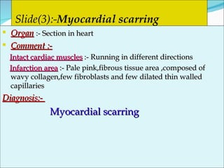
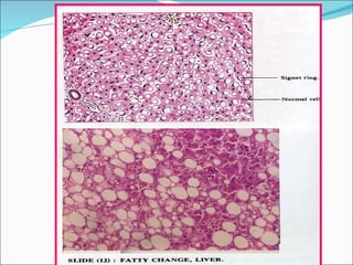
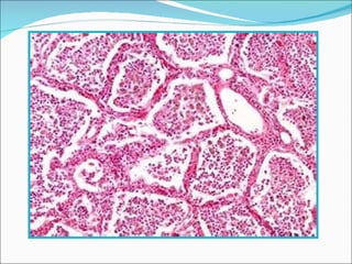
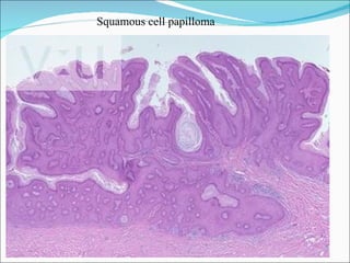
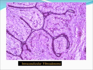
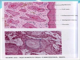
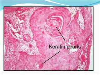
Pr.Dr. Magdy Ismael Ahmed
neutrophils lymphocyte Plasma cell macrophage
Mast cell eosinophil Giant  cell Fibroblast
Collagen  fibers
Inflammation   Acute inflammation :- e.g  Acute suppurative appendicitis. Chronic inflammation :- e.g  Ch.inflammation of skin. Repair  Myocardial  scarring
Appendicitis  Definition :-  Inflammation of appendix ( either acute or chronic) Etiology :-  Predisposed by:- 1-Obstruction of its lumen or hyperplasia of lymphoid follicles. 2- Fibrosis of the wall  Excited by  infection with Strep.E.coli or staph. Pathology :- 1-Acute catarrhal appendicitis (edema,congestion ,serous fluid) 2-Acute suppurative appendicitis(filled by pus)  3-Acute gangrenous appendicitis(black in colour and offensive). Complications :- 1-Perforation   septic peritonitis (diffuse or localized) 2-Appendicular mass :- Fibrosis  and adhesion with intestine and omentum 3-Appendicular abscess 4-Sinus & fistula 5-Portal pyemia
Pathology :- Gross picture  Comment  -The appendix    slightly enlarged ,congested and edematous . -The lumens are filled by inflammatory exudate -The serosa show dark spots of hemorrhages  Diagnosis:  Acute suppurative appendicitis.
Slide (1):- Acute suppurative appendicitis   Naked eye  :-Circular structure with bluish wall Organ :- T.S. of an appendix Comment :- 1-Lumen  :- Necrotic debris,intact and dead PNLs, 2-Mucosa :- Intact and ulcerated areas 3-Submucosa :- Edema ,dilated and congested blood vessels , hyperplastic lymphoid follicles and PNL,s may extends to musculosa or serosa  Diagnosis  :-  Acute suppurative appendicitis
 
Slide (2):-  Chronic inflammation Organ   : - Section in the skin Comment :- 1-Epidermis  :-  Thickened epidermis and keratin 2-Dermis  :- -Arterioles :- Thickened and narrowed (EAO) -Peri-vascular cuffing by (lymphocytes ,plasma cells and  macrophages) -Increased fibroblasts and collagen.  Diagnosis :-  Chronic inflammation in skin
 
Slide(3):- Myocardial scarring   Organ   :- Section in heart Comment :- Intact cardiac muscles  :- Running in different directions Infarction area  :- Pale pink,fibrous tissue area ,composed of wavy collagen,few fibroblasts and few dilated thin walled capillaries Diagnosis:-  Myocardial scarring
 
Cell injury (degeneration) Water  accumulation :- e.g  Cloudy swelling  of kidney  Fat  accumulation :- e.g  fatty change  of  liver
Cloudy swelling :-  Reversible cell  damage characterized by mild accumulation of water inside cells.  Oragns affected  :-Proximal convoluted tubules of kidney ,liver and heart Pathogenesis:- swollen mitochondria is fragmented with resultant decreased production of ATP and consequent distrubed Na/K pump   retention of Na and H2O inside cells . Pathology  Grossly :-  The organ size :- Increased  Colour :- Pale  Consistency:- Soft
Cloudy swelling of kidney
Section in the kidney :- Glomeruli :-  Normal Tubules :- - Lumen   :- Narrowed and star shaped - Lining epithelial cells  *Swollen *Conical (pyramidal ) shaped.Its apex directed  inward. *Pink and granular cytoplasm (mitochondrial damage.  *Intact rounded nuclei . Diagnosis  :-  Cloudy swelling of the kidney
 
Fatty change ,liver A condition characterized by accumulation of triglyceride inside hepatocytes Causes:- 1-Toxins->diphtheria  2-Chemicals  as phosphorus and CCL4 Etiology:- 1-Increased entrance of FFA  heptocytes 2-Increased synthesis of FA and decreasd its oxidation  3-Increased estrification of FA 4-Decreased formation of phospholipids 5-Decreased excretion of phospholipids.
Specimen   :- Section of liver  Size  :- enlarged Surface   :-Smooth Capsule   :-Thin ,stretched easily Streped Colour :-  Yellowish Borders  :- Rounded Consistency :- Soft  D13
Slide(5):-Fatty changes of liver   Section in liver :- Liver cells :-   Cytoplasm  :- Contain large clear vacuoles Nuclei :- Peripheral located nuclei(signet ring appearance. Some intact  liver cells  are seen. Diagnosis :- Fatty changes of liver.
 
Slide(6):-Recent thrombus  Naked eye   :-Rounded structure with intra-luminal thrombus (2 nd  rounded structure) Section in blood vessel :-  1- The lumen of blood vessel :-  -Filled by thrombus mass attached to the vessel wall at one point (head of the thrombus )  2-The thrombus mass containing :- pale pink lines radiating from the head ( lines of Zahn ) and contain in between meshwork of fibrin ,RBC,s and WBC,s. Diagnosis :-  Recent thrombus
2 nd  of circular lesions
Naked eye  :- 3 rd  rounded structure  Section in an artery :- Intima  :- Degenerated and elevated  Subintima  :- Fibro-fatty mass containing ,  needle like crystals of cholesterol. and haylinized C.T ± calcification  Elastic lamina  :-Fragmented Media opposite the lesion  :- Atrophic .  Diagnosis  :-  Atherosclerosis of an artery   Slide(7):-Atherosclerosis
Atheroma  Calcification  Media  crystals
Slide(8):-Lobar pneumonia  (grey hepatization)   Section in lung tissue  :- Alveolar  wall :- -Thickening of the alveolar wall. -Dilated congested blood capillaries Alveolar space:- -Fibrin mass entangling ,RBC,s,WBC,s and  large numbers of macrophages  -A retraction space is found separating the mass from the alveolar wall.  Diagnosis :- Lobar pneumonia grey hepatization
Fibrin mass   PNL,s   Retraction space   Lobar pneumonia grey hepatization
 
Slide(9):- Nasal polyp   Section in nasal polyp :-   The polyp composed of fibro-vascular C.T core covered by epithelium  . *The covering epithelium ;- -Pseudostratified columnar ciliated epithelium,with focal areas of ulceration or squamous metaplasia . *The core show :- -Stromal edema -Thin dilated blood vessels -Mixed inflammatory cellular infiltrates (PNL,s ,lymphocytes ,plasma cells ,macrophages and eosinophils) -Hyperplastic mucus secreting glands . Diagnosis :-  Nasal polyp
Blood vessels   Edema  Eosinophil Squamous metaplasia
 
Slide(10):- Bronchogenic carcinoma   Section in lung :- The lung tissue show :- -Area of intact lung tissue  -Infiltration of lung tissue by malignant cells are disposed in one of three patterns .  1-Squamous cell carcinoma  :-Nests of malignant squamous epithelium ± keratin pearls ( cell nests )  2- Or Adenocarcinoma   with well formed malignant acini 3-Or Aot cell carcinoma  :- Malignant ,darkly stained  small sized cells  with rounded ,oval or spindle shaped hyper-chromatic nuclei and scanty cytoplasm.  Diagnosis :-   Bronchogenic carcinoma
Squamous cell carcinoma   Fibrous septae   Aot cell carcinoma   Lung tissue
Bengin  tumors A-Tumors arise from the epithelium 1-Surface epithelium  :-  Squamous cell papilloma 2-Glandular epithelium  :- a- Adenoma of intestine  b- Fibroadenoma of the breast (intracanalicular and pericanalicular ) B-Tumors arises from mesenchymal tissue 1-Fibroma  2-Leiomyoma 3-Chondroma 5-Lipoma
Slide (11)  Squamous cell papilloma Definition  :- benign tumor of surface epithelium. Sites  :- Skin –lips –oral mucosa –cervix -pharynx -vagina and anal canal . Gross picture  :- Small sessile or pedunclated projection  Microscopic picture  :-  The core :- Composed of  fibro-vascular connective tissue  The cover :-Composed of hyperplastic stratified squamous epithelium showing:- - Hyperkeratosis :- Increased keratinous layers - Parakeratosis :- Nucleated keratin - Acanthosis :- Increased number of prickle cell layer
Squamous cell papilloma
Slide (12)  Adenoma  Definition  :-  A benign tumor of endocrine and exocrine glands and mucosal glands of GIT and endometrium. Gross picture  :- A well –defined capsulated ovoid mass  cut section : solid ,cystic or papillary cystic  Microscopic picture  :- Proliferated glands lined by cuboidal or columnar epithelium separated by a dense fibro-vascular connective tissue stroma (colon adenoma )
Adenoma
Adenoma
Slide (13)  Fibroadenoma of the breast Definition  :-  Mixed benign tumor composed of glandular and fibrous tissue elements Gross picture  :- Well circumscribed capsulated  tumor mass . Cut section :- Grayish white, firm and lobulated mass Microscopic picture  :- Proliferated fibrous tissue stroma and glandular tissues.The ducts are lined by double cell layers of benign ductal epithelial cells .  - Pericanalicular fibroadenoma  :-The ducts are  Patent  Intracanaliclar fibroadenoma  :-The ducts are closed .
Pericanalicular fibroadenoma
Intracanalicular  Fibroadenoma
Benign mesenchymal tumors Fibroma  Leiomyoma  Lipoma  Chondroma
Origin :  arise from the fibrous tissue. Sites:  Fibrous tissue stroma of organs (breast ,ovary and kidney) , intermuscular septa, submucosa, Grossly:  capsulated, oval, soft (cellular)  to hard and Cut section :- Whorly bundles grayish white (hard) or pinkish –white(cellular) in colour . Microscopically:  bundles of mature fibroblasts, collagenous tissue, and few capillaries.  If excess collagen  hard fibroma, if excess fibroblasts  soft fibroma.  Types: - Pure fibroma.-Mixed fibroma, with glands  fibroadenoma, with muscles   fibromyoma Slide (14) :  Fibroma
 
 
Slide (14) :  Leiomyoma Origin:  from smooth muscles. Sites:   uterus is the most common (fibroid) GIT, bladder. Grossly:  non capsulated (false capsule) , single or multiple, rounded, firm . C.S whorled pattern, composed of pinkish and whitish interlaced bundles .  Microscopically:  interlacing bundles of smooth muscle fibers separated by fibrovascular stroma.
 
Slide (15) :  lipoma Origin:   from fatty tissues. Sites:   Subcutaneous tissues of the arm, back, submucosa and retroperitoneum. Grossly:   capsulated, round or oval, lobulated soft  mass  C.S:-Bulging,pale- yellow and greasy Microscopically:   capsulated, with fine trabeculae  dividing the tumor into lobules that contain clusters of adult fat cells { clear cytoplasm with peripheral (signet ring) nuclei} and fewer clusters of embryonic fat cells ( granular  eosinophilic cytoplasm with central nuclei..


Slide (16) :  Chondroma   Origin:  Benign tumors of cartilage . Sites:   Short bones (hands & feets) , flat bones (sternum ,ribs ,scapula) and ends of long bones . Grossly:  Capsulated, rounded or oval, solitary or multiple mass .Cut section :- Sharp borders , lobulated  bluish  and translucent . Microscopically:  The fibrous capsule sends trabeculae that divide the tumor into lobules .Each lobule composed of  chondrocytes (vacuolated cytoplasm with central nuclei in lacunae) are arranged singly or in groups .The stroma is pale blue hyaline matrix .
 
Chondroma
Malignant tumors  Malignant epithelial tumors  :-  Carcinoma of surface epithelium  :- 1-Squamous cell carcinoma  2-Basal cell carcinoma  Carcinoma of glandular epithelium  :-  1-Adenocarcinoma  2-Mucoid adenocarcinoma  Malignant mesenchymal  Fibrosarcoma
Slide 17 :-  Squamous cell carcinoma  Definition :-  Malignant tumor of stratified squamous epithelium  Epidermis:-   Intact or partially ulcerated stratified squamous epithelial covering  Dermis :-  Groups of malignant squamous epithelial (prickle) cells with pale pink ill defined cytoplasm ,large size vesicular nuclei with prominent nucleoli .With or without central keratin  pearls (cell nests) .Few chronic inflammatory cells in between the nests. Hemorrhage and necrosis may be found.
 
SCC Cell nests  Keratin pearls  Lymphocytes
 
Slide 18  :  Basal cell carcinoma   (Rodent ulcer) Def:  -   Locally malignant tumor arising from the basal cell layer of epidermis in face skin exposed to sun rays  Gossly:-  starts as a firm reddish papule   nodule   finally ulcerate .It erodes surrounding structures through its spread ( rodent ulcer) .It characterized by   Margin : rounded or oval margin  Edge: raised inverted (rolled in ) . Floor: rough and necrotic . Base: indurated . Histologically:   invasion of the dermis by variable sized masses formed of malignant basaloid cells that form palisade appearance at the periphery. The surrounding stroma is retracted from them leaving spaces
 
Basaloid cells  Palisading  Retracted stroma  around the malignant masses
Slide 19 : Adenocarcinoma Origin:  glandular tissue. Sites:   endocrine, exocrine glands, mucus membranes, GIT, endometrium. Gross:  in solid organs  irregular infiltrative growth, in hollow organs  fungating, infiltrating or ulcerating growth. Microscopically:  The malignant glands are:  1-irregular in size and shape.  2- They are lined by malignant cells .  3-They are  present in abnormal location (in submucosa ,musculosa even reach serosa)  4-The glandular lumina are irregular or absent. Grading:  depends on architecture and cellular differentiation.
 
Adenocarcinoma colon Normal  mucosa  Malignant glands
Slide 20  :Mucoid carcinoma Origin:   from glandular epithelium producing mucin. Sites:  mostly in GIT (stomach & colon).rare in breast ,gall bladder and bronchi. Grossly:   appears as soft gelatinous mass . Microscopicaly:   Mucoid (colloid) carcinoma : adenocarcinoma    rupture of cells    release of large amount of extra-cellular mucin  Pools of mucin containing floating malignant cells. Signet cell carcinoma : cells are distended with intracellular mucin   Signet ring cells with minimal extracellular mucin.
 
Mucoid carcinoma, colon Normal  mucosa  Mucus pools
Slide 21 :Fibrosarcoma  Definition  :  malignant tumor of fibrous tissue Sites  :  subcutaneous,intermuscular and periosteal tissues Gross picture  :Large ,grayish white mass with foci of hemorrhage and necrosis. Microscopic picture :- Fasicles of malignant spindle cells separated by collagen (Herring –bone pattern)
Herring  bone
 
Thank  you

عملى الصيدلة

  • 1.
  • 2.
  • 3.
    Mast cell eosinophilGiant cell Fibroblast
  • 4.
  • 5.
    Inflammation Acute inflammation :- e.g Acute suppurative appendicitis. Chronic inflammation :- e.g Ch.inflammation of skin. Repair Myocardial scarring
  • 6.
    Appendicitis Definition:- Inflammation of appendix ( either acute or chronic) Etiology :- Predisposed by:- 1-Obstruction of its lumen or hyperplasia of lymphoid follicles. 2- Fibrosis of the wall Excited by infection with Strep.E.coli or staph. Pathology :- 1-Acute catarrhal appendicitis (edema,congestion ,serous fluid) 2-Acute suppurative appendicitis(filled by pus) 3-Acute gangrenous appendicitis(black in colour and offensive). Complications :- 1-Perforation  septic peritonitis (diffuse or localized) 2-Appendicular mass :- Fibrosis and adhesion with intestine and omentum 3-Appendicular abscess 4-Sinus & fistula 5-Portal pyemia
  • 7.
    Pathology :- Grosspicture Comment -The appendix  slightly enlarged ,congested and edematous . -The lumens are filled by inflammatory exudate -The serosa show dark spots of hemorrhages Diagnosis: Acute suppurative appendicitis.
  • 8.
    Slide (1):- Acutesuppurative appendicitis Naked eye :-Circular structure with bluish wall Organ :- T.S. of an appendix Comment :- 1-Lumen :- Necrotic debris,intact and dead PNLs, 2-Mucosa :- Intact and ulcerated areas 3-Submucosa :- Edema ,dilated and congested blood vessels , hyperplastic lymphoid follicles and PNL,s may extends to musculosa or serosa Diagnosis :- Acute suppurative appendicitis
  • 9.
  • 10.
    Slide (2):- Chronic inflammation Organ : - Section in the skin Comment :- 1-Epidermis :- Thickened epidermis and keratin 2-Dermis :- -Arterioles :- Thickened and narrowed (EAO) -Peri-vascular cuffing by (lymphocytes ,plasma cells and macrophages) -Increased fibroblasts and collagen. Diagnosis :- Chronic inflammation in skin
  • 11.
  • 12.
    Slide(3):- Myocardial scarring Organ :- Section in heart Comment :- Intact cardiac muscles :- Running in different directions Infarction area :- Pale pink,fibrous tissue area ,composed of wavy collagen,few fibroblasts and few dilated thin walled capillaries Diagnosis:- Myocardial scarring
  • 13.
  • 14.
    Cell injury (degeneration)Water accumulation :- e.g Cloudy swelling of kidney Fat accumulation :- e.g fatty change of liver
  • 15.
    Cloudy swelling :- Reversible cell damage characterized by mild accumulation of water inside cells. Oragns affected :-Proximal convoluted tubules of kidney ,liver and heart Pathogenesis:- swollen mitochondria is fragmented with resultant decreased production of ATP and consequent distrubed Na/K pump  retention of Na and H2O inside cells . Pathology Grossly :- The organ size :- Increased Colour :- Pale Consistency:- Soft
  • 16.
  • 17.
    Section in thekidney :- Glomeruli :- Normal Tubules :- - Lumen :- Narrowed and star shaped - Lining epithelial cells *Swollen *Conical (pyramidal ) shaped.Its apex directed inward. *Pink and granular cytoplasm (mitochondrial damage. *Intact rounded nuclei . Diagnosis :- Cloudy swelling of the kidney
  • 18.
  • 19.
    Fatty change ,liverA condition characterized by accumulation of triglyceride inside hepatocytes Causes:- 1-Toxins->diphtheria 2-Chemicals as phosphorus and CCL4 Etiology:- 1-Increased entrance of FFA  heptocytes 2-Increased synthesis of FA and decreasd its oxidation 3-Increased estrification of FA 4-Decreased formation of phospholipids 5-Decreased excretion of phospholipids.
  • 20.
    Specimen :- Section of liver Size :- enlarged Surface :-Smooth Capsule :-Thin ,stretched easily Streped Colour :- Yellowish Borders :- Rounded Consistency :- Soft D13
  • 21.
    Slide(5):-Fatty changes ofliver Section in liver :- Liver cells :- Cytoplasm :- Contain large clear vacuoles Nuclei :- Peripheral located nuclei(signet ring appearance. Some intact liver cells are seen. Diagnosis :- Fatty changes of liver.
  • 22.
  • 23.
    Slide(6):-Recent thrombus Naked eye :-Rounded structure with intra-luminal thrombus (2 nd rounded structure) Section in blood vessel :- 1- The lumen of blood vessel :- -Filled by thrombus mass attached to the vessel wall at one point (head of the thrombus ) 2-The thrombus mass containing :- pale pink lines radiating from the head ( lines of Zahn ) and contain in between meshwork of fibrin ,RBC,s and WBC,s. Diagnosis :- Recent thrombus
  • 24.
    2 nd of circular lesions
  • 25.
    Naked eye :- 3 rd rounded structure Section in an artery :- Intima :- Degenerated and elevated Subintima :- Fibro-fatty mass containing , needle like crystals of cholesterol. and haylinized C.T ± calcification Elastic lamina :-Fragmented Media opposite the lesion :- Atrophic . Diagnosis :- Atherosclerosis of an artery Slide(7):-Atherosclerosis
  • 26.
    Atheroma Calcification Media crystals
  • 27.
    Slide(8):-Lobar pneumonia (grey hepatization) Section in lung tissue :- Alveolar wall :- -Thickening of the alveolar wall. -Dilated congested blood capillaries Alveolar space:- -Fibrin mass entangling ,RBC,s,WBC,s and large numbers of macrophages -A retraction space is found separating the mass from the alveolar wall. Diagnosis :- Lobar pneumonia grey hepatization
  • 28.
    Fibrin mass PNL,s Retraction space Lobar pneumonia grey hepatization
  • 29.
  • 30.
    Slide(9):- Nasal polyp Section in nasal polyp :- The polyp composed of fibro-vascular C.T core covered by epithelium . *The covering epithelium ;- -Pseudostratified columnar ciliated epithelium,with focal areas of ulceration or squamous metaplasia . *The core show :- -Stromal edema -Thin dilated blood vessels -Mixed inflammatory cellular infiltrates (PNL,s ,lymphocytes ,plasma cells ,macrophages and eosinophils) -Hyperplastic mucus secreting glands . Diagnosis :- Nasal polyp
  • 31.
    Blood vessels Edema Eosinophil Squamous metaplasia
  • 32.
  • 33.
    Slide(10):- Bronchogenic carcinoma Section in lung :- The lung tissue show :- -Area of intact lung tissue -Infiltration of lung tissue by malignant cells are disposed in one of three patterns . 1-Squamous cell carcinoma :-Nests of malignant squamous epithelium ± keratin pearls ( cell nests ) 2- Or Adenocarcinoma with well formed malignant acini 3-Or Aot cell carcinoma :- Malignant ,darkly stained small sized cells with rounded ,oval or spindle shaped hyper-chromatic nuclei and scanty cytoplasm. Diagnosis :- Bronchogenic carcinoma
  • 34.
    Squamous cell carcinoma Fibrous septae Aot cell carcinoma Lung tissue
  • 35.
    Bengin tumorsA-Tumors arise from the epithelium 1-Surface epithelium :- Squamous cell papilloma 2-Glandular epithelium :- a- Adenoma of intestine b- Fibroadenoma of the breast (intracanalicular and pericanalicular ) B-Tumors arises from mesenchymal tissue 1-Fibroma 2-Leiomyoma 3-Chondroma 5-Lipoma
  • 36.
    Slide (11) Squamous cell papilloma Definition :- benign tumor of surface epithelium. Sites :- Skin –lips –oral mucosa –cervix -pharynx -vagina and anal canal . Gross picture :- Small sessile or pedunclated projection Microscopic picture :- The core :- Composed of fibro-vascular connective tissue The cover :-Composed of hyperplastic stratified squamous epithelium showing:- - Hyperkeratosis :- Increased keratinous layers - Parakeratosis :- Nucleated keratin - Acanthosis :- Increased number of prickle cell layer
  • 37.
  • 38.
  • 39.
  • 40.
    Slide (12) Adenoma Definition :- A benign tumor of endocrine and exocrine glands and mucosal glands of GIT and endometrium. Gross picture :- A well –defined capsulated ovoid mass cut section : solid ,cystic or papillary cystic Microscopic picture :- Proliferated glands lined by cuboidal or columnar epithelium separated by a dense fibro-vascular connective tissue stroma (colon adenoma )
  • 41.
  • 42.
  • 43.
    Slide (13) Fibroadenoma of the breast Definition :- Mixed benign tumor composed of glandular and fibrous tissue elements Gross picture :- Well circumscribed capsulated tumor mass . Cut section :- Grayish white, firm and lobulated mass Microscopic picture :- Proliferated fibrous tissue stroma and glandular tissues.The ducts are lined by double cell layers of benign ductal epithelial cells . - Pericanalicular fibroadenoma :-The ducts are Patent Intracanaliclar fibroadenoma :-The ducts are closed .
  • 44.
  • 45.
  • 46.
    Benign mesenchymal tumorsFibroma Leiomyoma Lipoma Chondroma
  • 47.
    Origin : arise from the fibrous tissue. Sites: Fibrous tissue stroma of organs (breast ,ovary and kidney) , intermuscular septa, submucosa, Grossly: capsulated, oval, soft (cellular) to hard and Cut section :- Whorly bundles grayish white (hard) or pinkish –white(cellular) in colour . Microscopically: bundles of mature fibroblasts, collagenous tissue, and few capillaries. If excess collagen  hard fibroma, if excess fibroblasts  soft fibroma. Types: - Pure fibroma.-Mixed fibroma, with glands  fibroadenoma, with muscles  fibromyoma Slide (14) : Fibroma
  • 48.
  • 49.
  • 50.
    Slide (14) : Leiomyoma Origin: from smooth muscles. Sites: uterus is the most common (fibroid) GIT, bladder. Grossly: non capsulated (false capsule) , single or multiple, rounded, firm . C.S whorled pattern, composed of pinkish and whitish interlaced bundles . Microscopically: interlacing bundles of smooth muscle fibers separated by fibrovascular stroma.
  • 51.
  • 52.
    Slide (15) : lipoma Origin: from fatty tissues. Sites: Subcutaneous tissues of the arm, back, submucosa and retroperitoneum. Grossly: capsulated, round or oval, lobulated soft mass C.S:-Bulging,pale- yellow and greasy Microscopically: capsulated, with fine trabeculae dividing the tumor into lobules that contain clusters of adult fat cells { clear cytoplasm with peripheral (signet ring) nuclei} and fewer clusters of embryonic fat cells ( granular eosinophilic cytoplasm with central nuclei..
  • 53.
  • 54.
  • 55.
  • 56.
    Slide (16) : Chondroma Origin: Benign tumors of cartilage . Sites: Short bones (hands & feets) , flat bones (sternum ,ribs ,scapula) and ends of long bones . Grossly: Capsulated, rounded or oval, solitary or multiple mass .Cut section :- Sharp borders , lobulated bluish and translucent . Microscopically: The fibrous capsule sends trabeculae that divide the tumor into lobules .Each lobule composed of chondrocytes (vacuolated cytoplasm with central nuclei in lacunae) are arranged singly or in groups .The stroma is pale blue hyaline matrix .
  • 57.
  • 58.
  • 59.
    Malignant tumors Malignant epithelial tumors :- Carcinoma of surface epithelium :- 1-Squamous cell carcinoma 2-Basal cell carcinoma Carcinoma of glandular epithelium :- 1-Adenocarcinoma 2-Mucoid adenocarcinoma Malignant mesenchymal Fibrosarcoma
  • 60.
    Slide 17 :- Squamous cell carcinoma Definition :- Malignant tumor of stratified squamous epithelium Epidermis:- Intact or partially ulcerated stratified squamous epithelial covering Dermis :- Groups of malignant squamous epithelial (prickle) cells with pale pink ill defined cytoplasm ,large size vesicular nuclei with prominent nucleoli .With or without central keratin pearls (cell nests) .Few chronic inflammatory cells in between the nests. Hemorrhage and necrosis may be found.
  • 61.
  • 62.
    SCC Cell nests Keratin pearls Lymphocytes
  • 63.
  • 64.
    Slide 18 : Basal cell carcinoma (Rodent ulcer) Def: - Locally malignant tumor arising from the basal cell layer of epidermis in face skin exposed to sun rays Gossly:- starts as a firm reddish papule  nodule  finally ulcerate .It erodes surrounding structures through its spread ( rodent ulcer) .It characterized by Margin : rounded or oval margin Edge: raised inverted (rolled in ) . Floor: rough and necrotic . Base: indurated . Histologically: invasion of the dermis by variable sized masses formed of malignant basaloid cells that form palisade appearance at the periphery. The surrounding stroma is retracted from them leaving spaces
  • 65.
  • 66.
    Basaloid cells Palisading Retracted stroma around the malignant masses
  • 67.
    Slide 19 :Adenocarcinoma Origin: glandular tissue. Sites: endocrine, exocrine glands, mucus membranes, GIT, endometrium. Gross: in solid organs  irregular infiltrative growth, in hollow organs  fungating, infiltrating or ulcerating growth. Microscopically: The malignant glands are: 1-irregular in size and shape. 2- They are lined by malignant cells . 3-They are present in abnormal location (in submucosa ,musculosa even reach serosa) 4-The glandular lumina are irregular or absent. Grading: depends on architecture and cellular differentiation.
  • 68.
  • 69.
    Adenocarcinoma colon Normal mucosa Malignant glands
  • 70.
    Adenocarcinoma colon Normal mucosa Malignant glands
  • 71.
    Slide 20 :Mucoid carcinoma Origin: from glandular epithelium producing mucin. Sites: mostly in GIT (stomach & colon).rare in breast ,gall bladder and bronchi. Grossly: appears as soft gelatinous mass . Microscopicaly: Mucoid (colloid) carcinoma : adenocarcinoma  rupture of cells  release of large amount of extra-cellular mucin  Pools of mucin containing floating malignant cells. Signet cell carcinoma : cells are distended with intracellular mucin  Signet ring cells with minimal extracellular mucin.
  • 72.
  • 73.
    Mucoid carcinoma, colonNormal mucosa Mucus pools
  • 74.
    Slide 21 :Fibrosarcoma Definition : malignant tumor of fibrous tissue Sites : subcutaneous,intermuscular and periosteal tissues Gross picture :Large ,grayish white mass with foci of hemorrhage and necrosis. Microscopic picture :- Fasicles of malignant spindle cells separated by collagen (Herring –bone pattern)
  • 75.
  • 76.
  • 77.