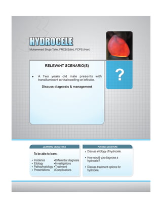
Hydrocele is a collection of fluid in the scrotum that results from a defect or irritation in the tunica vaginalis. It is usually painless and causes swelling of the scrotum. A hydrocele can be diagnosed based on physical exam findings like transillumination of the swollen area and ultrasound findings of a cystic mass around the testicle. While most hydroceles are benign, further investigation is needed to rule out other causes for scrotal swelling like hernia or testicular torsion that require urgent treatment.
This document discusses hydroceles, which is an abnormal fluid collection in the scrotum. Hydroceles in infants are usually caused by incomplete closure of the processus vaginalis during development. For older boys and men, hydroceles can be idiopathic or caused by inflammation or injury in the scrotum. The document describes the causes, risk factors, signs and symptoms, diagnosis, and treatment of hydroceles. Treatment typically involves draining the fluid through a minor operation, with complications being rare.
Pilonidal sinus is a chronic inflammation in one or more sinuses in the midline of the natal cleft caused by hair and debris. It is more common in overweight males between puberty and age 40. Risk factors include obesity, local trauma, sitting for long periods, and a deep natal cleft. Acquired pilonidal sinus occurs when hair breaks the skin in the natal cleft, collects debris, and grows deeper causing infection. Symptoms include intermittent pain and drainage in the natal cleft area. Diagnosis is usually clinical based on examination finding sinuses, pits, or abscesses in the natal cleft. Treatment depends on severity, with incision and drainage for abscesses and
A hydrocele is a collection of fluid in the sac surrounding a testicle. It is usually congenital but can also be caused by injury or infection. On physical exam, a hydrocele feels like a smooth, cystic mass that can be moved above the testicle. While often asymptomatic, larger hydroceles can cause discomfort. Treatment involves surgery to remove or drain the fluid, with risks including bleeding and infection. Without treatment, potential complications include infection, calcification, or testicular atrophy.
Digital Rectal Examination for Surgical Traineeshosam hamza
Digital Rectal Examination (DRE) is an important procedure in surgical practice used to examine the rectum and surrounding structures. It involves visual inspection of the external anal area and digital palpation of the internal rectum. The 12 key steps of a DRE are outlined, including introducing the procedure to the patient, inspecting externally, lubricating the finger, inserting the finger to palpate internal structures, and communicating findings to the patient. DRE allows examination of the prostate, cervix, and other pelvic structures to detect abnormalities like masses, hemorrhoids, or tenderness that can indicate various diseases.
Femoral hernia is the third common hernia after inguinal and incisional hernias. The swelling in femoral hernia is below and lateral to pubic tubercle. It is more common in females. Strangulation is very common in this hernia.
This document discusses Priapism, which is a prolonged and sometimes painful erection that lasts over 4 hours and is unrelated to sexual stimulation. It defines two types, ischemic (low blood flow) and non-ischemic (high blood flow). The case presented is of a 64-year old man who developed an erection after injecting his penis with medications for erectile dysfunction. His priapism was determined to be ischemic based on history and blood gas analysis. He underwent drainage of blood from his penis, injection of the drug phenylephrine, and follow up with urology.
This document provides information about orchitis, including its causes, symptoms, diagnosis, and treatment. Orchitis is an inflammation of the testicles that is usually caused by a virus like mumps or bacteria that spreads from an associated epididymitis. Common symptoms include pain and tenderness in the testicles and scrotum. Diagnosis involves examination of the testicles and scrotum along with potential laboratory tests. While viral orchitis cannot be cured, it will resolve on its own. Bacterial orchitis is treated with antibiotics and anti-inflammatory medications.
This document provides information about Peyronie's disease, including its symptoms, causes, diagnosis, and treatment options. It defines Peyronie's disease as the formation of scar tissue plaques within the penis that can cause penile curvature and pain during erections. Common symptoms are pain and curvature of the penis to one side. While small, asymptomatic cases may not require treatment, injection of medications into plaques or surgery to correct curvature may be options for more severe cases. The document also reviews normal penile anatomy and the erectile process.
This document discusses bladder exstrophy and epispadias complex. It begins by introducing the moderators and provides an overview of the spectrum of anomalies from glanular epispadias to cloacal exstrophy. Skeletal anomalies including rotational and dimensional pelvic anomalies are described. Pelvic floor defects and abdominal wall defects associated with bladder exstrophy are summarized. Genitourinary defects in males including a shortened penis and in females including a shorter vagina are outlined. Urinary defects from maturational delay in bladder development are also noted.
This document discusses the management and treatment of hydrocele. It outlines the necessary investigations which may include blood tests, urine tests, and chest x-rays. Ultrasound is helpful for determining testis position and abnormalities. Fluid aspiration can indicate different conditions. Surgical procedures like lord's plication and Jaboulay's operation are described for fixing different types of hydroceles. Post-operative care and potential complications are also covered. The document provides an overview of evaluating and treating hydroceles.
Hydrocele is a collection of fluid within the processus vaginalis that causes swelling in the scrotum. It is usually caused by a failure of the processus vaginalis to completely fuse during development. Hydroceles can be communicating or non-communicating depending on whether the processus vaginalis remains open to the abdominal cavity. The standard treatment is surgical repair to prevent the fluid from reaccumulating. Surgery involves ligating and removing the hydrocele sac to restore normal drainage from the scrotum. Complications are rare if the surgery is performed properly.
Testicular torsion occurs when the spermatic cord twists, reducing blood flow to the testicle. There are two main types - intravaginal torsion which is more common and often seen at puberty due to a "bell-clapper" deformity, and extravaginal torsion which occurs in newborns without this deformity. Risk factors include a history of cryptorchidism or an abnormally long spermatic cord. Physical exam may reveal an elevated, tender testicle with absent cremasteric reflex. Ultrasound can show reduced blood flow, but surgery should not be delayed for imaging. Manual detorsion can be attempted in some cases but all patients require surgical exploration to detorse
This document discusses hydroceles, which are abnormal collections of serous fluid in the processus vaginalis of the scrotum. There are congenital and acquired types. Congenital hydroceles occur when the processus vaginalis remains patent, while acquired hydroceles develop secondary to conditions like epididymo-orchitis or testicular tumors. Hydroceles are usually painless swellings that surround the testis and epididymis, making them difficult to palpate. Treatment involves surgical excision of the hydrocele sac. Complications can arise if hydroceles are not treated, such as rupture or testicular atrophy.
This document describes a case report of a congenital vaginal hydrocele in a 4-year old boy. A hydrocele is a collection of fluid within the tunica vaginalis that often presents as an enlarged scrotum in infants. This boy had swelling of the right scrotum since birth. Examination confirmed a right-sided hydrocele. Ultrasound also showed fluid in the right scrotum. The hydrocele was diagnosed as congenital and communicating. It was surgically corrected through a hydrocelectomy procedure to drain the fluid and evert and stitch the tunica vaginalis. Complications can include infection, blood clots, or recurrence of the hydrocele. Most hydroce
seminar (Undescended testes)
Define the undescended testes.
Differentiate between the undescended testes , retractile testes .
Etiology and complication of the undescended testes.
Work up and management plan for the undescended testes .
Define the undescended testes.
Differentiate between the undescended testes , retractile testes .
Etiology and complication of the undescended testes.
Work up and management plan for the undescended testes .
Define the undescended testes.
Differentiate between the undescended testes , retractile testes .
Etiology and complication of the undescended testes.
Work up and management plan for the undescended testes .
Define the undescended testes.
Differentiate between the undescended testes , retractile testes .
Etiology and complication of the undescended testes.
Work up and management plan for the undescended testes Majmaah University
The document provides tips for using a PowerPoint presentation (PPT) for active learning sessions on medical topics. It recommends:
1) Freely editing, modifying, and adding your name to the PPT.
2) Noting that half the slides are blank except for the title to elicit student responses.
3) Showing blank slides, asking students what they know, and then showing the content slide.
4) Repeating this process of blank slide + student response + content slide at the end for reinforcement.
5) This active learning approach can be repeated over three sessions for effective learning.
Testicular torsion refers to twisting of the spermatic cord and loss of blood supply to the testicle. It is a urological emergency as early diagnosis and treatment are needed to save the testicle. Ultrasound with Doppler is the primary imaging method and shows absent or decreased blood flow in the affected testicle compared to the normal side. Prompt surgical detorsion and orchioplexy are the definitive treatments.
A pancreatic pseudocyst is a fluid collection containing pancreatic enzymes that usually forms after pancreatitis. Symptoms include abdominal pain and bloating. Pseudocysts are diagnosed using CT scans, MRI, x-rays, or ultrasounds. Small pseudocysts may resolve on their own, but large or symptomatic ones often require surgery to drain the fluid by creating a connection between the cyst and stomach, intestine, or duodenum. Complications can include infection, bleeding, obstruction, or rupture.
This document discusses congenital inguinal hernia and hydrocele. It notes that a congenital inguinal hernia occurs when the processus vaginalis fails to close after testicular descent in fetal development, allowing abdominal contents to enter the scrotum. It has a high rate in premature infants and is more common in males. A hydrocele occurs when the processus vaginalis remains fluid-filled but no abdominal contents enter. Both often resolve spontaneously in infants but may require surgery if persisting past 2 years. The hernia repair procedure involves high ligation of the hernia sac at the internal ring through an inguinal incision.
Undescended testis, or cryptorchidism, refers to the absence of one or both testes from the normal scrotal position. It occurs in 1-4% of full-term and 1-45% of preterm newborn boys. The specific cause is often unknown, but risk factors include prematurity and maternal estrogen exposure. Diagnosis involves examination to locate any undescended testes. Early surgical treatment before age 18 months is recommended to preserve fertility and reduce cancer risks. Options include inguinal or abdominal orchidopexy. Long term risks include reduced sperm counts and increased testicular cancer risk.
1. Vesicoureteral reflux (VUR) occurs when urine flows backward from the bladder into the ureters and kidneys. This document discusses the history, demography, causes, diagnosis, evaluation, natural history and management of VUR.
2. VUR is more common in younger children, males, and those with urinary tract infections. The prevalence is higher in siblings of children with VUR.
3. VUR can be primary due to structural issues or secondary due to bladder dysfunction. Grading depends on extent of reflux during cystography.
4. Management focuses on antibiotics to prevent infections while allowing for spontaneous resolution, which is more likely for lower grades of
This document discusses urinary obstruction, including its pathophysiology, causes, effects on renal physiology and function, histological changes, clinical impact, and renal recovery after relief of obstruction. It provides an overview of how urinary obstruction can lead to permanent kidney damage depending on the severity, chronicity, and baseline kidney condition. Both unilateral and bilateral obstruction are examined, along with the triphasic response and changes in renal blood flow, filtration, and tubular transport that occur.
Rectal bleeding has many potential causes, both minor and major. Minor bleeding may be due to hemorrhoids or fissures, while more severe bleeding requires emergency treatment. In cases of massive bleeding, initial steps include admission to the hospital, insertion of IV lines, monitoring of vitals, and blood transfusions as needed to stabilize the patient. Further tests such as colonoscopy or angiography aim to locate the source of bleeding so it can be addressed through methods like cauterization or surgery. Surgical intervention may be needed if other measures do not stop severe or persistent bleeding.
1) Priapism is a prolonged, painful erection that persists without sexual stimulation. It is caused by a failure of the penis to return to its flaccid state after erection and can lead to permanent erectile dysfunction if not properly treated.
2) There are two main types - low-flow (ischemic/veno-occlusive) priapism which is painful due to a lack of blood flow, and high-flow (non-ischemic) priapism which is usually painless. Low-flow priapism is more common and a medical emergency requiring treatment to prevent tissue damage.
3) Treatment depends on the type of priapism and involves initially aspirating blood
Dr. Shirish Silwal provides a summary of different types of hernias including inguinal, umbilical, paraumbilical, incisional, epigastric, spigelian, and lumbar hernias. The document discusses the history, anatomy, causes, presentations, complications, and management approaches for each hernia type. Meshes are recommended for repair when there is a large defect size, multiple defects, or lax abdominal walls to create a tension-free repair and reduce recurrence rates.
Hydronephrosis is the dilation of the renal pelvis and calyces caused by obstruction of urine flow from the kidney. It can be caused by issues in the ureter, bladder, or urethra that limit urine outflow. Unilateral hydronephrosis may cause dull flank pain while bilateral obstruction can lead to decreased urine output. Left untreated, hydronephrosis can damage kidney tissue and impair renal function. Diagnosis is made through imaging tests like intravenous pyelogram. Treatment focuses on resolving the underlying cause of obstruction.
Hydrocele is a collection of fluid around one or both testicles, causing swelling of the scrotum or groin area. It commonly occurs in men over 40. There are two types of hydrocele: noncommunicating, where the fluid sac closes but the body does not absorb the fluid; and communicating, where the sac surrounding the testicle does not close completely, allowing fluid to flow in and out. Symptoms include swelling of the scrotum, pain, redness, and a feeling of pressure at the base of the penis. Diagnosis involves physical examination, transillumination to identify structures beneath the skin, and ultrasound to check for fluid in the scrotum. Treatment options include surgery, needle aspiration
Hydrocele Seminar - A comprehensive review of literatureHarmandeep Jabbal
- Hydrocele is an abnormal fluid collection between the layers of the tunica vaginalis that surrounds the testis. It can be congenital if there is a failure of closure of the processus vaginalis, or acquired due to various causes like infection, trauma, or tumors.
- Physical exam reveals a smooth, cystic mass surrounding the testis that transilluminates and can be lifted above the swelling. Surgery is the definitive treatment and involves excising the sac (hydrocelectomy) or suturing it behind the testis (Jaboulay procedure). Complications are rare but include injury to surrounding structures or recurrence.
This document provides an overview of hydrocele, including:
- Hydrocele is an abnormal collection of fluid between layers of the tunica vaginalis that causes painless scrotal swelling.
- It most commonly affects men over 40 and can also affect newborns. There are various types including primary/idiopathic, secondary, congenital, and infantile hydroceles.
- Signs include fluctuant, transilluminant scrotal swelling. Surgical treatments include tapping, evacuation/eversion, and excision of the hydrocele sac. Ayurveda views it as caused by suppressed urges to urinate.
This document provides an overview of hydrocele, including:
1) Hydrocele is an abnormal collection of serous fluid in the tunica vaginalis that commonly presents as a painless scrotal swelling.
2) There are several types of hydrocele including primary/idiopathic, secondary, congenital, and infantile varieties.
3) Evaluation involves physical exam findings like transillumination and fluctuation testing while treatment options range from conservative management to surgical procedures.
Hydrocele- All types & treatment optionsPawanKurliye
This document provides an overview of hydrocele, including its definition, developmental anatomy, risk factors, classification, signs and symptoms, diagnosis, complications, differential diagnosis, management, and a clinical case scenario. A hydrocele is an abnormal collection of fluid between layers of the tunica vaginalis that can be congenital or acquired. It presents as a painless scrotal swelling that is transilluminant and fluctuant. Management involves surgical excision via techniques like Lord's plication or Jaboulay's procedure to prevent recurrence of the fluid collection.
This document discusses the anatomy, clinical features, and management of various anorectal conditions including rectal prolapse, pilonidal disease, and perianal fistula. It begins with the anatomy of the rectum and anal canal. For rectal prolapse, it describes the types, risk factors, clinical features, and surgical management including procedures like Delormes and Altemeier's operation. Pilonidal disease and its pathogenesis, risk factors, clinical exam, and treatments are outlined. Perianal fistula is defined and the Goodsall rule, Park's classification system, investigations, and common surgical interventions like fistulotomy are summarized.
This document provides information about hydrocele, which is an abnormal fluid collection in the scrotum. It discusses that hydroceles are most common in infants and can be caused by incomplete closure of the tunica vaginalis. Signs include swelling of the scrotum. Diagnosis involves physical exam and sometimes ultrasound. Treatment options include aspiration of the fluid or surgical repair through incision or excision of the tunica vaginalis. Surgery is a minor procedure done as an outpatient to drain the fluid and prevent reaccumulation.
This document discusses various disorders of the scrotum and testes that can be evaluated with ultrasound imaging. It covers congenital anomalies like undescended testes, acquired conditions like hydrocele and epididymitis, and traumatic injuries. Ultrasound is described as the preferred method for diagnosing these conditions by identifying features like cysts, swelling, blood flow changes, and ruptures. Timely ultrasound exams are important for urgent issues like testicular torsion.
The acute scrotum is the painful, swollen scrotum or its contents of sudden onset. The “acute scrotum” may be viewed as the urologist’s equivalent to the general surgeon’s “acute abdomen.” Scrotal emergencies are rare but potentially life and fertility threatening. The most common causes of acute scrotal pain in adults are testicular torsion and epididymitis.
Patients with scrotal pain less than the age of 16 have torsion until proven otherwise. Scrotal pain with nausea & vomiting is specific for torsion.
A small but real, negative exploration rate is acceptable to minimize the risk of missing a critical surgical diagnosis. TIME IS TESTICLE
This document discusses various non-neoplastic diseases of the salivary glands. It covers common and uncommon infectious diseases like viral and bacterial sialadenitis. It also discusses specific infections like mumps which causes parotid gland swelling. Other conditions covered include Sjögren's syndrome, sialolithiasis (salivary stones), drug-induced salivary gland disorders, sialosis (non-inflammatory swelling) and bulimia nervosa related salivary gland enlargement. The document provides details on clinical features, investigations, diagnosis and treatment of these salivary gland conditions.
The document discusses the anatomy, development, disorders, and tumors of the parotid gland. It begins by describing the location and structures of the parotid gland. It then discusses various inflammatory and infectious disorders that can affect the gland such as mumps, bacterial infections, and HIV-associated sialadenitis. Obstructive disorders from stones, strictures, or papillary obstruction are also reviewed. The majority of the document focuses on tumors of the parotid gland, describing the classification and most common tumor types as well as their presentation, investigations, and surgical management.
This document summarizes the use of ultrasound in evaluating acute scrotal pain. It describes the normal anatomy and vascular supply of the scrotum and then discusses various pathologies that can cause acute scrotal pain like epididymo-orchitis, testicular torsion, varicocele, trauma and tumors. Epididymo-orchitis is the most common cause of acute scrotum. Ultrasound is useful for diagnosing the specific condition causing pain by identifying features like enlarged epididymis, lack of blood flow in a twisted testis, or intratesticular fluid in an abscess. Color Doppler ultrasound in particular helps evaluate blood flow and make an accurate diagnosis to guide appropriate treatment.
A hydrocele is a collection of fluid within the processus vaginalis that causes swelling in the scrotum. It can be primary (idiopathic) or secondary to diseases like tuberculosis. The processus vaginalis normally closes after birth but failure of closure can lead to a communicating hydrocele. Surgery is usually not needed for small hydroceles in infants but may be required for larger or symptomatic hydroceles to prevent complications. Common surgical techniques include excision or plication of the hydrocele sac. Postoperative follow up is needed to ensure complete recovery. Recurrence after surgery is rare.
- Applies anterior traction on the gland
- Identifies and protects facial nerve branches
- Aspirates blood to maintain clear field
Surgeon:
- Develops plane between gland and overlying fascia
- Identifies and protects facial nerve branches
- Ligates vessels as encountered
- Completes mobilization of gland
- Identification of facial nerve:
Main trunk is usually identified in the upper part of the gland posterior to the upper pole vessels.
It is dissected proximally and distally using nerve stimulator and magnification.
Branches are identified and preserved.
- Removal of gland:
Gland is delivered by dividing its attachments to the surrounding tissues.
Haemost
Approah to a child / adult presenting with acute scrotum - testicular pain.
The acute scrotum – definition and causes with differential diagnosis
Management of the acute scrotum
Testicular torsion
Torsion of a testicular or epididymal appendage
Epididymitis or epididymo-orchitis
Idiopathic scrotal oedema
Fat necrosis of the scrotum
Case Discussion
This document provides information on intussusception in children. It discusses that intussusception is the telescoping of one segment of intestine into another and is most common in children under 1 year old. It can be idiopathic or have a pathological lead point such as Meckel's diverticulum. Diagnosis is usually made clinically or with ultrasound or contrast enema. Treatment involves non-operative reduction with hydrostatic or pneumatic enema, which has a high success rate. Surgery is needed if reduction fails or there are complications like perforation. Prognosis is generally excellent with prompt treatment.
The document discusses various radiological findings seen in different clinical conditions. It describes the spinnaker sign seen on neonatal chest x-rays indicating pneumomediastinum. It also discusses nutcracker syndrome characterized by compression of the left renal vein resulting in hematuria, varicocele and hypertension. Further, it summarizes Kartagener's syndrome seen as a classic triad of situs inversus, sinusitis and bronchiectasis. It also briefly describes findings of ranula seen as a cystic neck lesion, bunionette seen as a prominence over the 5th metatarsal head, and esophageal candidiasis seen as irregular plaque-like filling defects on barium swallow in immunocomprom
Toxoplasmosis is caused by the protozoan Toxoplasma gondii. It most commonly causes retinochoroiditis which presents as focal areas of retinal inflammation and necrosis near preexisting chorioretinal scars. While most cases are asymptomatic, it can also cause systemic symptoms like fever and lymphadenopathy. Congenital infection is a major concern as it can lead to vision loss, hydrocephalus or brain damage in infants if acquired during pregnancy. Immunocompromised individuals are also at high risk for severe toxoplasmosis affecting the brain, lungs or heart.
The document summarizes key information about hydatid cyst of the liver caused by the larva of the dog tapeworm Echinococcus granulosus. It describes the parasite's lifecycle between dogs and intermediate hosts like sheep and humans. Symptoms arise when the slow-growing cyst presses on organs or complications occur. Investigations include serology, ultrasound, CT and MRI. Treatment involves surgery to remove the cyst along with scolicidal agents or percutaneous drainage preceded by albendazole therapy. Complications can include biliary leakage, infection and recurrence.
Nano-gold for Cancer Therapy chemistry investigatory projectSIVAVINAYAKPK
chemistry investigatory project
The development of nanogold-based cancer therapy could revolutionize oncology by providing a more targeted, less invasive treatment option. This project contributes to the growing body of research aimed at harnessing nanotechnology for medical applications, paving the way for future clinical trials and potential commercial applications.
Cancer remains one of the leading causes of death worldwide, prompting the need for innovative treatment methods. Nanotechnology offers promising new approaches, including the use of gold nanoparticles (nanogold) for targeted cancer therapy. Nanogold particles possess unique physical and chemical properties that make them suitable for drug delivery, imaging, and photothermal therapy.
NAVIGATING THE HORIZONS OF TIME LAPSE EMBRYO MONITORING.pdfRahul Sen
Time-lapse embryo monitoring is an advanced imaging technique used in IVF to continuously observe embryo development. It captures high-resolution images at regular intervals, allowing embryologists to select the most viable embryos for transfer based on detailed growth patterns. This technology enhances embryo selection, potentially increasing pregnancy success rates.
Breast cancer: Post menopausal endocrine therapyDr. Sumit KUMAR
Breast cancer in postmenopausal women with hormone receptor-positive (HR+) status is a common and complex condition that necessitates a multifaceted approach to management. HR+ breast cancer means that the cancer cells grow in response to hormones such as estrogen and progesterone. This subtype is prevalent among postmenopausal women and typically exhibits a more indolent course compared to other forms of breast cancer, which allows for a variety of treatment options.
Diagnosis and Staging
The diagnosis of HR+ breast cancer begins with clinical evaluation, imaging, and biopsy. Imaging modalities such as mammography, ultrasound, and MRI help in assessing the extent of the disease. Histopathological examination and immunohistochemical staining of the biopsy sample confirm the diagnosis and hormone receptor status by identifying the presence of estrogen receptors (ER) and progesterone receptors (PR) on the tumor cells.
Staging involves determining the size of the tumor (T), the involvement of regional lymph nodes (N), and the presence of distant metastasis (M). The American Joint Committee on Cancer (AJCC) staging system is commonly used. Accurate staging is critical as it guides treatment decisions.
Treatment Options
Endocrine Therapy
Endocrine therapy is the cornerstone of treatment for HR+ breast cancer in postmenopausal women. The primary goal is to reduce the levels of estrogen or block its effects on cancer cells. Commonly used agents include:
Selective Estrogen Receptor Modulators (SERMs): Tamoxifen is a SERM that binds to estrogen receptors, blocking estrogen from stimulating breast cancer cells. It is effective but may have side effects such as increased risk of endometrial cancer and thromboembolic events.
Aromatase Inhibitors (AIs): These drugs, including anastrozole, letrozole, and exemestane, lower estrogen levels by inhibiting the aromatase enzyme, which converts androgens to estrogen in peripheral tissues. AIs are generally preferred in postmenopausal women due to their efficacy and safety profile compared to tamoxifen.
Selective Estrogen Receptor Downregulators (SERDs): Fulvestrant is a SERD that degrades estrogen receptors and is used in cases where resistance to other endocrine therapies develops.
Combination Therapies
Combining endocrine therapy with other treatments enhances efficacy. Examples include:
Endocrine Therapy with CDK4/6 Inhibitors: Palbociclib, ribociclib, and abemaciclib are CDK4/6 inhibitors that, when combined with endocrine therapy, significantly improve progression-free survival in advanced HR+ breast cancer.
Endocrine Therapy with mTOR Inhibitors: Everolimus, an mTOR inhibitor, can be added to endocrine therapy for patients who have developed resistance to aromatase inhibitors.
Chemotherapy
Chemotherapy is generally reserved for patients with high-risk features, such as large tumor size, high-grade histology, or extensive lymph node involvement. Regimens often include anthracyclines and taxanes.
The skin is the largest organ and its health plays a vital role among the other sense organs. The skin concerns like acne breakout, psoriasis, or anything similar along the lines, finding a qualified and experienced dermatologist becomes paramount.
Pictorial and detailed description of patellar instability with sign and symptoms and how to diagnose , what investigations you should go with and how to approach with treatment options . I have presented this slide in my 2nd year junior residency in orthopedics at LLRM medical college Meerut and got good reviews for it
After getting it read you will definitely understand the topic.
Summer is a time for fun in the sun, but the heat and humidity can also wreak havoc on your skin. From itchy rashes to unwanted pigmentation, several skin conditions become more prevalent during these warmer months.
These lecture slides, by Dr Sidra Arshad, offer a simplified look into the mechanisms involved in the regulation of respiration:
Learning objectives:
1. Describe the organisation of respiratory center
2. Describe the nervous control of inspiration and respiratory rhythm
3. Describe the functions of the dorsal and respiratory groups of neurons
4. Describe the influences of the Pneumotaxic and Apneustic centers
5. Explain the role of Hering-Breur inflation reflex in regulation of inspiration
6. Explain the role of central chemoreceptors in regulation of respiration
7. Explain the role of peripheral chemoreceptors in regulation of respiration
8. Explain the regulation of respiration during exercise
9. Integrate the respiratory regulatory mechanisms
10. Describe the Cheyne-Stokes breathing
Study Resources:
1. Chapter 42, Guyton and Hall Textbook of Medical Physiology, 14th edition
2. Chapter 36, Ganong’s Review of Medical Physiology, 26th edition
3. Chapter 13, Human Physiology by Lauralee Sherwood, 9th edition
Osvaldo Bernardo Muchanga-GASTROINTESTINAL INFECTIONS AND GASTRITIS-2024.pdfOsvaldo Bernardo Muchanga
GASTROINTESTINAL INFECTIONS AND GASTRITIS
Osvaldo Bernardo Muchanga
Gastrointestinal Infections
GASTROINTESTINAL INFECTIONS result from the ingestion of pathogens that cause infections at the level of this tract, generally being transmitted by food, water and hands contaminated by microorganisms such as E. coli, Salmonella, Shigella, Vibrio cholerae, Campylobacter, Staphylococcus, Rotavirus among others that are generally contained in feces, thus configuring a FECAL-ORAL type of transmission.
Among the factors that lead to the occurrence of gastrointestinal infections are the hygienic and sanitary deficiencies that characterize our markets and other places where raw or cooked food is sold, poor environmental sanitation in communities, deficiencies in water treatment (or in the process of its plumbing), risky hygienic-sanitary habits (not washing hands after major and/or minor needs), among others.
These are generally consequences (signs and symptoms) resulting from gastrointestinal infections: diarrhea, vomiting, fever and malaise, among others.
The treatment consists of replacing lost liquids and electrolytes (drinking drinking water and other recommended liquids, including consumption of juicy fruits such as papayas, apples, pears, among others that contain water in their composition).
To prevent this, it is necessary to promote health education, improve the hygienic-sanitary conditions of markets and communities in general as a way of promoting, preserving and prolonging PUBLIC HEALTH.
Gastritis and Gastric Health
Gastric Health is one of the most relevant concerns in human health, with gastrointestinal infections being among the main illnesses that affect humans.
Among gastric problems, we have GASTRITIS AND GASTRIC ULCERS as the main public health problems. Gastritis and gastric ulcers normally result from inflammation and corrosion of the walls of the stomach (gastric mucosa) and are generally associated (caused) by the bacterium Helicobacter pylor, which, according to the literature, this bacterium settles on these walls (of the stomach) and starts to release urease that ends up altering the normal pH of the stomach (acid), which leads to inflammation and corrosion of the mucous membranes and consequent gastritis or ulcers, respectively.
In addition to bacterial infections, gastritis and gastric ulcers are associated with several factors, with emphasis on prolonged fasting, chemical substances including drugs, alcohol, foods with strong seasonings including chilli, which ends up causing inflammation of the stomach walls and/or corrosion. of the same, resulting in the appearance of wounds and consequent gastritis or ulcers, respectively.
Among patients with gastritis and/or ulcers, one of the dilemmas is associated with the foods to consume in order to minimize the sensation of pain and discomfort.
5-hydroxytryptamine or 5-HT or Serotonin is a neurotransmitter that serves a range of roles in the human body. It is sometimes referred to as the happy chemical since it promotes overall well-being and happiness.
It is mostly found in the brain, intestines, and blood platelets.
5-HT is utilised to transport messages between nerve cells, is known to be involved in smooth muscle contraction, and adds to overall well-being and pleasure, among other benefits. 5-HT regulates the body's sleep-wake cycles and internal clock by acting as a precursor to melatonin.
It is hypothesised to regulate hunger, emotions, motor, cognitive, and autonomic processes.
Are you looking for a long-lasting solution to your missing tooth?
Dental implants are the most common type of method for replacing the missing tooth. Unlike dentures or bridges, implants are surgically placed in the jawbone. In layman’s terms, a dental implant is similar to the natural root of the tooth. It offers a stable foundation for the artificial tooth giving it the look, feel, and function similar to the natural tooth.
Computer in pharmaceutical research and development-Mpharm(Pharmaceutics)MuskanShingari
Statistics- Statistics is the science of collecting, organizing, presenting, analyzing and interpreting numerical data to assist in making more effective decisions.
A statistics is a measure which is used to estimate the population parameter
Parameters-It is used to describe the properties of an entire population.
Examples-Measures of central tendency Dispersion, Variance, Standard Deviation (SD), Absolute Error, Mean Absolute Error (MAE), Eigen Value
“Psychiatry and the Humanities”: An Innovative Course at the University of Mo...Université de Montréal
“Psychiatry and the Humanities”: An Innovative Course at the University of Montreal Expanding the medical model to embrace the humanities. Link: https://www.psychiatrictimes.com/view/-psychiatry-and-the-humanities-an-innovative-course-at-the-university-of-montreal
Demystifying Fallopian Tube Blockage- Grading the Differences and Implication...
31 uro-hydrocele
1.
2. HYDROCELE 02
HYDROCELE
Shuja Tahir, FRCS(Edin), FCPS (Hon)
ETIOLOGY & PATHOPHYSIOLOGY
Hydrocole is collection of fluid in persistant tunica than 40 years.
vaginalis in males. It is collection of serous fluid
resulting from a defect or irritation in the tunica Hydrocele is bilateral in 7-10% of cases. Hydrocele
vaginalis of the scrotum. Hydroceles also may arise often is associated with hernia, especially on the
in the spermatic cord in males or the canal of Nuck in right side of the body in infants and children.
females.
ETIOLOGY
FREQUENCY Most pediatric hydroceles are congenital; however,
Hydrocele is estimated to affect 1% of adult men. malignancy, infection, and circulatory compromise
More than 80% of newborn boys have a patent are possible causes of hydrocele.
processus vaginalis, but most close spontaneously
within 18 months of age. Hydrocele of the cord is associated with pathologic
closure of the distal processus vaginalis, which
Most hydroceles are congenital and are noted in allows fluid pooling in the mid portion of the
children aged 1-2 years of age. The incidence of spermatic cord.
hydrocele is rising with the increasing survival rate of
premature infants and with increasing use of the Communicating hydrocele is caused by failed
peritoneal cavity for ventriculoperitoneal (VP) closure of the processus vaginalis at the internal ring.
shunts, dialysis, and renal transplants. Hydrocele is Noncommunicating hydrocele results from
a disease observed only in males. Chronic or pathologic closure of the processus vaginalis and
secondary hydroceles usually occur in men older trapping of peritoneal fluid.
Hydrocele (trans illumination test) Hydrocele (ultrasound scan) Hydrocele (ultrasound scan)
SURGERY - UROLOGICAL PROBLEMS 258
3. HYDROCELE 03
Adult-onset hydrocele may be secondary to orchitis closure through infancy and childhood. Hydroceles
or epididymitis. Hydrocele also can be caused by are classified into three principal types.
malignancy, tuberculosis and by tropical infections
such as filariasis. CONGENITAL
These are also called a communicating (congenital)
Testicular torsion may cause a reactive hydrocele in hydroceles. Patent processus vaginalis permits flow
20% of cases. The clinician may be mis-led by of peritoneal fluid into the scrotum. Indirect inguinal
focusing on the hydrocele, which delays the hernias are associated with this type of hydrocele.
diagnosis of torsion. Tumor, especially germ cell ACQUIRED
tumors or tumors of the testicular adnexa may cause It is also called noncommunicating hydrocele. Patent
hydrocele. Traumatic (ie, hemorrhagic) hydroceles processus vaginalis is present, but no communi-
are common. Ipsilateral hydrocele occurs in as many cation with the peritoneal cavity occurs.
as 70% of patients after renal transplantation.
Radiation therapy is associated with cases of HYDROCELE OF CORD
hydrocele. Closure of the tunica vaginalis is defective. The distal
end of the processus vaginalis closes correctly, but
Exstrophy of the bladder may lead to hydrocele. the mid portion of the processus remains patent. The
Hydrocele may arise from Ehlers-Danlos syndrome. proximal end may be open or closed in this type of
Hydrocele may result from a change in the type or hydrocele.
volume of peritoneal fluid, like in patients undergoing
peritoneal dialysis and those with a ventri- SECONDARY HYDROCELE
culoperitoneal shunt. Adult hydroceles are usually late-onset (secondary).
Late-onset hydroceles may present acutely following
PATHOPHYSIOLOGY local injury, infections, and radiotherapy; these may
Embryologically, the processus vaginalis is a present chronically from gradual fluid accumulation.
diverticulum of the peritoneal cavity. It descends with Morbidity may result from chronic infection after
the testes into the scrotum via the inguinal canal surgical repair. This type of hydrocele can adversely
around the 28th gestational week with gradual affect fertility.
PRESENTATIONS
ASYMPHTOMATIC SCROTAL DISCOMFORT
Most hydroceles are asymptomatic or subclinical. Sensation of heaviness, fullness, or dragging may be
Onset, duration, and severity of signs and symptoms felt by the patient. Patients occasionally report mild
are evaluated. Relevant genitourinary (GU) history, discomfort radiating along the inguinal area to the
sexual history, recent trauma, exercise, or systemic mid portion of the back.
illnesses are identified.
PAIN
SCROTAL SWEELING Hydrocele usually is not painful; pain may be an
Most common presentation is a painless enlarged indication of an accompanying acute epididymal
scrotum. infection. The size may decrease with recumbency
SURGERY - UROLOGICAL PROBLEMS 259
4. HYDROCELE 04
or increase in the upright position. Chronically erythema or scrotal discoloration is observed.
formed hydroceles appear to be larger in size than
acutely formed ones. Transillumination
A light source shined through the scrotum causes the
SYSTEMIC SYMPTOMS hydrocele to illuminate. The bowel also may
Fever, chills, nausea, or vomiting are absent in transilluminate; thus, positive transillumination
uncomplicated hydrocele. GU symptoms are absent findings are not diagnostic of hydrocele. Positive
in uncomplicated hydrocele. transillumination findings should not stop the
clinician from investigating serious causes or co-
Hydroceles are located superior and anterior to the morbid conditions that may be associated with
testis, in contrast to spermatoceles, which lie secondary hydrocele. This procedure is not reliable
superior and posterior to the testis. for final diagnosis.
The size and the palpable consistency of hydroceles Transillumination test is usually positive.
can vary with position. Hydrocele usually becomes
smaller and softer after lying down, it usually A light source shines brightly through a hydrocele.
becomes larger and tenser after prolonged standing. Transillumination is common, and diagnostic for
Systemic signs of toxicity are absent. The patient is hydrocele. Transillumination may be observed with
usually afebrile with normal vital signs. Abdominal or other etiologies of scrotal swelling (eg, hernia).
testicular tenderness is absent. No abdominal DIFFERENTIAL DIAGNOSIS
distension is present. Bowel sounds cannot be Indirect inguinal hernia
auscultated in the scrotum unless an associated Epididymitis
hernia is present. Traumatic injury to the testicle
Unless an infection causes an acute hydrocele, no
INVESTIGATIONS
BLOOD EXAMINATION bowel.
A CBC with differential count may indicate the
existence of an inflammatory process. Urinalysis Hydrocele appears as a cystic mass within the
may detect proteinuria or pyuria. spermatic cord (hydrocele of the cord) or as mass
surrounding the testicle.
ULTRASOUND SCAN
It is used to confirm the diagnosis. It may be useful to DOPPLER ULTRASOUND FLOW STUDY
identify abnormalities in the testis, complex cystic This study is recommended to assess perfusion,
masses, tumors, appendages, spermatocele, or even if an acute scrotum is clinically unlikely. This
associated hernia. In the context of pain or testicular must be performed urgently if there is suspicion of
bleeding after trauma, an imaging test can testicular torsion or of traumatic hemorrhage into a
differentiate between a hydrocele and incarcerated hydrocele or testes. Sensitivity of Doppler ultrasound
SURGERY - UROLOGICAL PROBLEMS 260
5. HYDROCELE 05
is 86-100%; specificity is up to 100%. incomplete torsion, and following detorsion.
Specificity for torsion can be 90%, but it is decreased
Limited availability of this test within a clinically useful in the presence of scrotal fluid collections (such as
period reduces its usefulness. hydrocele, hernia, abscess and hematocele).
TESTICULAR SCINTIGRAPHY X-RAY ABDOMEN
This nuclear scan is particularly useful, especially in Abdominal x-ray findings usually are normal in
children, if testicular torsion is suspected. Decreased patients with hydrocele. If films demonstrate an
or absent flow to one testis or a testicular pole obstructive gas pattern, they may help to
indicates torsion. Sensitivity for torsion can be 90%, differentiate between incarcerated hernia and
but it is decreased with infancy, early torsion, hydrocele.
TREATMENT
ASPIRATION hydrocele.
Aspiration of a hydrocele reveals a clear amber fluid. ! Ischemic testicle in children
Aspiration is not therapeutic because the fluid
generally reaccumulates quickly. Aspiration of SURGICAL TREATMENT
hydroceles is not recommended because it is Hydrocele is treated through inguinal incisions with
associated with a high rate of immediate recurrence high ligation of the patent processus vaginalis
and with a risk of introducing an infection. If an (herniotomy) and excision of the distal sac.
associated hernia is present, risk of perforating a ! Herniotomy
loop of bowel also exists. ! Eversion of sac
! Lord’s operation
EMERGENCY CARE
Differentiating between a hydrocele and an acute Spontaneous closure is unlikely in children older
scrotum (eg, testicular torsion, strangulated hernia) than 1 year. Infants with hydrocele are observed for
is important. As many as 50% of acute scrotum 1-2 years. Surgical treatment is offered afterwards.
cases are initially misdiagnosed.
COMPLICATIONS
Transillumination is not diagnostic and cannot rule An extremely large hydrocele may impinge on the
out an acute scrotum. testicular blood supply. The resulting ischemia can
cause testicular atrophy and subsequent impairment
Ultrasound examination, imaging and Doppler of fertility.
evaluation of testicular blood flow is indicated when
an acute scrotum is suspected. Hemorrhage into the hydrocele can result from
testicular trauma.
ACUTE SCROTAL PROBLEMS
! Traumatic hemorrhage into a hydrocele or Incarceration or strangulation of an associated
testes hernia may occur.
! Testicular torsion with or without a secondary
SURGERY - UROLOGICAL PROBLEMS 261
6. HYDROCELE 06
SURGICAL COMPLICATIONS later in life depends upon the etiology of the
Accidental injury to the vas deferens can occur hydrocele.
during inguinal surgery for hydrocele.
Adult-onset hydrocele is not uncommonly
Postoperative wound infections occur in 2% of associated with an underlying malignancy.
patients undergoing surgery for hydrocele.
MISCELLANEOUS
Postoperative hemorrhagic hydrocele is not Medical/Legal Pitfalls
uncommon, but it usually resolves spontaneously. In a patient with signs and symptoms of an acute
scrotum, the presence of a hydrocele and a finding of
Direct injury to the spermatic vessels may occur. positive transilluminance does not rule out testicular
torsion. Immediate definitive tests are indicated to
PROGNOSIS rule out torsion because testicular survival is poor
The prognosis for congenital hydrocele after surgery after 4 hours of ischemia. A reasonable search for
is excellent. possible etiologies should be documented.
Most congenital cases resolve by the end of the first SPECIAL CONCERNS
year of life. Pediatric: Most cases resolve without intervention.
Geriatric: Hydroceles in this group rarely resolve
Persistent congenital hydrocele is readily corrected without surgical intervention.
surgically. The prognosis of hydrocele presenting
REFERENCES
1. Blaivas M, Brannam L. Testicular ultrasound. Emerg Med 4. Schul MW, Keating MA. The acute pediatric scrotum. J
Clin North Am. Aug 2004;22(3):723-48, ix. [Medline]. Emerg Med. Sep-Oct 1993;11(5):565-77. [Medline].
2. Jayanthi VR. Adolescent urology. Adolesc Med Clin. Oct 5. Skoog SJ, Conlin MJ. Pediatric hernias and hydroceles.
2004;15(3):521-34. [Medline]. The urologist''s perspective. Urol Clin North Am. Feb
1995;22(1):119-30. [Medline].
3. McAchran SE, Dogra V, Resnick MI. Office urologic
ultrasound. Urol Clin North Am. Aug 2005;32(3):337-52, 6. Tanagho EA, McAninch JW. Disorders of the spermatic
vii. cord. In: Smith's General Urology. 1992;620-3. [Medline].
SURGERY - UROLOGICAL PROBLEMS 262