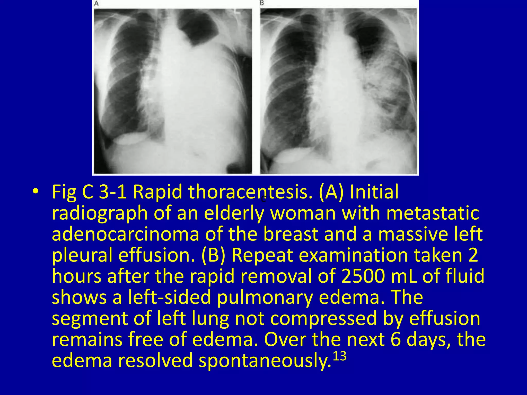
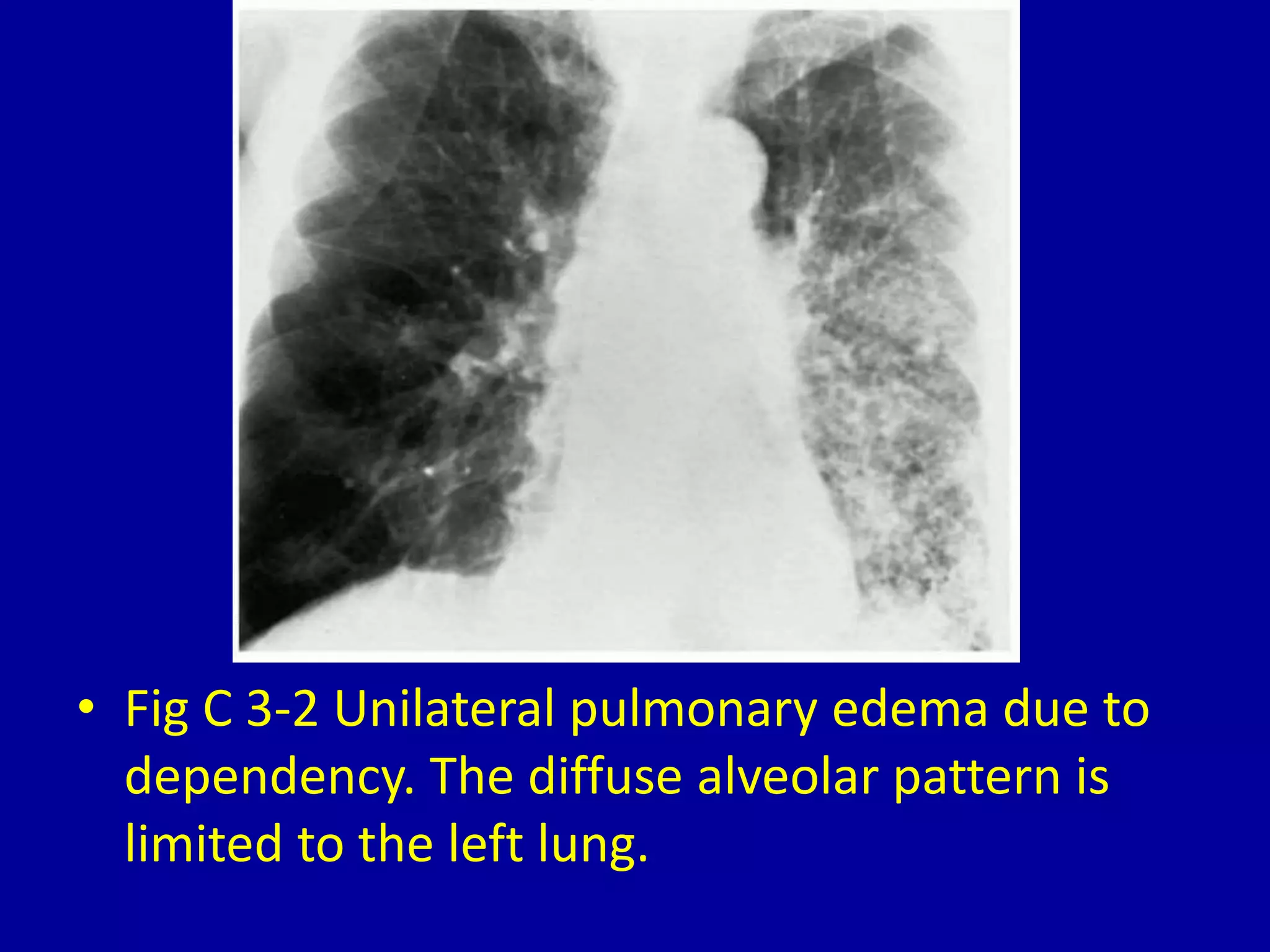
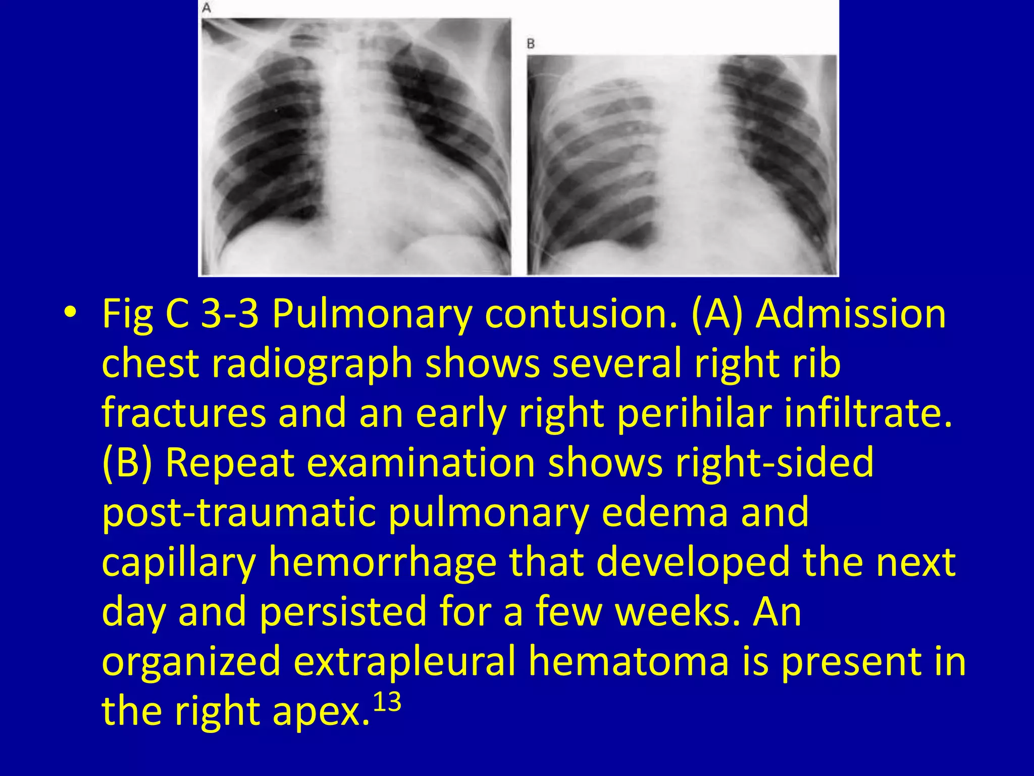
3 Unilateral Pulmonary Edema
Pattern
CLINICAL IMAGAGING
AN ATLAS OF DIFFERENTIAL DAIGNOSIS
EISENBERG
DR. Muhammad Bin Zulfiqar
PGR-FCPS III SIMS/SHL
• Fig C 3-1 Rapid thoracentesis. (A) Initial
radiograph of an elderly woman with metastatic
adenocarcinoma of the breast and a massive left
pleural effusion. (B) Repeat examination taken 2
hours after the rapid removal of 2500 mL of fluid
shows a left-sided pulmonary edema. The
segment of left lung not compressed by effusion
remains free of edema. Over the next 6 days, the
edema resolved spontaneously.13
c
• Fig C 3-2 Unilateral pulmonary edema due to
dependency. The diffuse alveolar pattern is
limited to the left lung.
• Fig C 3-3 Pulmonary contusion. (A) Admission
chest radiograph shows several right rib
fractures and an early right perihilar infiltrate.
(B) Repeat examination shows right-sided
post-traumatic pulmonary edema and
capillary hemorrhage that developed the next
day and persisted for a few weeks. An
organized extrapleural hematoma is present in
the right apex.13
• Fig C 3-4 Edema contralateral to pulmonary embolization. (A) Chest
radiograph in an acutely dyspneic older man shows moderate
cardiomegaly and early right-sided edema that spares the base. (B) An
angiogram obtained at the bedside shows nonopacification of all left
pulmonary branches and a few segmental right lower lobe branches. (C)
Radionuclide scan shows complete lack of perfusion in the left lung and
poor perfusion of the right lower lobe.13
3 unilateral pulmonary edema pattern

3 unilateral pulmonary edema pattern

  • 1.
  • 2.
    CLINICAL IMAGAGING AN ATLASOF DIFFERENTIAL DAIGNOSIS EISENBERG DR. Muhammad Bin Zulfiqar PGR-FCPS III SIMS/SHL
  • 3.
    • Fig C3-1 Rapid thoracentesis. (A) Initial radiograph of an elderly woman with metastatic adenocarcinoma of the breast and a massive left pleural effusion. (B) Repeat examination taken 2 hours after the rapid removal of 2500 mL of fluid shows a left-sided pulmonary edema. The segment of left lung not compressed by effusion remains free of edema. Over the next 6 days, the edema resolved spontaneously.13 c
  • 4.
    • Fig C3-2 Unilateral pulmonary edema due to dependency. The diffuse alveolar pattern is limited to the left lung.
  • 5.
    • Fig C3-3 Pulmonary contusion. (A) Admission chest radiograph shows several right rib fractures and an early right perihilar infiltrate. (B) Repeat examination shows right-sided post-traumatic pulmonary edema and capillary hemorrhage that developed the next day and persisted for a few weeks. An organized extrapleural hematoma is present in the right apex.13
  • 6.
    • Fig C3-4 Edema contralateral to pulmonary embolization. (A) Chest radiograph in an acutely dyspneic older man shows moderate cardiomegaly and early right-sided edema that spares the base. (B) An angiogram obtained at the bedside shows nonopacification of all left pulmonary branches and a few segmental right lower lobe branches. (C) Radionuclide scan shows complete lack of perfusion in the left lung and poor perfusion of the right lower lobe.13