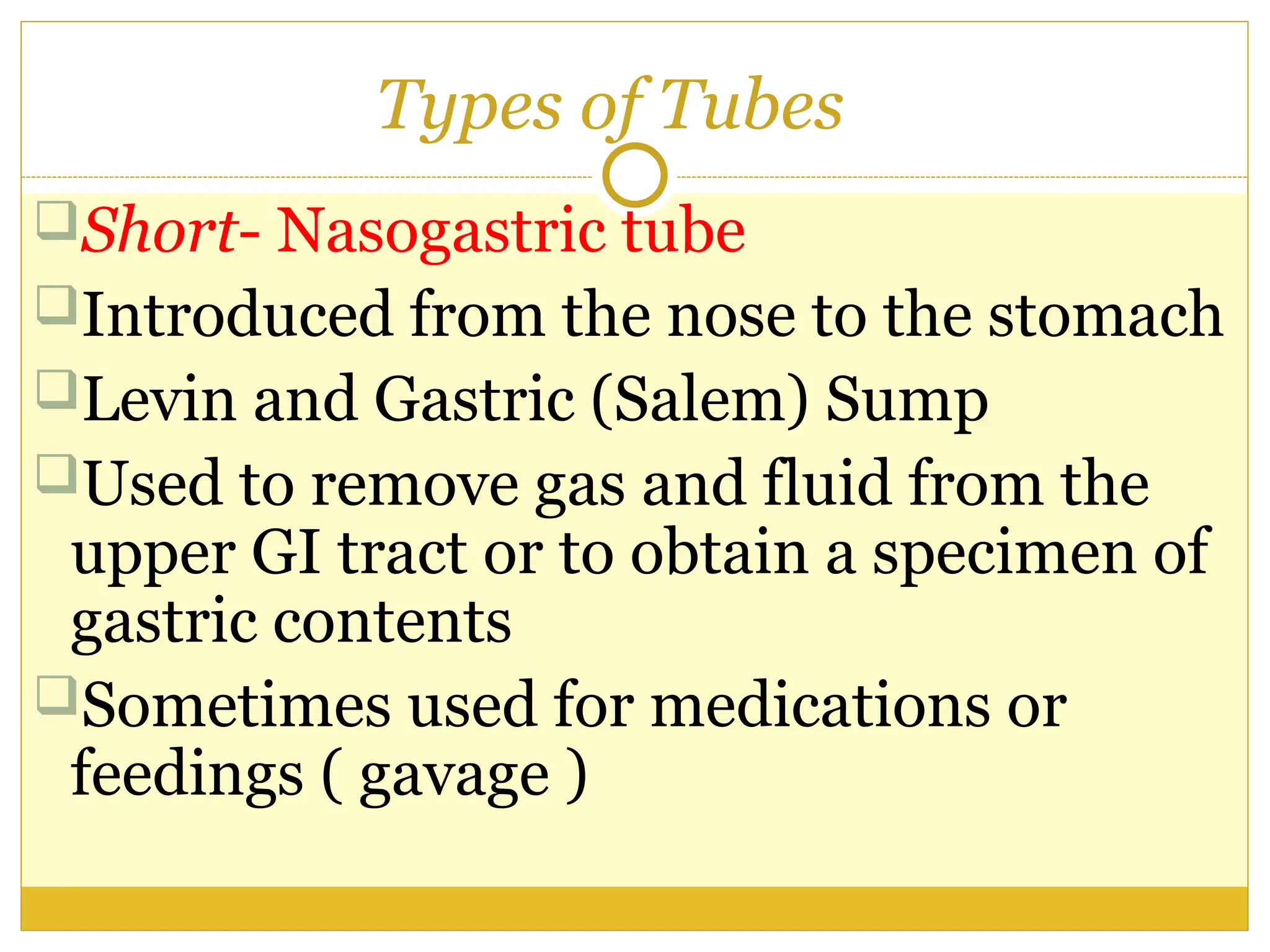
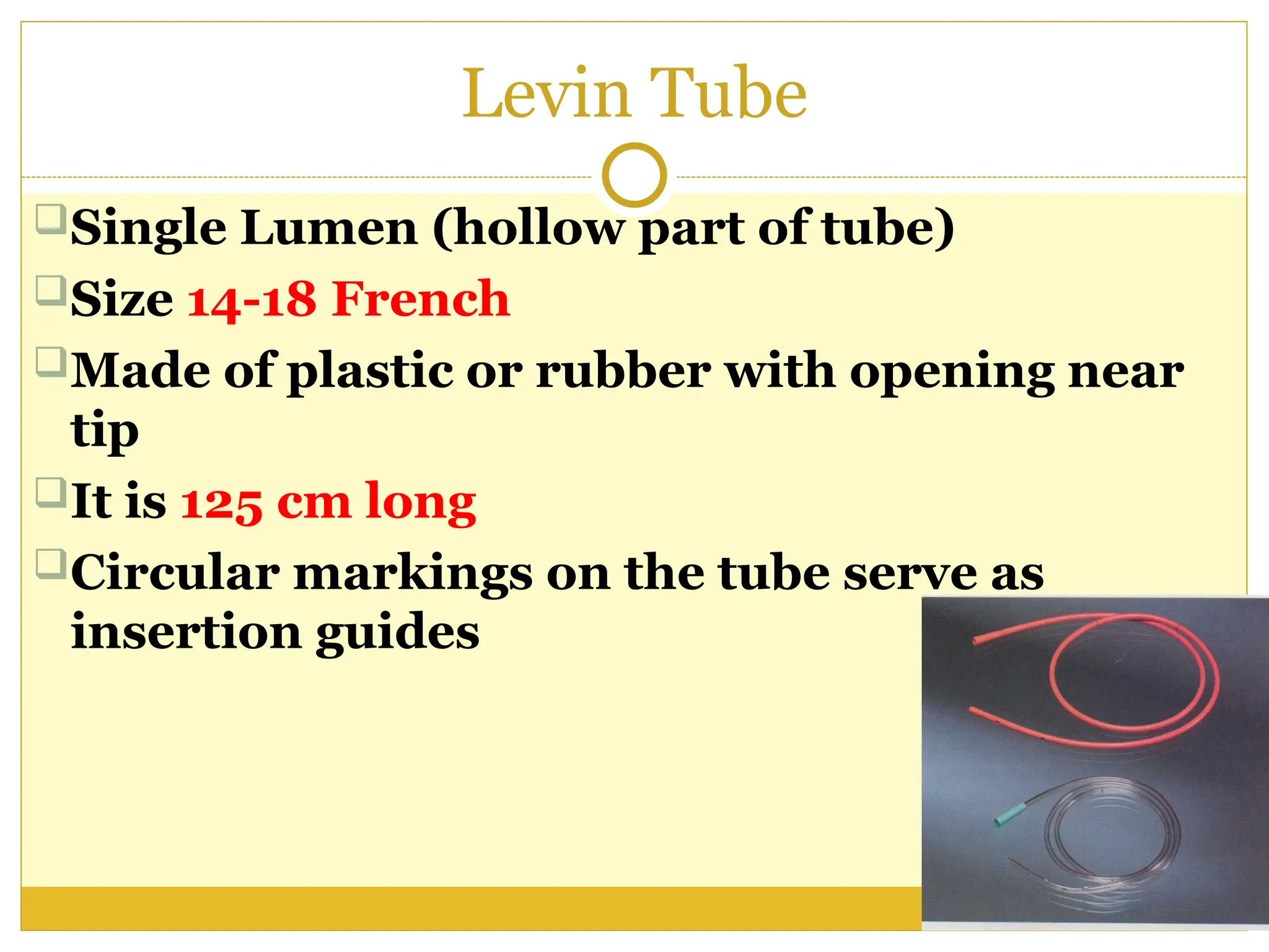
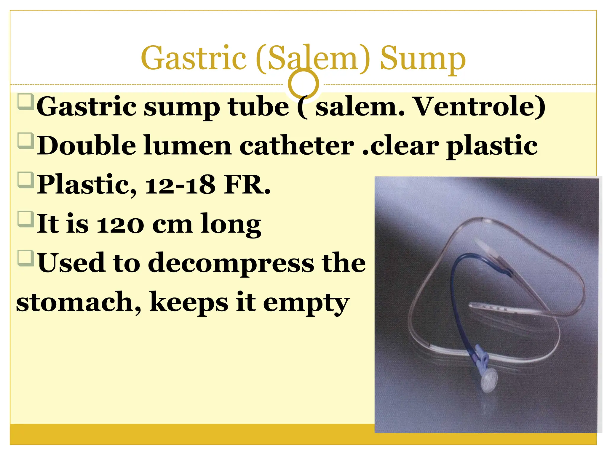
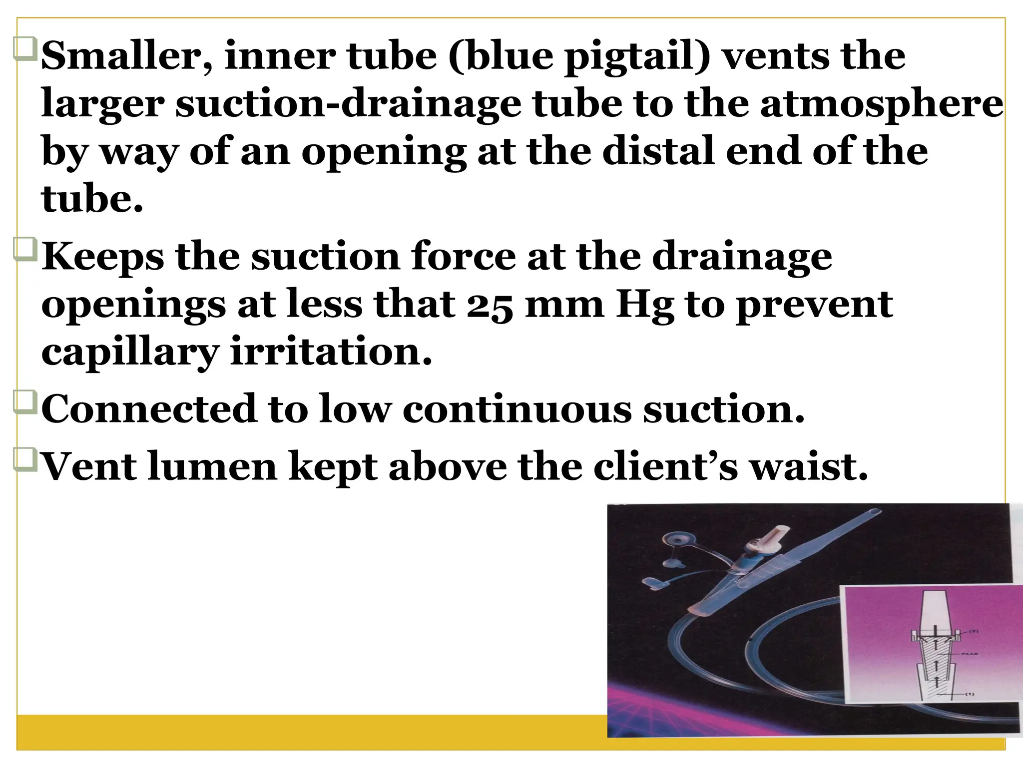
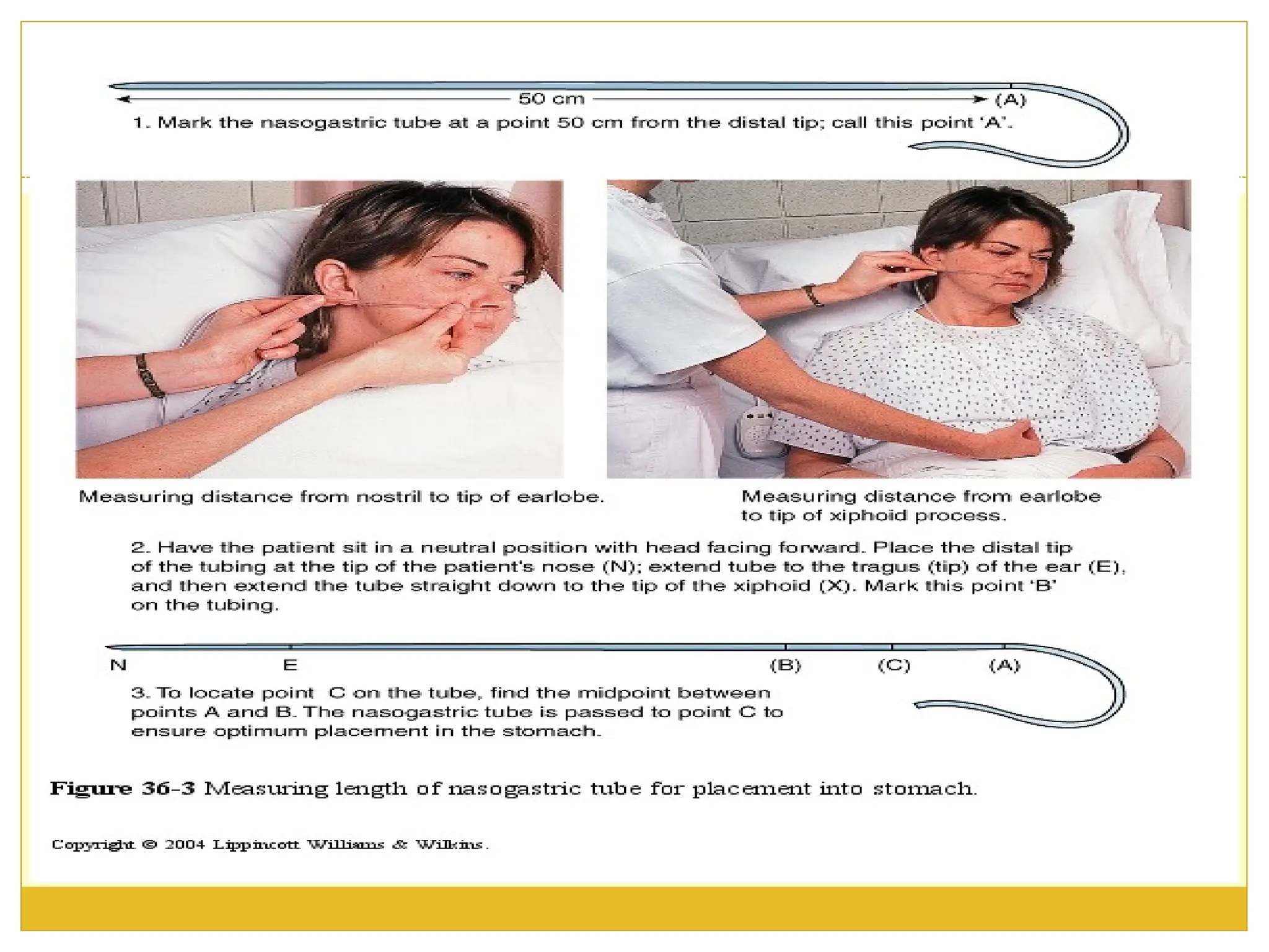
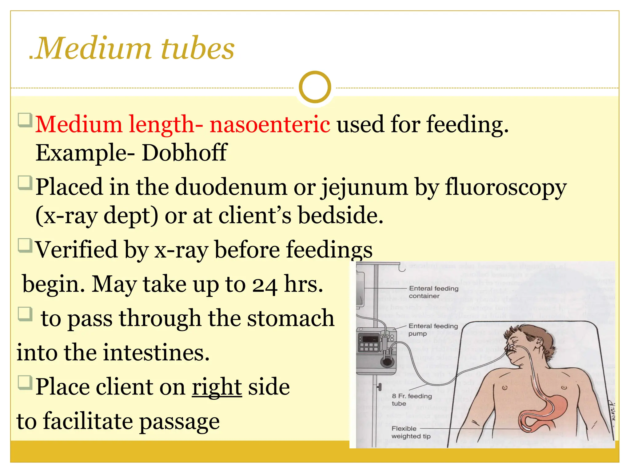
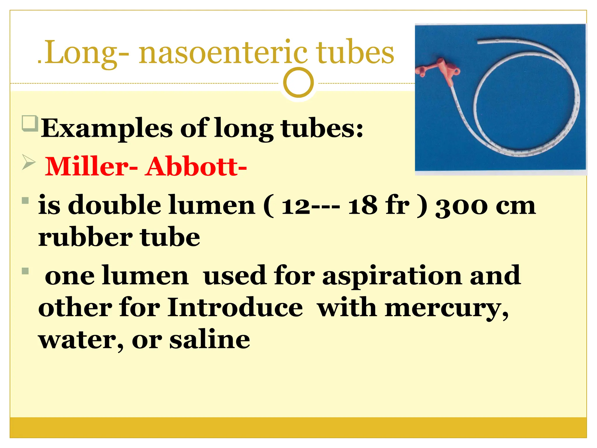
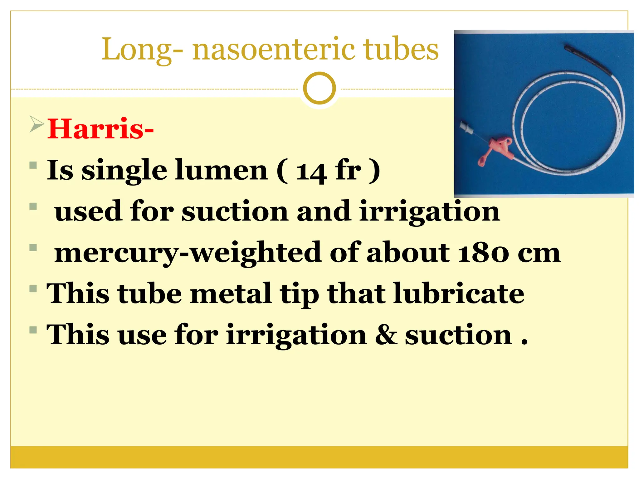
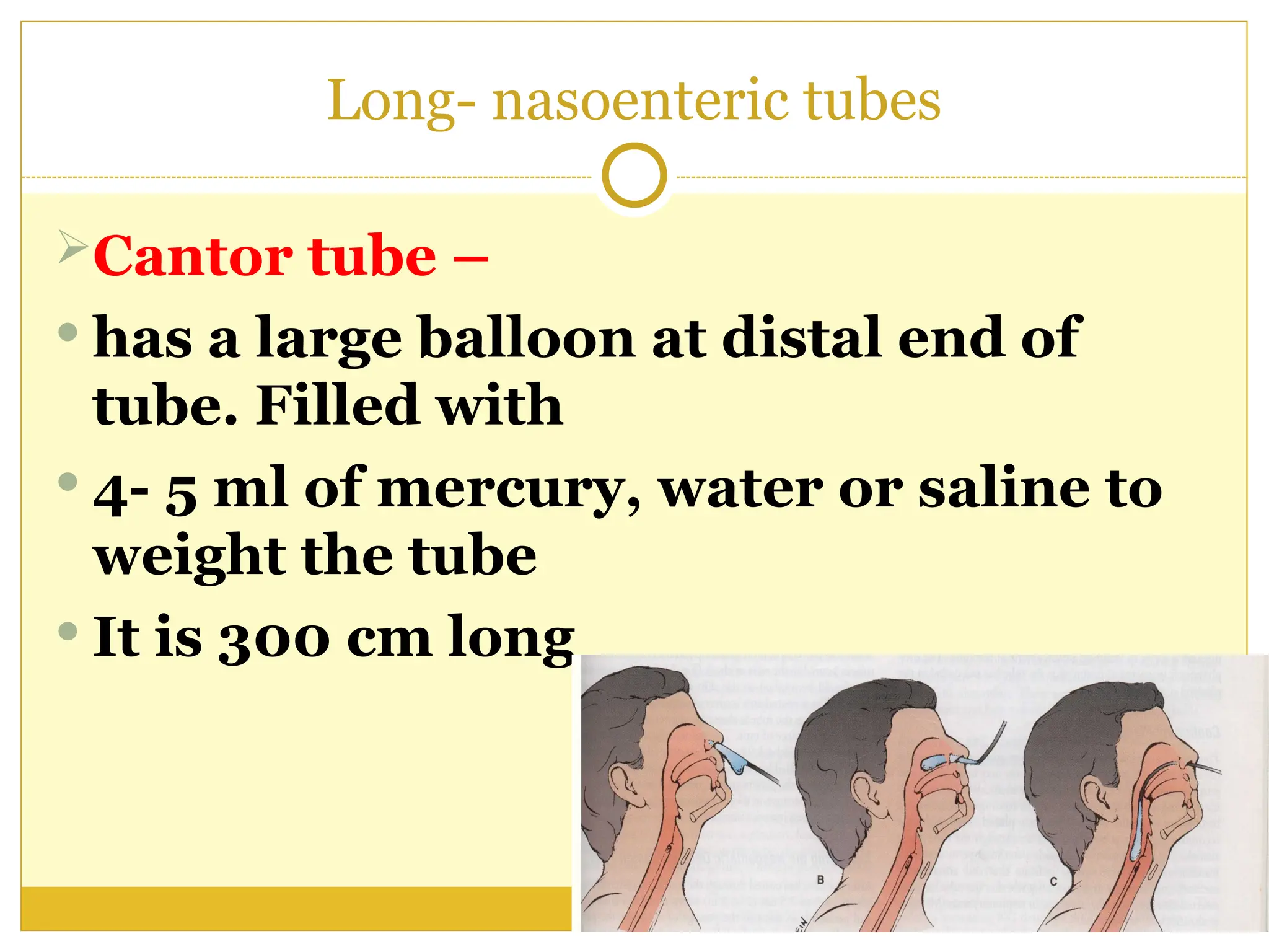
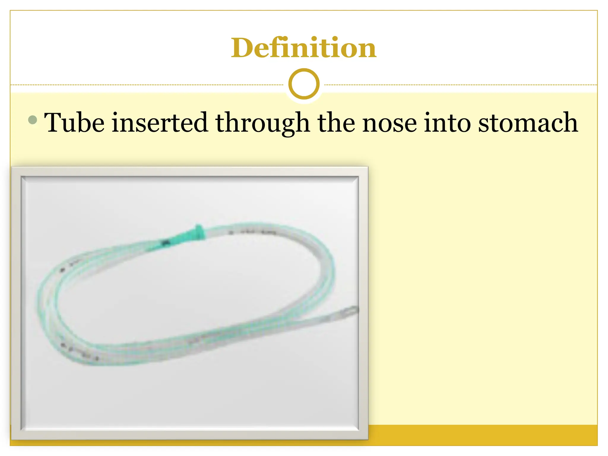
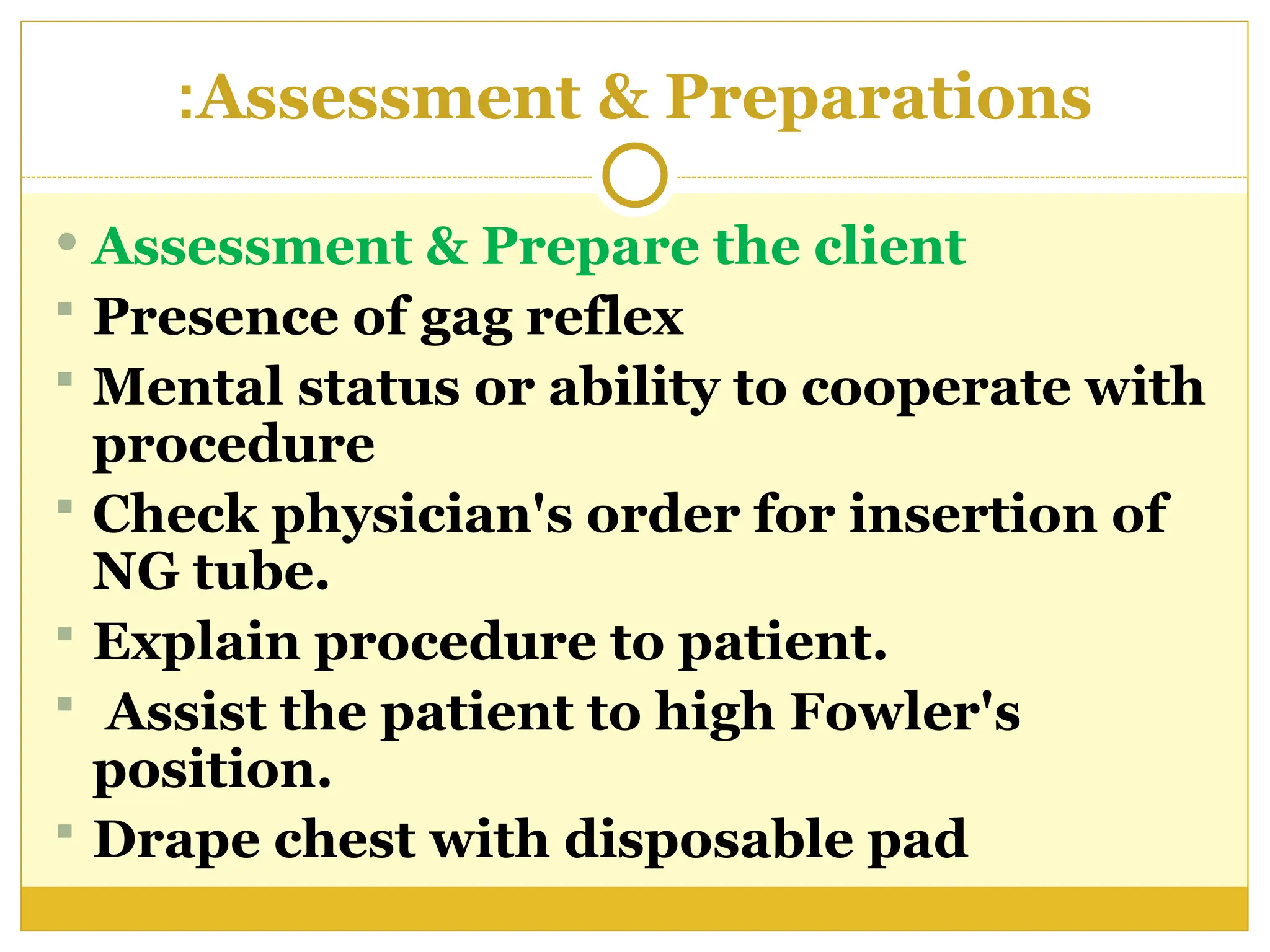
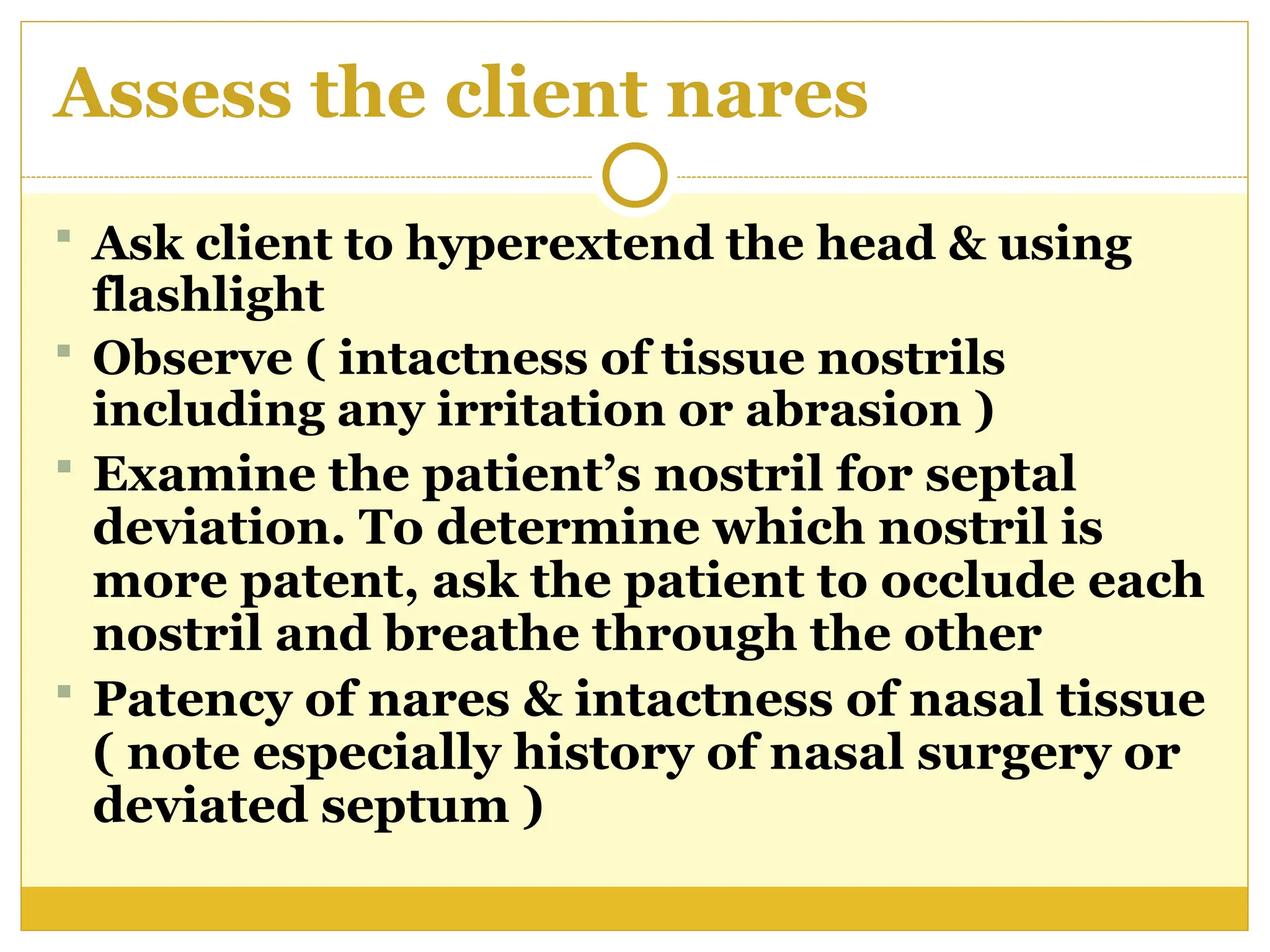
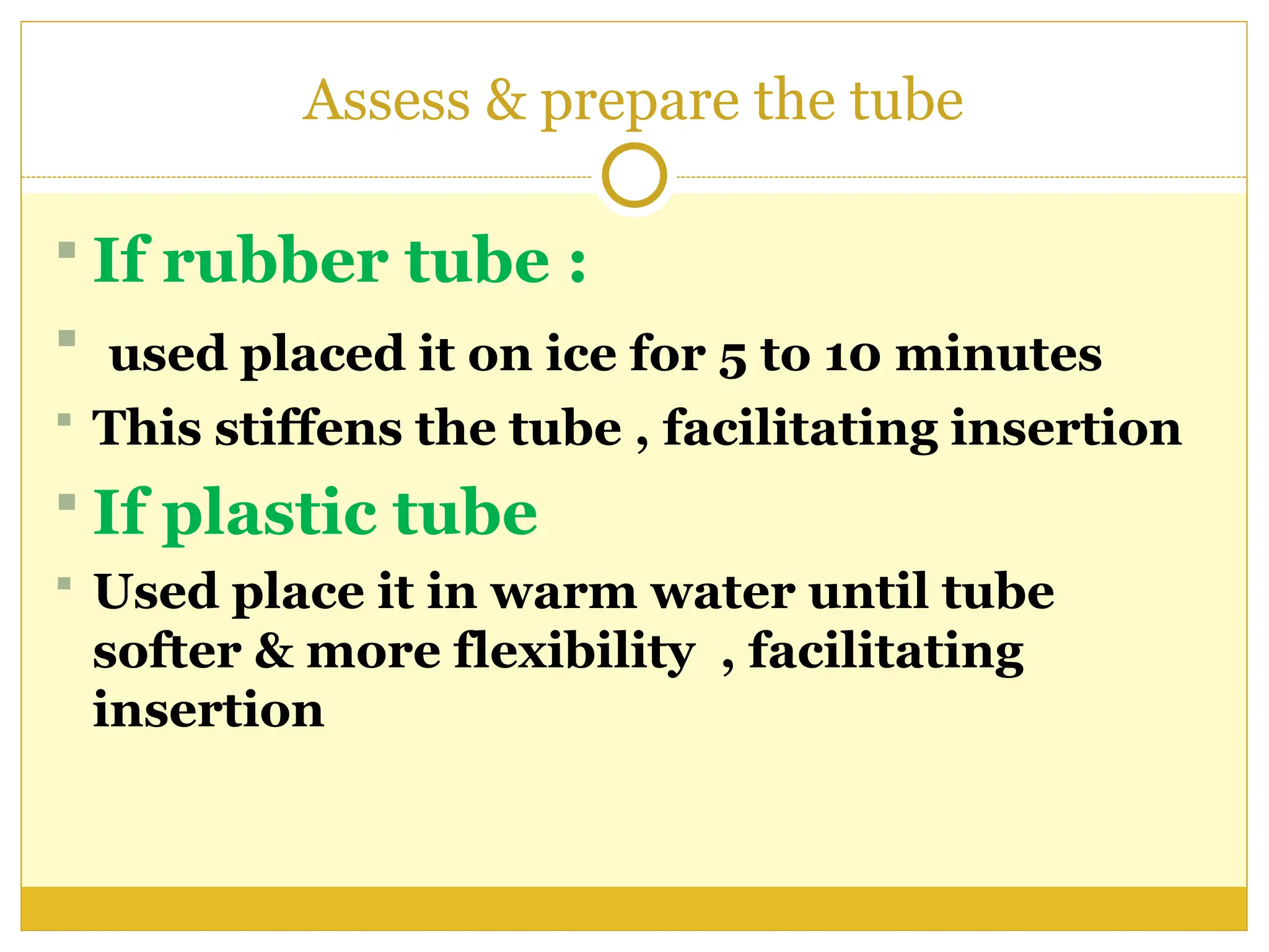
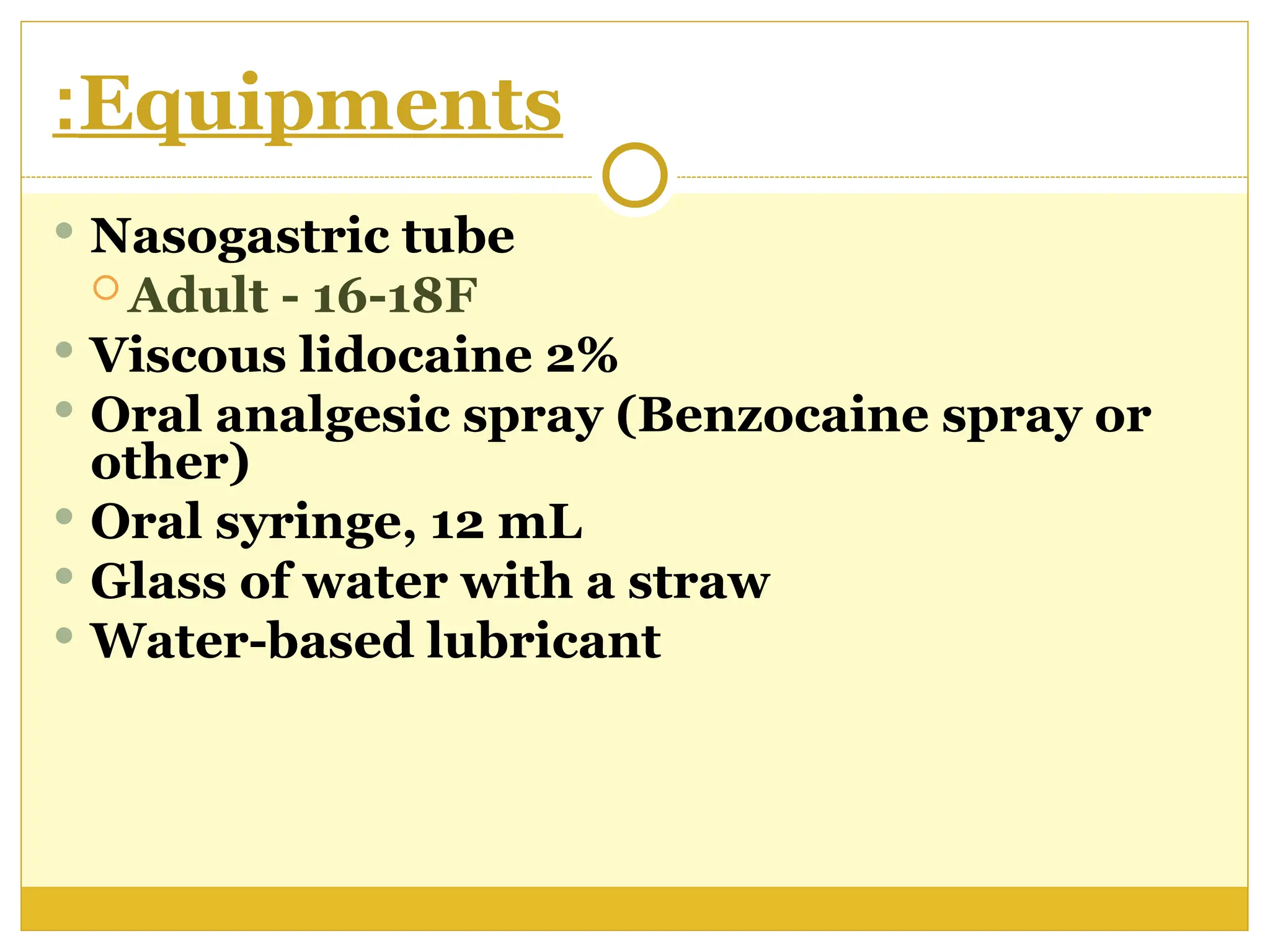
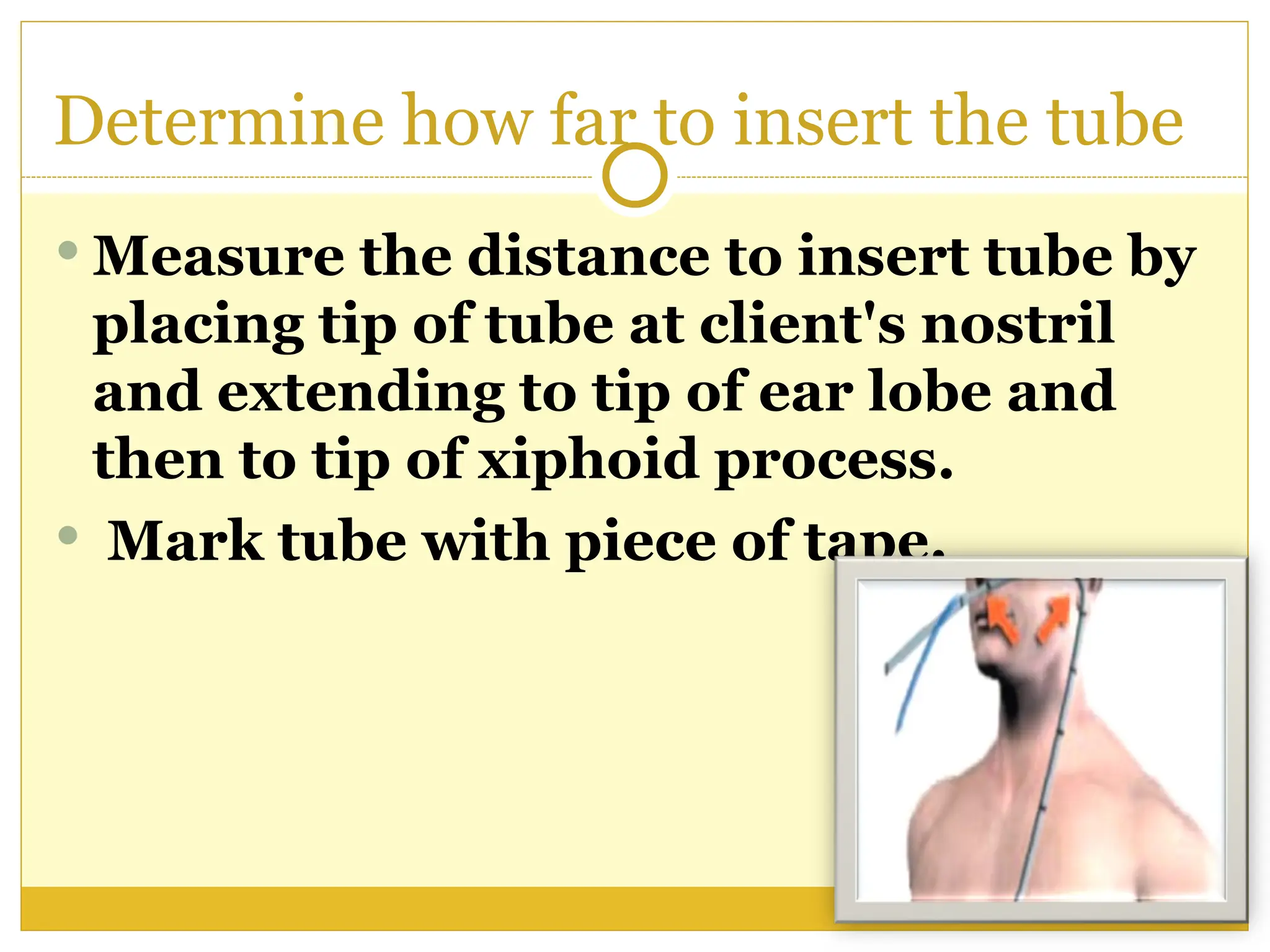
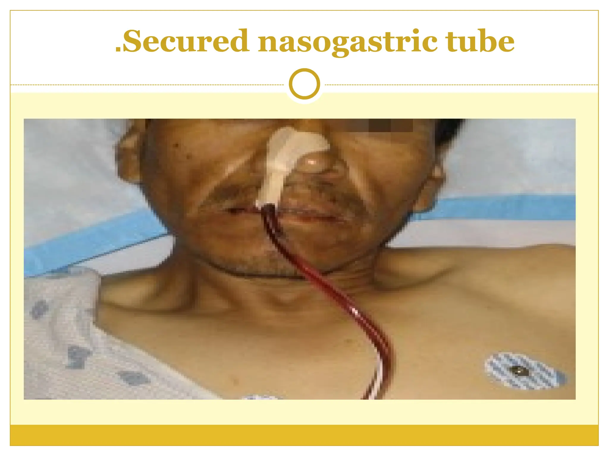
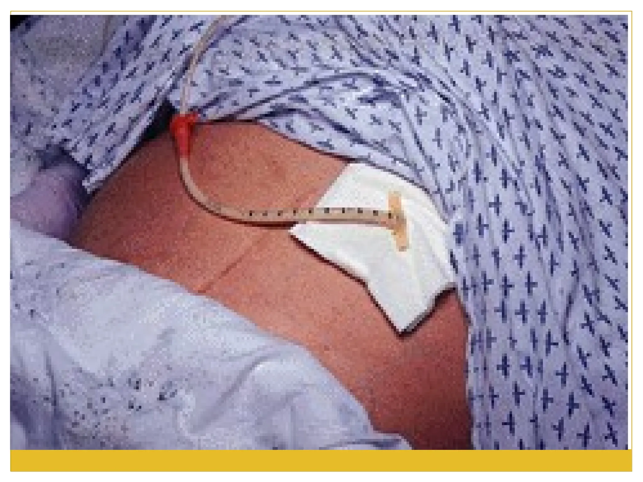
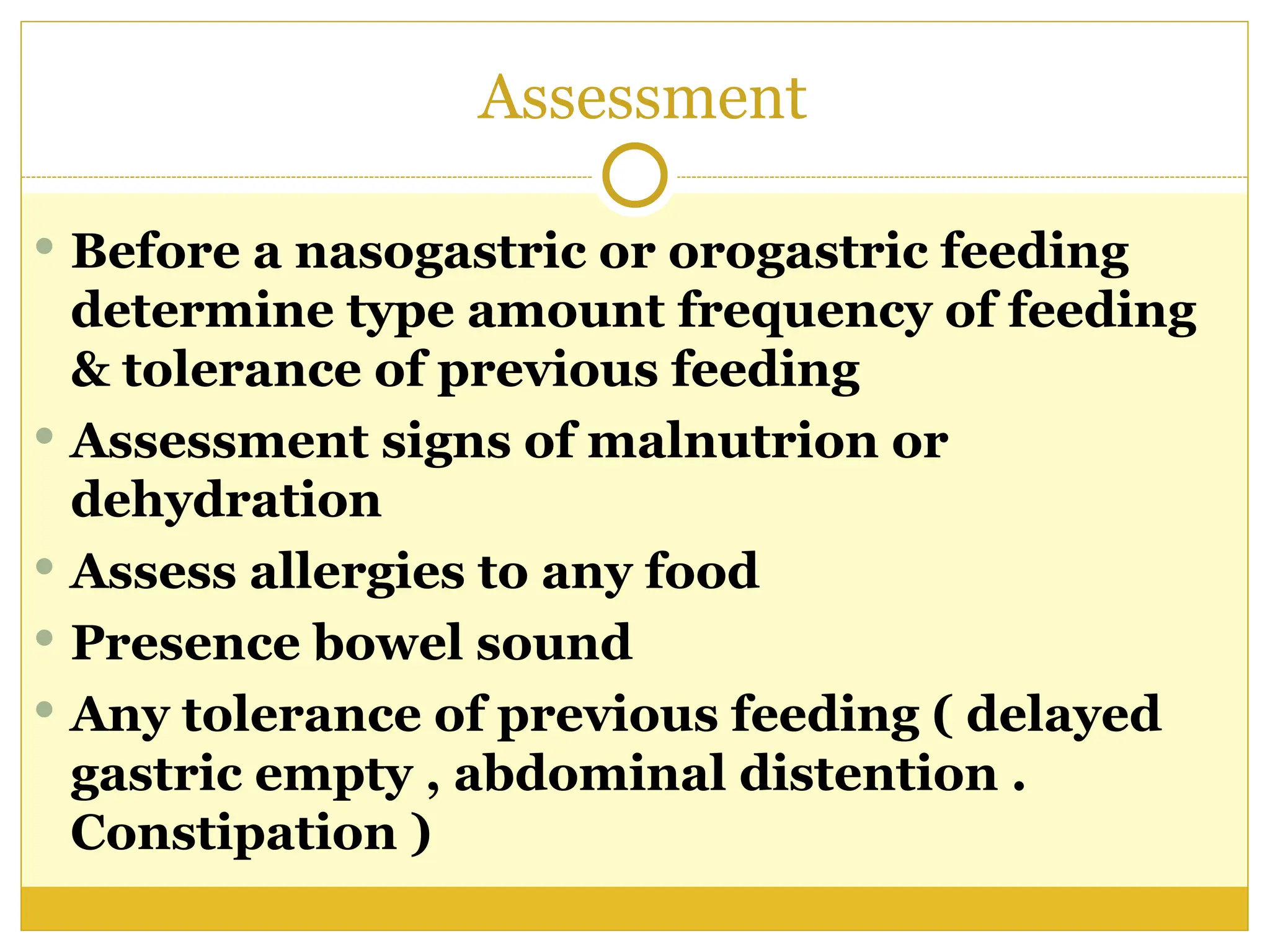
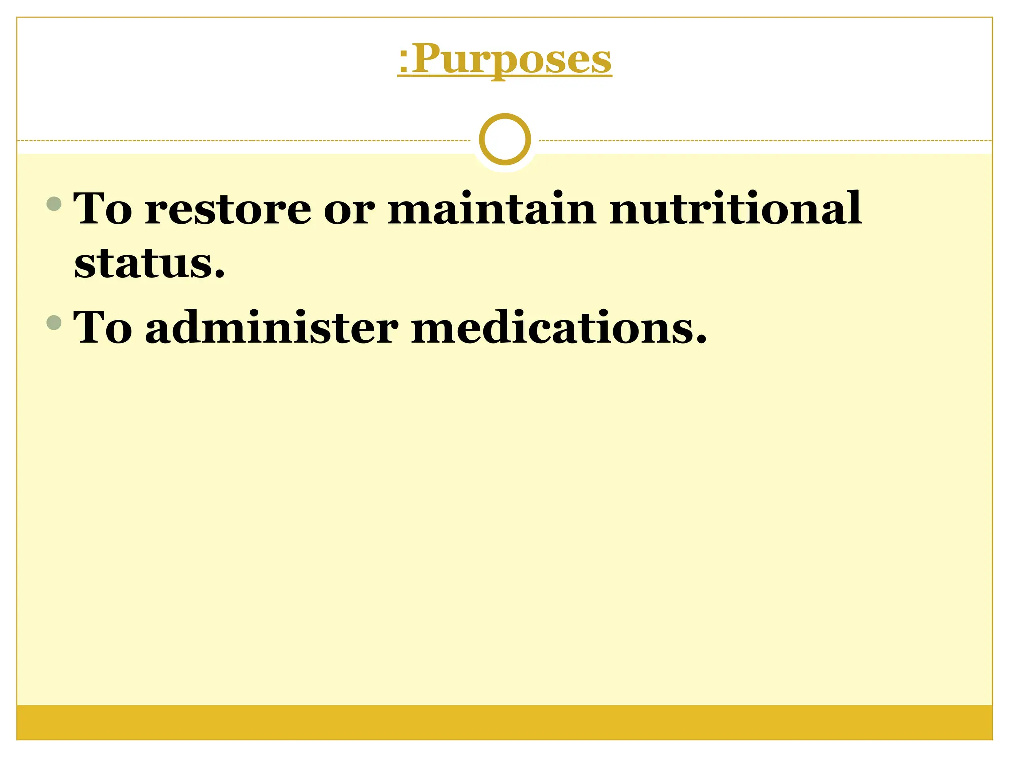
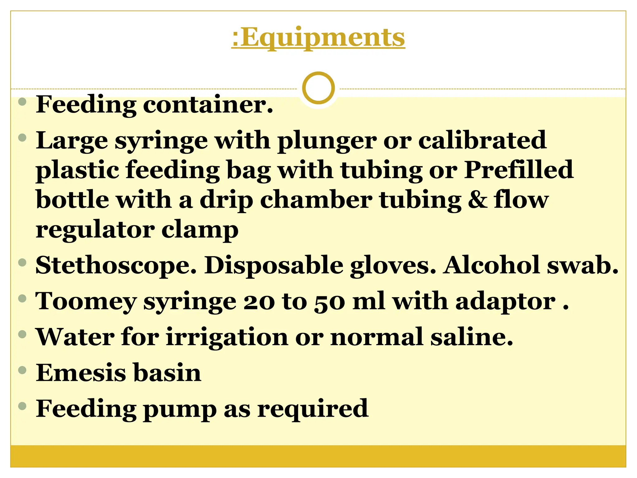
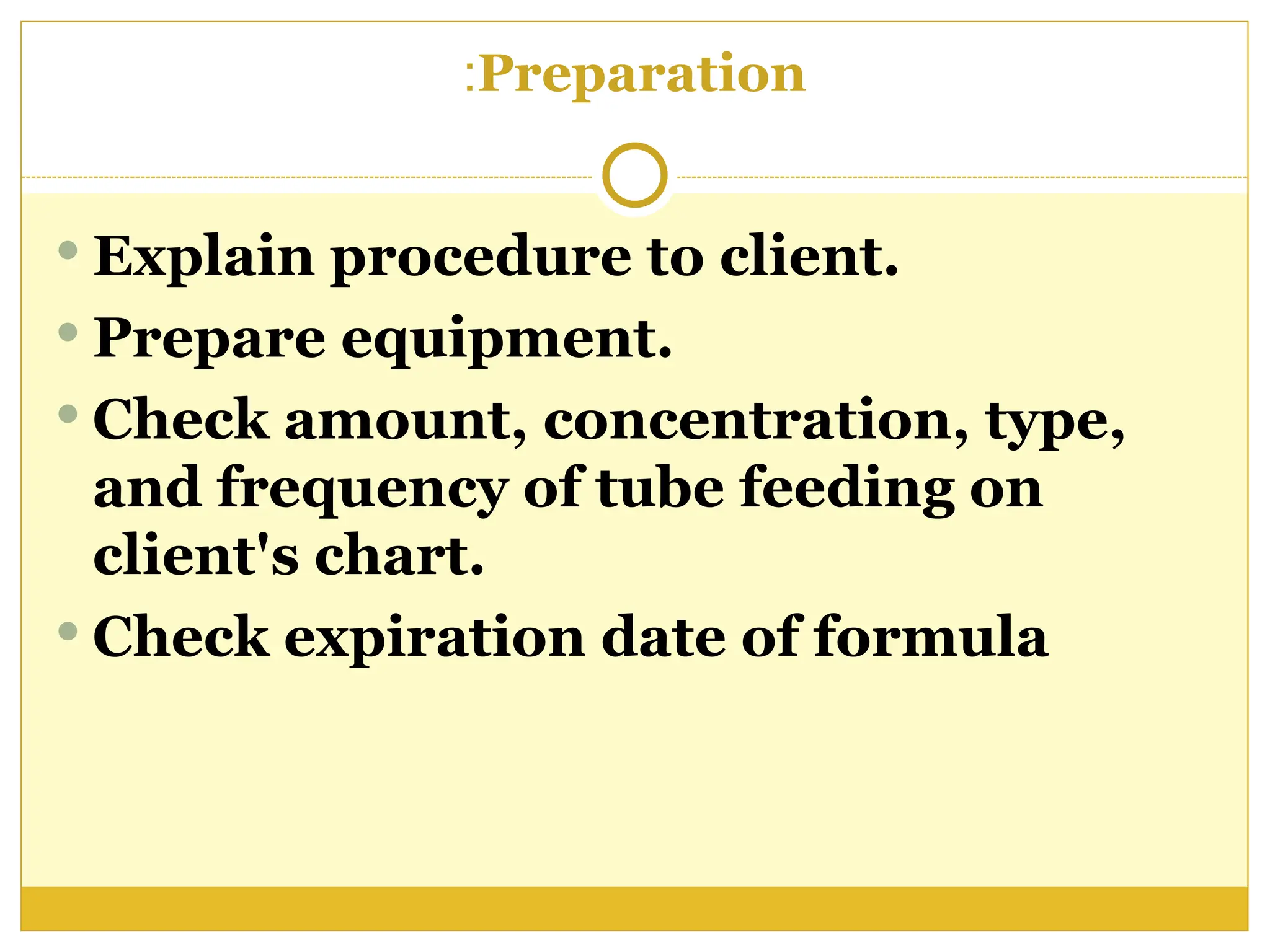
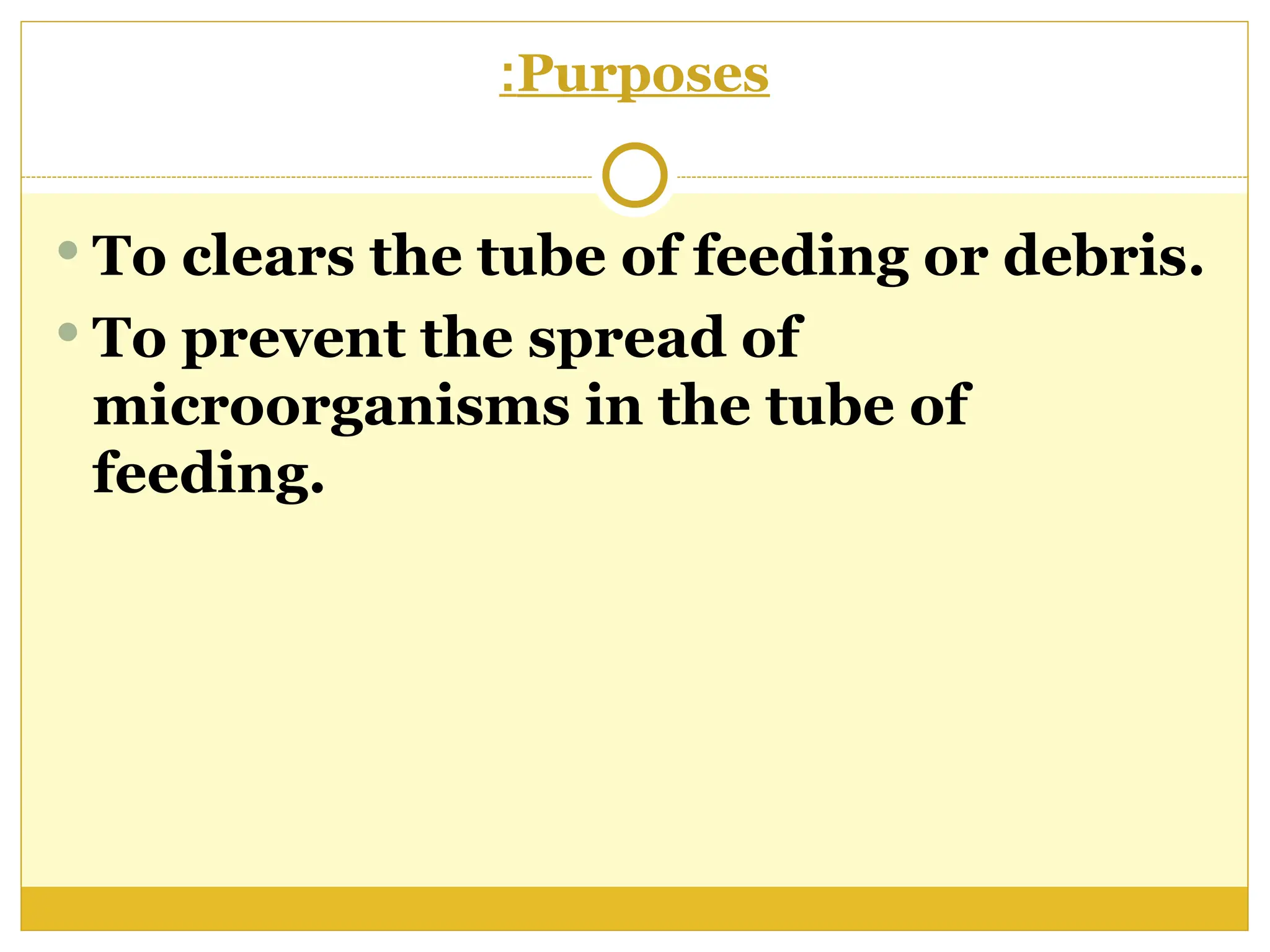
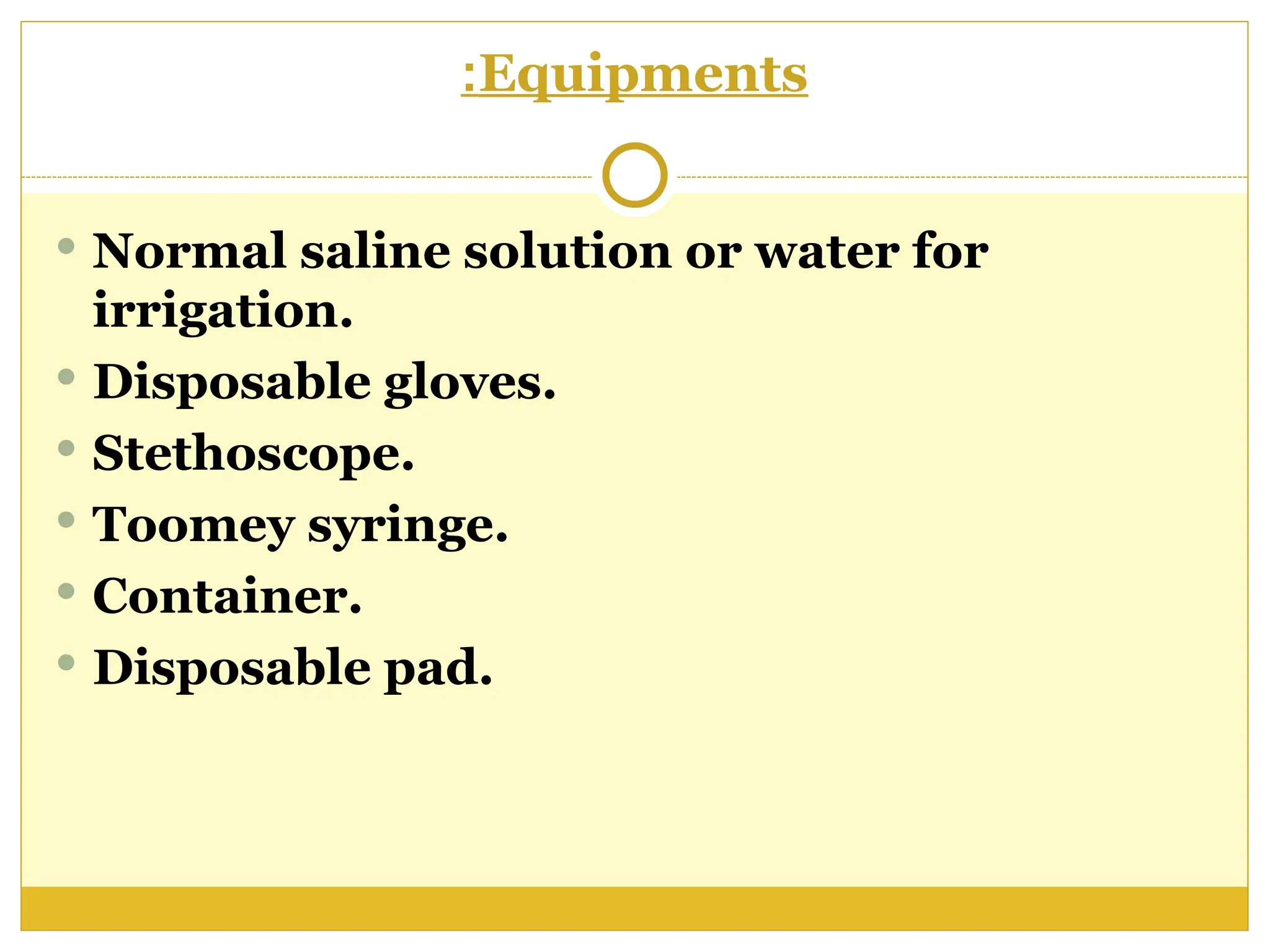
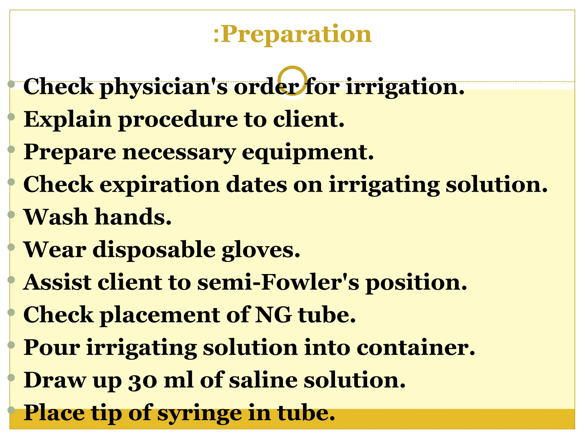
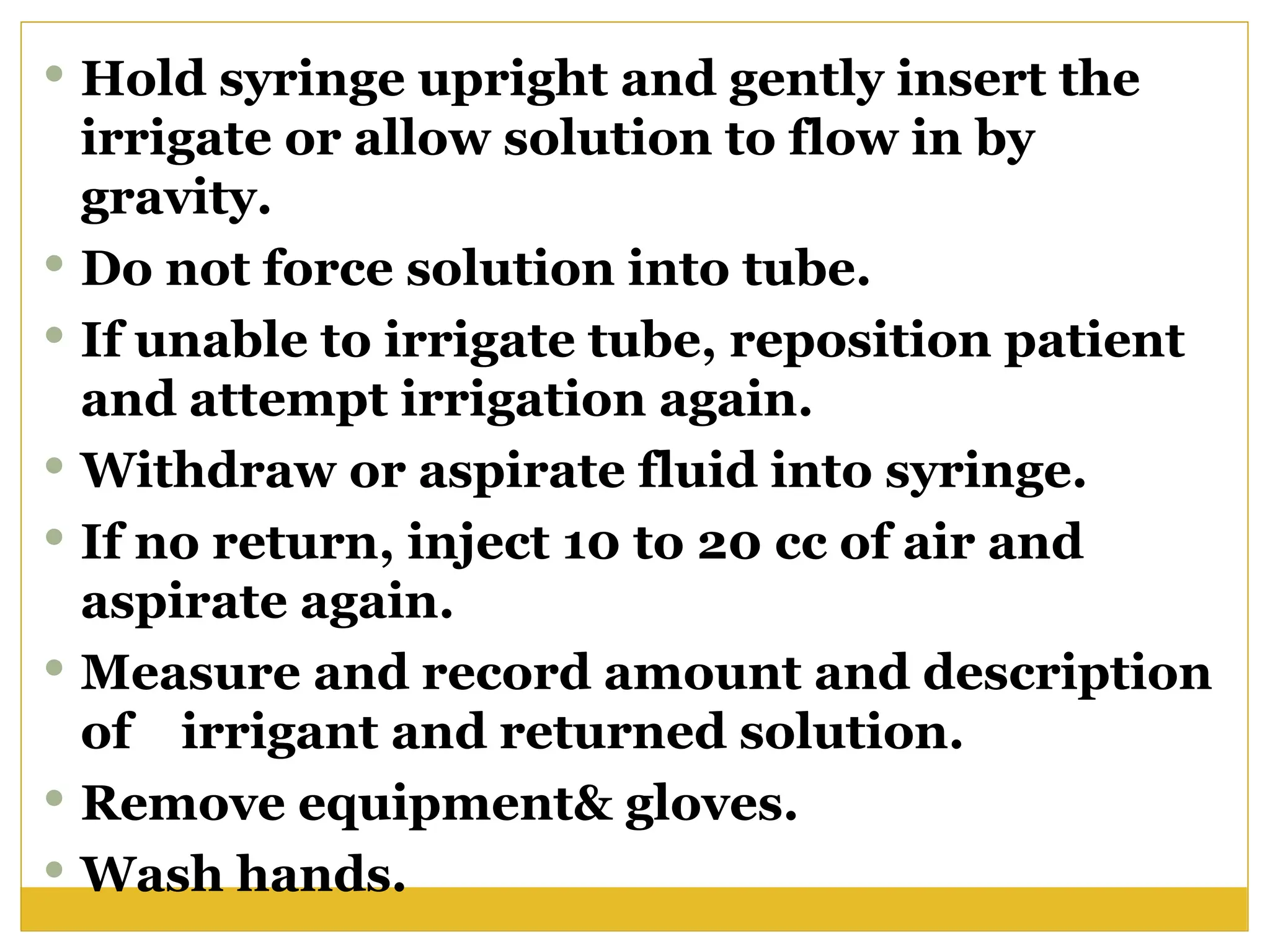
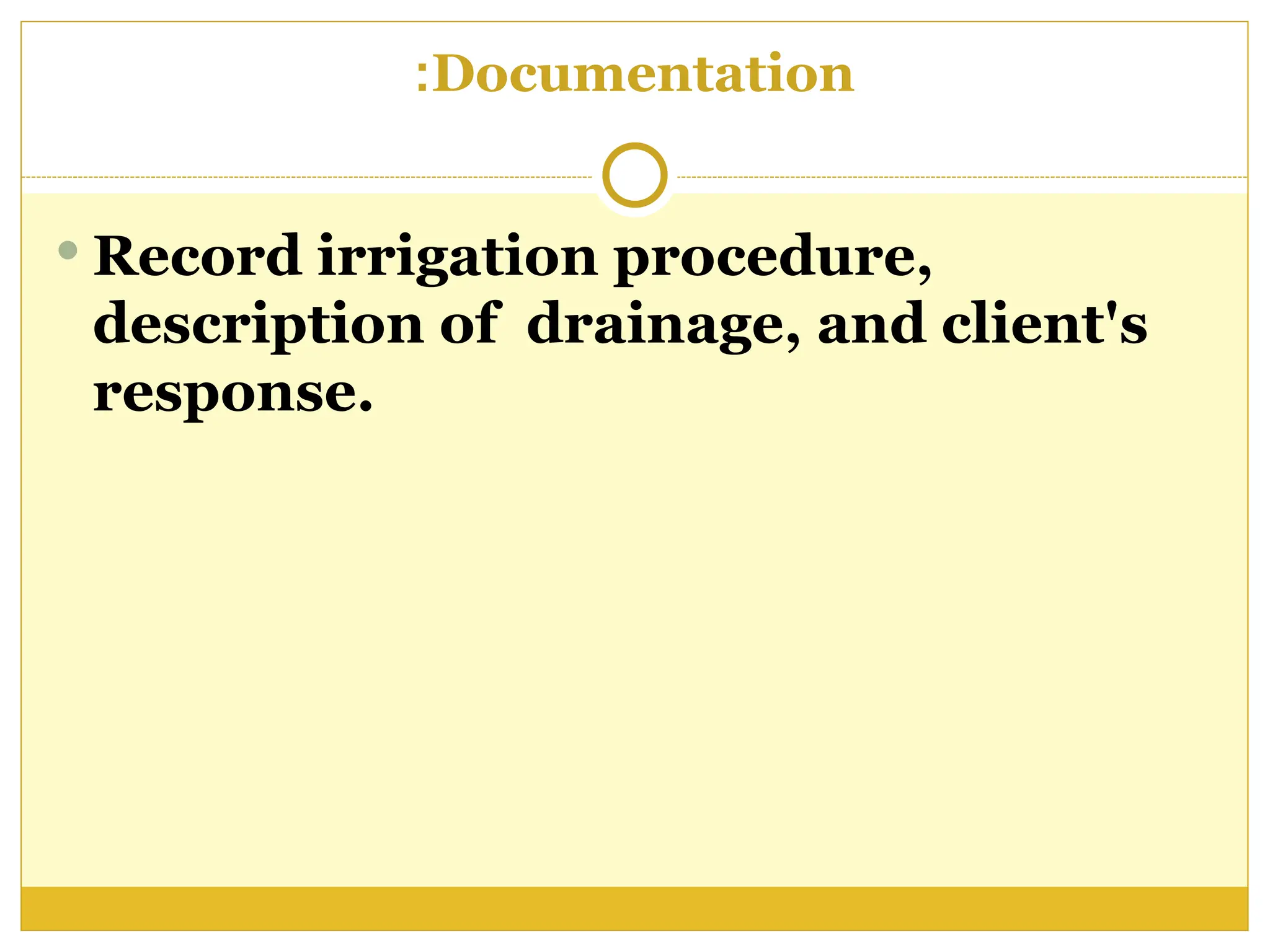
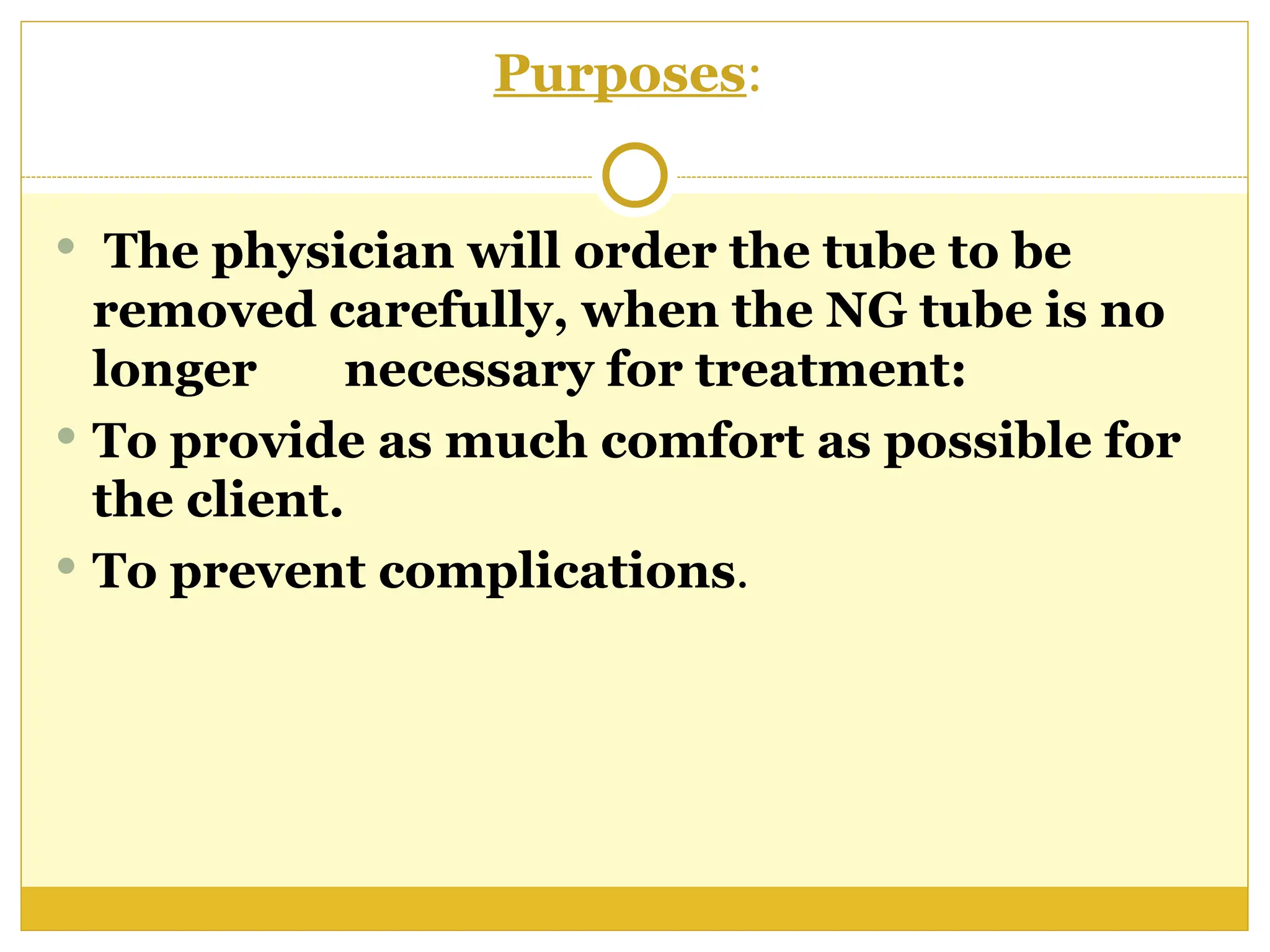
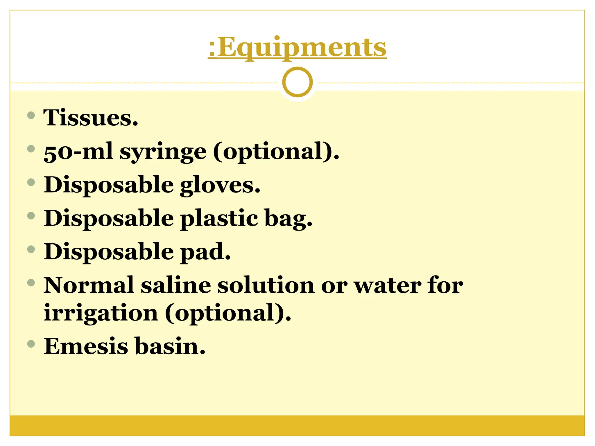
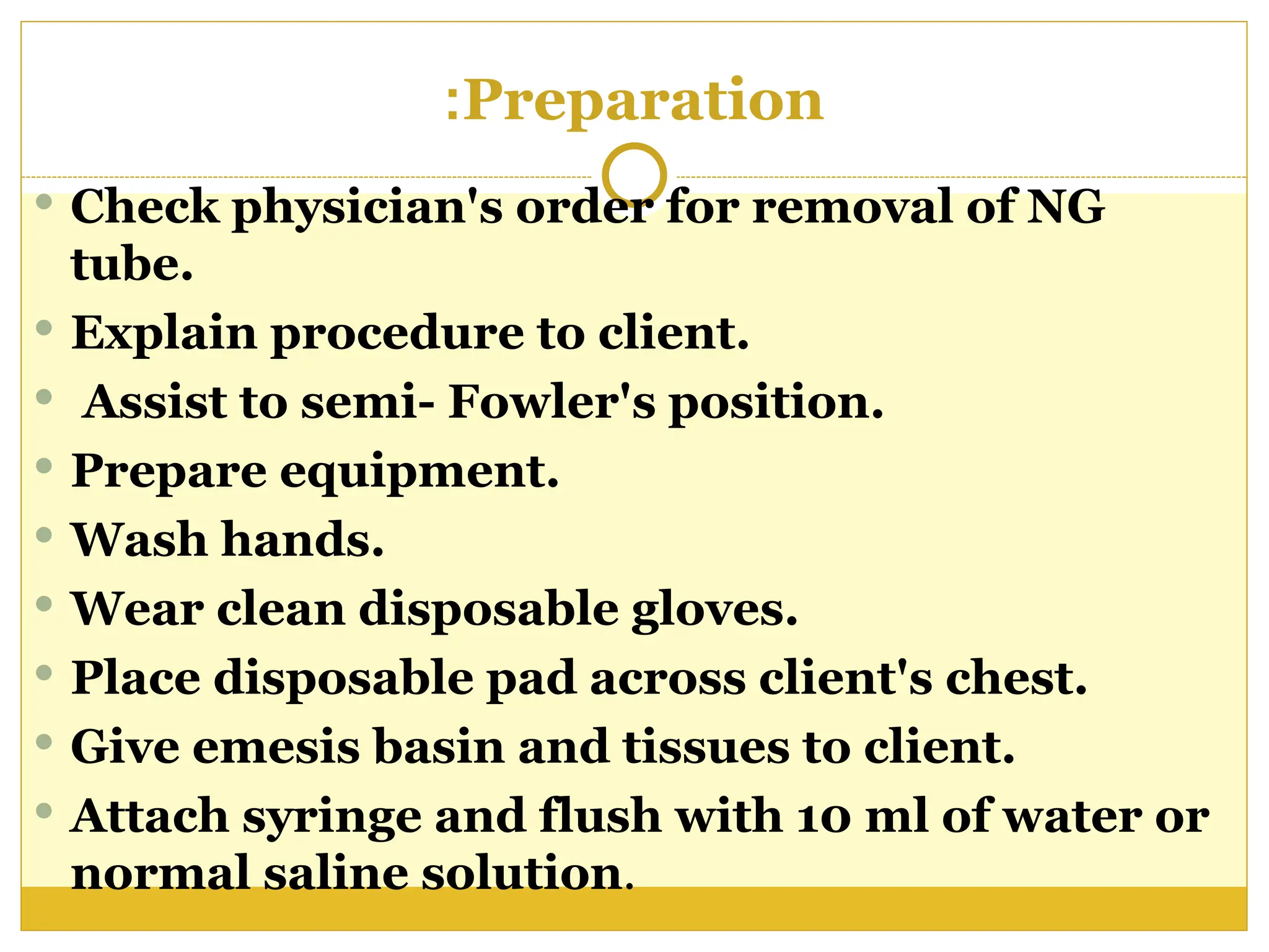
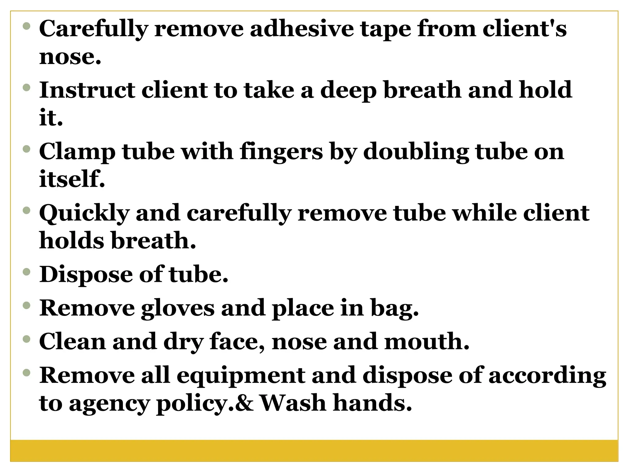
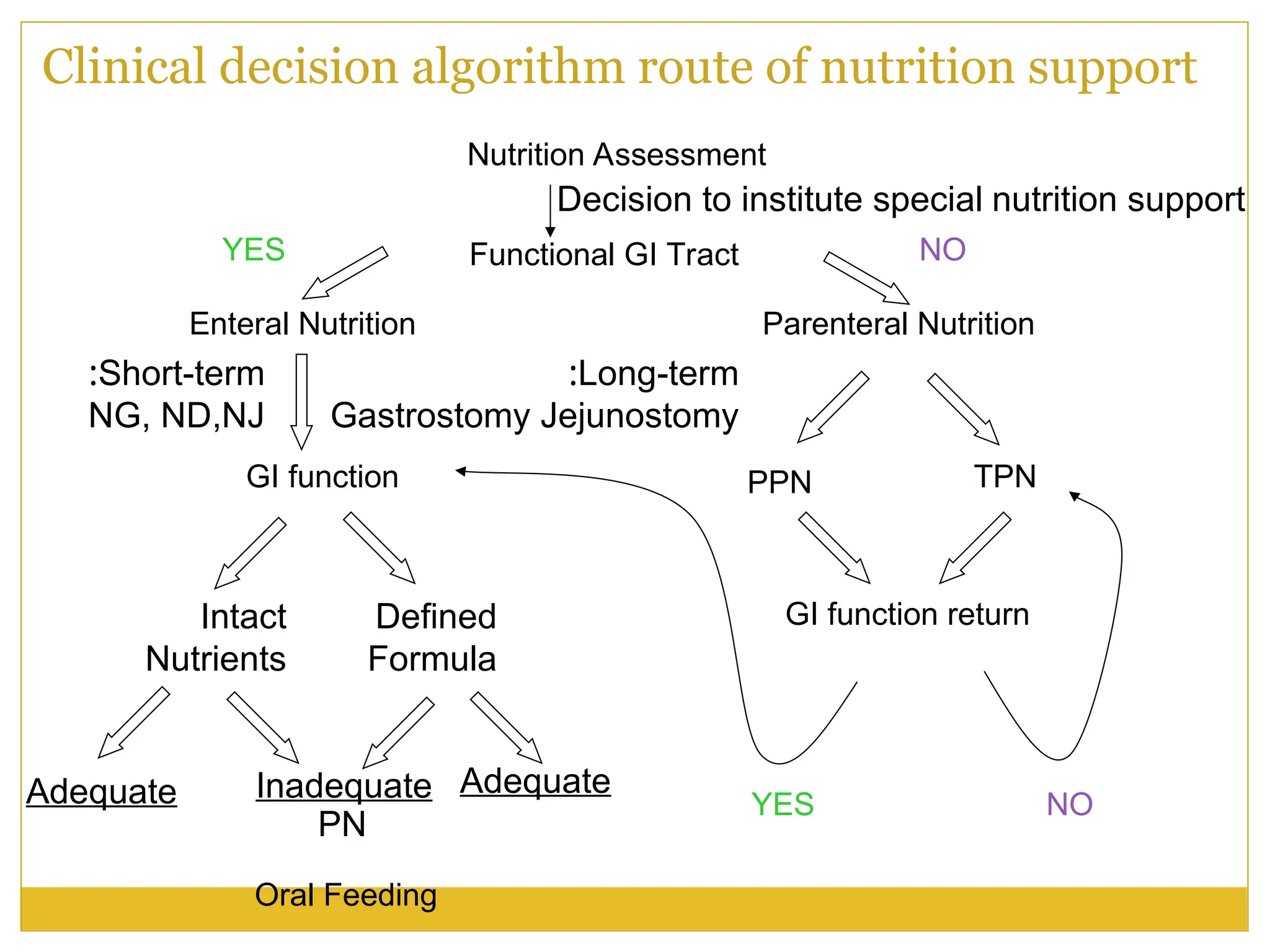
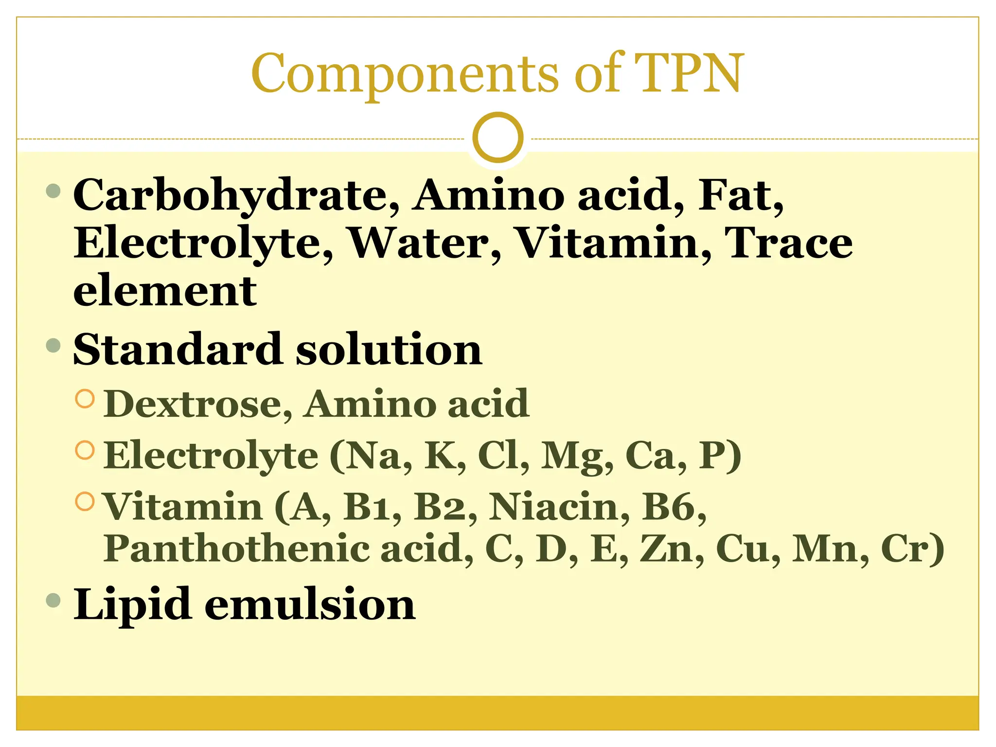
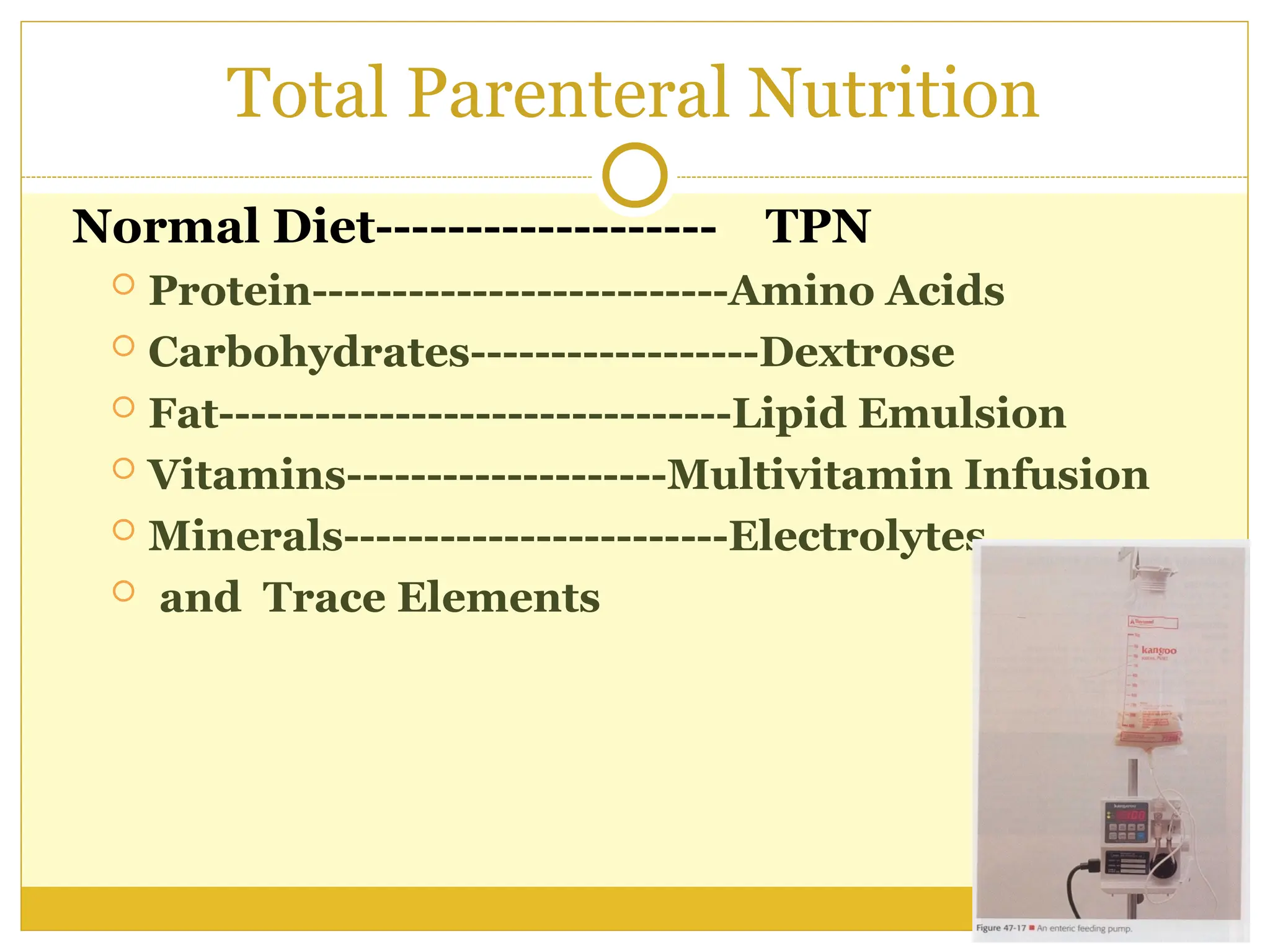
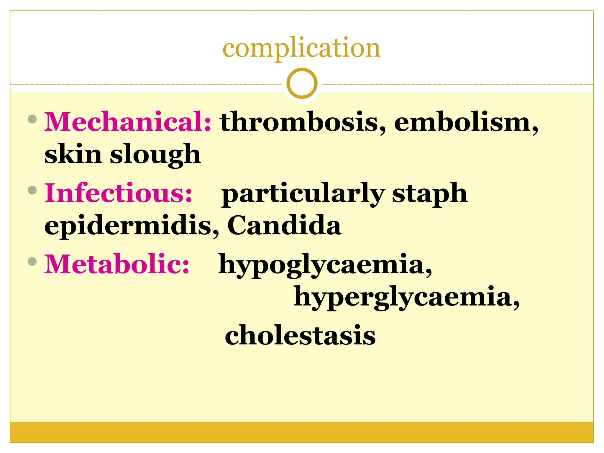
Out line
 Define the nasogastric tube
 Discuss the types of nasogastric tube .
 List the purpose of using the nasogastric tube
 Discuss insertion nasogastric tube
 Discuss removing nasogastric tube
 Discuss administering a tube feeding
 Discuss Irrigating Nasogastric Tube
 Explain the procedure.
 List the potential complications of Nasogastric Tube.
 Demonstrate the procedure.
Introduction
Gastrointestinal intubation is
inserting of rubber or plastic tube into
the stomach , duodenum or intestinal
The tube inserted through
mouth .nose , or abdominal
( gastrostomy .jejunostomy )
The tube short , medium , long
Types of Tubes
Short- Nasogastric tube
Introduced from the nose to the stomach
Levin and Gastric (Salem) Sump
Used to remove gas and fluid from the
upper GI tract or to obtain a specimen of
gastric contents
Sometimes used for medications or
feedings ( gavage )
Levin Tube
Single Lumen (hollow part of tube)
Size 14-18 French
Made of plastic or rubber with opening near
tip
It is 125 cm long
Circular markings on the tube serve as
insertion guides
Gastric (Salem) Sump
Gastric sump tube ( salem. Ventrole)
Double lumen catheter .clear plastic
Plastic, 12-18 FR.
It is 120 cm long
Used to decompress the
stomach, keeps it empty
Smaller, inner tube (blue pigtail) vents the
larger suction-drainage tube to the atmosphere
by way of an opening at the distal end of the
tube.
Keeps the suction force at the drainage
openings at less that 25 mm Hg to prevent
capillary irritation.
Connected to low continuous suction.
Vent lumen kept above the client’s waist.
Medium tubes
.
Medium length- nasoenteric used for feeding.
Example- Dobhoff
Placed in the duodenum or jejunum by fluoroscopy
(x-ray dept) or at client’s bedside.
Verified by x-ray before feedings
begin. May take up to 24 hrs.
 to pass through the stomach
into the intestines.
Place client on right side
to facilitate passage
Long- nasoenteric tubes
.
Long- nasoenteric tubes introduced through
the nose and passed through the esophagus
and stomach into the intestinal tract.
Used to aspirate intestinal contents-ie. gas and
fluid
Used to (Decompression) to prevent intestinal
obstruction.
Due to  peristalsis, prevents vomiting,
reduces tension at the incision line and
prevents obstruction.
Long- nasoenteric tubes
.
Examples of long tubes:
 Miller- Abbott-
 is double lumen ( 12--- 18 fr ) 300 cm
rubber tube
 one lumen used for aspiration and
other for Introduce with mercury,
water, or saline
Long- nasoenteric tubes
Harris-
 Is single lumen ( 14 fr )
 used for suction and irrigation
 mercury-weighted of about 180 cm
 This tube metal tip that lubricate
 This use for irrigation & suction .
Long- nasoenteric tubes
Cantor tube –
 has a large balloon at distal end of
tube. Filled with
 4- 5 ml of mercury, water or saline to
weight the tube
 It is 300 cm long
Procedure of Inserting
nasogastric tube
Definition
 Tube inserted through the nose into stomach
Purposes
:
 To administer tube feedings and medications
to clients unable to eat by mouth or swallow a
sufficient diet without aspirating food or fluids
into the lungs
 To establish a means for suctioning stomach
contents to prevent gastric distention ,nausea,
and vomiting.
 To remove stomach contents for laboratory
analysis
 To lavage(wash)the stomach in case of
poisoning or overdose of medications.
Purposes
 To drain fluid or air from the
stomach.
 To promote healing after bowel
surgery.
 To monitor bleeding in the
gastrointestinal (GI) tract.
 To help treat an intestinal
obstruction.
Assessment & Preparations
:
 Assessment & Prepare the client
 Presence of gag reflex
 Mental status or ability to cooperate with
procedure
 Check physician's order for insertion of
NG tube.
 Explain procedure to patient.
 Assist the patient to high Fowler's
position.
 Drape chest with disposable pad
Assess the client nares
 Ask client to hyperextend the head & using
flashlight
 Observe ( intactness of tissue nostrils
including any irritation or abrasion )
 Examine the patient’s nostril for septal
deviation. To determine which nostril is
more patent, ask the patient to occlude each
nostril and breathe through the other
 Patency of nares & intactness of nasal tissue
( note especially history of nasal surgery or
deviated septum )
Assess & prepare the tube
 If rubber tube :
 used placed it on ice for 5 to 10 minutes
 This stiffens the tube , facilitating insertion
 If plastic tube
 Used place it in warm water until tube
softer & more flexibility , facilitating
insertion
Equipments
:
 Nasogastric tube
 Adult - 16-18F
 Viscous lidocaine 2%
 Oral analgesic spray (Benzocaine spray or
other)
 Oral syringe, 12 mL
 Glass of water with a straw
 Water-based lubricant
Equipments
:
Non allergenic adhesive Tape 2,5 cm wide
Emesis basin or plastic bag
Wall suction, set to low intermittent suction
Suction tubing and container
Flashlight .
Stethoscope.
Toomey syringe (20 to 50 ml) .
Tissues
Disposable pad & gloves . .
Tongue blade .
Normal saline solution (for irrigation only).
Procedure
:
Note
 A nasogastric (NG) tube is used for
the procedure. The placement of an
NG tube can be uncomfortable for the
patient if the patient is not adequately
prepared with anesthesia to the nasal
passages and specific instructions on
how to cooperate with the operator
during the procedure
Determine how far to insert the tube
 Measure the distance to insert tube by
placing tip of tube at client's nostril
and extending to tip of ear lobe and
then to tip of xiphoid process.
 Mark tube with piece of tape.
Nasogastric tube lubrication with water-based
lubricant
.
Estimation of nasogastric tube length
from nostril to stomach
Insert the tube
 Prepare equipment.
 Wash hands.
 Wear disposable gloves.
 Instill 10 mL of viscous lidocaine 2% (for oral
use) down the more patent nostril with the
head tilted backwards, and ask the patient to
sniff and swallow to anesthetize
 Lubricate tip of tube with water soluble
lubricant.
 Ask client to lift head, and insert tube into
nostril while directing tube upward and
backward.
Aspiration of viscous lidocaine into an
oral syringe
insert of viscous lidocaine 2%
Cont
,,
 If client gag when tube reaches
pharynx, provide tissues for tearing
or watering of eyes.
 When pharynx is reached, instruct
client to touch chin to chest.
 Encourage client to sip water through
a straw or swallow even if no fluids
are permitted.
Patient flexing
his neck and
drinking water
while a
nasogastric
tube is
inserted
.
 Advance tube in downward and backward
direction when client swallows.
 Stop when client breathes.
 If gagging and coughing persist, check
placement of tube with tongue blade and flash
light.
 Keep advancing tube until tape marking is
reached.
 Do not use force, rotate tube if it meets
resistance.
 Discontinue procedure and remove tube if
there are signs of distress, such as gasping,
coughing, cyanosis, and inability to speak or
hum.
Confirming Placement
Tube placement is confirmed prior to any use
of the tube for suction, irrigation, medication
admin. or feedings.
Initially, an x-ray should be ordered to confirm
placement of weighted feeding tubes (Dobhoff).
Verify NG or Salem Sump tubes by auscultation
of an injected air bolus over the epigastrium or
aspirate stomach contents.
Measurement of tube length, visual inspection
and measuring of the aspirate pH is also
recommended.
Auscultation over the stomach
Nasogastric tube in lung
.
Securing the GI tube
Use a skin barrier to prep the skin
Use NG strip or place a piece of tape
under the tube at the nose and secure
to the skin, place another piece of
tape over the first piece.
Secure tube to client’s gown with a
safety pin.
Secured nasogastric tube
.
Document
Document: Tube type and size
Drainage or aspirate (residuals)
amount, color and consistency
Irrigation type and amount
Suction- type and level (i.e. low
intermittent)
Feeding- type and amount
Patient tolerance
Patient/ Family education and response
NG Suction
Tube for decompression will be attached
to Intermittent Suction- keep suction
between 20-80mm Hg.
Continuous suction greater than 25mm
Hg can cause damage to the gastric
mucosa.
Do not clamp or plug the vent lumen.
A soft hissing sound will be heard from
the vent lumen if it’s patent.
Record amt. on I&O.
Conte
,,,
 Remove disposable gloves.
 Wash hands.
 Remove all equipment.
 Keep the client at comfortable
position.
 Assist with or provide oral hygiene at
regular intervals.
Complications
 The main complications of NG tube
insertion :-
 aspiration and tissue trauma.
 Placement of the catheter can induce
gagging or vomiting, Patient discomfort
 Epistaxis
 Pulmonary complication
 Esophageal perforation
Contraindications
 Absolute contraindications
 Severe mid face trauma
 Recent nasal surgery
 Relative contraindications
 Coagulation abnormality
 Esophageal varicose or stricture
 Alkaline ingestion
Procedure of Administering a
Tube Feeding
.
Tube Feedings
Meet nutritional needs when oral
intake not possible
Advantageous over TPN
GI integrity is preserved
Normal insulin/glucagon ratios are
maintained
Admin. intermittent, continuous
Accessed by nasogastric, nasoenteric,
gastrostomy or jejunostomy tube
Assessment
 Before a nasogastric or orogastric feeding
determine type amount frequency of feeding
& tolerance of previous feeding
 Assessment signs of malnutrion or
dehydration
 Assess allergies to any food
 Presence bowel sound
 Any tolerance of previous feeding ( delayed
gastric empty , abdominal distention .
Constipation )
Purposes
:
 To restore or maintain nutritional
status.
 To administer medications.
Equipments
:
 Feeding container.
 Large syringe with plunger or calibrated
plastic feeding bag with tubing or Prefilled
bottle with a drip chamber tubing & flow
regulator clamp
 Stethoscope. Disposable gloves. Alcohol swab.
 Toomey syringe 20 to 50 ml with adaptor .
 Water for irrigation or normal saline.
 Emesis basin
 Feeding pump as required
Procedure
:
Preparation
:
 Explain procedure to client.
 Prepare equipment.
 Check amount, concentration, type,
and frequency of tube feeding on
client's chart.
 Check expiration date of formula
Procedure
 Use stethoscope to assess bowel
sounds.
 Wash hands.
 Wear disposable gloves.
 Position client with head of bed
elevated at least 30 degrees or as near
normal position for eating as
possible. Fowlers position
Performance
:
 Check to see that the NG tube is properly
located in the stomach.
 Flush tube with 30 ml of water for
irrigation.
 Disconnect syringe from tubing.
 Warm feeding to room temperature
 Assess residual feeding content
 Aspirate all stomach content & measure a
mount prior to administering the feeding
Feeding bag Open system
)
)
 Cleanse top of feeding container with alcohol
before opening it.
 Pour formula into feeding bag and allow
solution to run through tubing.
 Close clamp.
 Attach feeding setup to feeding tube.
 Open clamp.
 Regulate drip according to physician's order,
or allow feeding to run in over 30 minutes.
Feeding bag Open system
)
)
 Add 30 to 60 ml of water for irrigation
to feeding bag when feeding is almost
completed and allow it to run through
the tube.
 Clamp tubing immediately after water
has been instilled.
 Disconnect from feeding tube.
 Clamp tube and cover end.
Open system ) ) Syringe feeding
 Remove plunger from 30- or 60-ml syringe.
 Open clamp.
 Attach syringe to feeding tube.
 Pour amount of tube feeding into syringe.
 Allow food to enter tube.
 Regulate rate, by height of the syringe.
 Do not push formula with syringe plunger.
 When syringe has emptied, hold syringe
high.
Syringe feeding
 Add 30 to 60 ml of water for irrigation to syringe when
feeding is almost completed, and allow it to run
through the tube.
 Clamp tube .Disconnect from tube
 Cover end of tube.
 Observe the client's response during and after tube
feeding.
 Keep client in upright position for
at least 30 minutes to 1 hour after feeding.
 Remove gloves. Wash hands
Documentation
:
Record type and amount of feeding,
residual amount ,and client's
response, monitor blood glucose
level, if ordered by physician.
Procedure of Irrigating Nasogastric
Tube
Purposes
:
 To clears the tube of feeding or debris.
 To prevent the spread of
microorganisms in the tube of
feeding.
Equipments
:
 Normal saline solution or water for
irrigation.
 Disposable gloves.
 Stethoscope.
 Toomey syringe.
 Container.
 Disposable pad.
Procedure
:
Preparation
:
 Check physician's order for irrigation.
 Explain procedure to client.
 Prepare necessary equipment.
 Check expiration dates on irrigating solution.
 Wash hands.
 Wear disposable gloves.
 Assist client to semi-Fowler's position.
 Check placement of NG tube.
 Pour irrigating solution into container.
 Draw up 30 ml of saline solution.
 Place tip of syringe in tube.
 Hold syringe upright and gently insert the
irrigate or allow solution to flow in by
gravity.
 Do not force solution into tube.
 If unable to irrigate tube, reposition patient
and attempt irrigation again.
 Withdraw or aspirate fluid into syringe.
 If no return, inject 10 to 20 cc of air and
aspirate again.
 Measure and record amount and description
of irrigant and returned solution.
 Remove equipment& gloves.
 Wash hands.
Documentation
:
 Record irrigation procedure,
description of drainage, and client's
response.
Procedure of Removing a
Nasogastric Tube
Purposes:
 The physician will order the tube to be
removed carefully, when the NG tube is no
longer necessary for treatment:
 To provide as much comfort as possible for
the client.
 To prevent complications.
Equipments
:
 Tissues.
 50-ml syringe (optional).
 Disposable gloves.
 Disposable plastic bag.
 Disposable pad.
 Normal saline solution or water for
irrigation (optional).
 Emesis basin.
Procedure
:
Preparation
:
 Check physician's order for removal of NG
tube.
 Explain procedure to client.
 Assist to semi- Fowler's position.
 Prepare equipment.
 Wash hands.
 Wear clean disposable gloves.
 Place disposable pad across client's chest.
 Give emesis basin and tissues to client.
 Attach syringe and flush with 10 ml of water or
normal saline solution.
 Carefully remove adhesive tape from client's
nose.
 Instruct client to take a deep breath and hold
it.
 Clamp tube with fingers by doubling tube on
itself.
 Quickly and carefully remove tube while client
holds breath.
 Dispose of tube.
 Remove gloves and place in bag.
 Clean and dry face, nose and mouth.
 Remove all equipment and dispose of according
to agency policy.& Wash hands.
(
Total parental nutrition( TPN
Definition of Parenteral Nutrition
The administration of complete and
balanced nutrition by intravenous
infusion in order to support
anabolism, body weight maintenance
or gain, and nitrogen balance, when
oral or enteral nutrition are not
feasible or are inadequate
Indications for TPN
Mall absorption syndromes, such as
short bowel syndrome
Conditions requiring complete bowel
rest for prolonged periods
Pre and post-operative support in
patients with pre-existing malnutrition,
in who GI function is impaired
Malignancy undergoing treatment,
surgery, radiation, chemo who are
unable to obtain adequate nutrition by an
enteral route
TPN is generally NOT indicated
…
 When an inpatient has a functioning GI
tract
 TPN therapy is expected to be less than 5
days
 Prognosis does not warrant aggressive
nutrition support
Source of Nutrition
Eternal nutrition
Parenteral nutrition
 Central parenteral nutrition (CPN=TPN)
 Peripheral parenteral nutrition (PPN)
 Long-term home parenteral
nutrition (HPN)
Clinical decision algorithm route of nutrition support
Decision to institute special nutrition support
Oral Feeding
Nutrition Assessment
Functional GI Tract
Enteral Nutrition Parenteral Nutrition
GI function PPN TPN
GI function return
Intact
Nutrients
Defined
Formula
Adequate Inadequate
PN
Short-term
:
NG, ND,NJ
Long-term
:
Gastrostomy Jejunostomy
YES NO
NO
YES
Adequate
Components of TPN
 Carbohydrate, Amino acid, Fat,
Electrolyte, Water, Vitamin, Trace
element
 Standard solution
 Dextrose, Amino acid
 Electrolyte (Na, K, Cl, Mg, Ca, P)
 Vitamin (A, B1, B2, Niacin, B6,
Panthothenic acid, C, D, E, Zn, Cu, Mn, Cr)
 Lipid emulsion
Total Parenteral Nutrition
Normal Diet------------------- TPN
 Protein--------------------------Amino Acids
 Carbohydrates------------------Dextrose
 Fat--------------------------------Lipid Emulsion
 Vitamins--------------------Multivitamin Infusion
 Minerals------------------------Electrolytes
 and Trace Elements
complication
 Mechanical: thrombosis, embolism,
skin slough
 Infectious: particularly staph
epidermidis, Candida
 Metabolic: hypoglycaemia,
hyperglycaemia,
cholestasis
259153869-Procedure-of-Inserting-nasogastric-tube-ppt.ppt

259153869-Procedure-of-Inserting-nasogastric-tube-ppt.ppt

  • 2.
    Out line  Definethe nasogastric tube  Discuss the types of nasogastric tube .  List the purpose of using the nasogastric tube  Discuss insertion nasogastric tube  Discuss removing nasogastric tube  Discuss administering a tube feeding  Discuss Irrigating Nasogastric Tube  Explain the procedure.  List the potential complications of Nasogastric Tube.  Demonstrate the procedure.
  • 3.
    Introduction Gastrointestinal intubation is insertingof rubber or plastic tube into the stomach , duodenum or intestinal The tube inserted through mouth .nose , or abdominal ( gastrostomy .jejunostomy ) The tube short , medium , long
  • 4.
    Types of Tubes Short-Nasogastric tube Introduced from the nose to the stomach Levin and Gastric (Salem) Sump Used to remove gas and fluid from the upper GI tract or to obtain a specimen of gastric contents Sometimes used for medications or feedings ( gavage )
  • 5.
    Levin Tube Single Lumen(hollow part of tube) Size 14-18 French Made of plastic or rubber with opening near tip It is 125 cm long Circular markings on the tube serve as insertion guides
  • 6.
    Gastric (Salem) Sump Gastricsump tube ( salem. Ventrole) Double lumen catheter .clear plastic Plastic, 12-18 FR. It is 120 cm long Used to decompress the stomach, keeps it empty
  • 7.
    Smaller, inner tube(blue pigtail) vents the larger suction-drainage tube to the atmosphere by way of an opening at the distal end of the tube. Keeps the suction force at the drainage openings at less that 25 mm Hg to prevent capillary irritation. Connected to low continuous suction. Vent lumen kept above the client’s waist.
  • 9.
    Medium tubes . Medium length-nasoenteric used for feeding. Example- Dobhoff Placed in the duodenum or jejunum by fluoroscopy (x-ray dept) or at client’s bedside. Verified by x-ray before feedings begin. May take up to 24 hrs.  to pass through the stomach into the intestines. Place client on right side to facilitate passage
  • 10.
    Long- nasoenteric tubes . Long-nasoenteric tubes introduced through the nose and passed through the esophagus and stomach into the intestinal tract. Used to aspirate intestinal contents-ie. gas and fluid Used to (Decompression) to prevent intestinal obstruction. Due to  peristalsis, prevents vomiting, reduces tension at the incision line and prevents obstruction.
  • 11.
    Long- nasoenteric tubes . Examplesof long tubes:  Miller- Abbott-  is double lumen ( 12--- 18 fr ) 300 cm rubber tube  one lumen used for aspiration and other for Introduce with mercury, water, or saline
  • 12.
    Long- nasoenteric tubes Harris- Is single lumen ( 14 fr )  used for suction and irrigation  mercury-weighted of about 180 cm  This tube metal tip that lubricate  This use for irrigation & suction .
  • 13.
    Long- nasoenteric tubes Cantortube –  has a large balloon at distal end of tube. Filled with  4- 5 ml of mercury, water or saline to weight the tube  It is 300 cm long
  • 14.
  • 15.
    Definition  Tube insertedthrough the nose into stomach
  • 16.
    Purposes :  To administertube feedings and medications to clients unable to eat by mouth or swallow a sufficient diet without aspirating food or fluids into the lungs  To establish a means for suctioning stomach contents to prevent gastric distention ,nausea, and vomiting.  To remove stomach contents for laboratory analysis  To lavage(wash)the stomach in case of poisoning or overdose of medications.
  • 17.
    Purposes  To drainfluid or air from the stomach.  To promote healing after bowel surgery.  To monitor bleeding in the gastrointestinal (GI) tract.  To help treat an intestinal obstruction.
  • 18.
    Assessment & Preparations : Assessment & Prepare the client  Presence of gag reflex  Mental status or ability to cooperate with procedure  Check physician's order for insertion of NG tube.  Explain procedure to patient.  Assist the patient to high Fowler's position.  Drape chest with disposable pad
  • 19.
    Assess the clientnares  Ask client to hyperextend the head & using flashlight  Observe ( intactness of tissue nostrils including any irritation or abrasion )  Examine the patient’s nostril for septal deviation. To determine which nostril is more patent, ask the patient to occlude each nostril and breathe through the other  Patency of nares & intactness of nasal tissue ( note especially history of nasal surgery or deviated septum )
  • 20.
    Assess & preparethe tube  If rubber tube :  used placed it on ice for 5 to 10 minutes  This stiffens the tube , facilitating insertion  If plastic tube  Used place it in warm water until tube softer & more flexibility , facilitating insertion
  • 21.
    Equipments :  Nasogastric tube Adult - 16-18F  Viscous lidocaine 2%  Oral analgesic spray (Benzocaine spray or other)  Oral syringe, 12 mL  Glass of water with a straw  Water-based lubricant
  • 22.
    Equipments : Non allergenic adhesiveTape 2,5 cm wide Emesis basin or plastic bag Wall suction, set to low intermittent suction Suction tubing and container Flashlight . Stethoscope. Toomey syringe (20 to 50 ml) . Tissues Disposable pad & gloves . . Tongue blade . Normal saline solution (for irrigation only).
  • 24.
  • 25.
    Note  A nasogastric(NG) tube is used for the procedure. The placement of an NG tube can be uncomfortable for the patient if the patient is not adequately prepared with anesthesia to the nasal passages and specific instructions on how to cooperate with the operator during the procedure
  • 26.
    Determine how farto insert the tube  Measure the distance to insert tube by placing tip of tube at client's nostril and extending to tip of ear lobe and then to tip of xiphoid process.  Mark tube with piece of tape.
  • 27.
    Nasogastric tube lubricationwith water-based lubricant .
  • 28.
    Estimation of nasogastrictube length from nostril to stomach
  • 29.
    Insert the tube Prepare equipment.  Wash hands.  Wear disposable gloves.  Instill 10 mL of viscous lidocaine 2% (for oral use) down the more patent nostril with the head tilted backwards, and ask the patient to sniff and swallow to anesthetize  Lubricate tip of tube with water soluble lubricant.  Ask client to lift head, and insert tube into nostril while directing tube upward and backward.
  • 30.
    Aspiration of viscouslidocaine into an oral syringe
  • 31.
    insert of viscouslidocaine 2%
  • 32.
    Cont ,,  If clientgag when tube reaches pharynx, provide tissues for tearing or watering of eyes.  When pharynx is reached, instruct client to touch chin to chest.  Encourage client to sip water through a straw or swallow even if no fluids are permitted.
  • 33.
    Patient flexing his neckand drinking water while a nasogastric tube is inserted .
  • 34.
     Advance tubein downward and backward direction when client swallows.  Stop when client breathes.  If gagging and coughing persist, check placement of tube with tongue blade and flash light.  Keep advancing tube until tape marking is reached.  Do not use force, rotate tube if it meets resistance.  Discontinue procedure and remove tube if there are signs of distress, such as gasping, coughing, cyanosis, and inability to speak or hum.
  • 35.
    Confirming Placement Tube placementis confirmed prior to any use of the tube for suction, irrigation, medication admin. or feedings. Initially, an x-ray should be ordered to confirm placement of weighted feeding tubes (Dobhoff). Verify NG or Salem Sump tubes by auscultation of an injected air bolus over the epigastrium or aspirate stomach contents. Measurement of tube length, visual inspection and measuring of the aspirate pH is also recommended.
  • 36.
  • 37.
  • 38.
    Securing the GItube Use a skin barrier to prep the skin Use NG strip or place a piece of tape under the tube at the nose and secure to the skin, place another piece of tape over the first piece. Secure tube to client’s gown with a safety pin.
  • 39.
  • 40.
    Document Document: Tube typeand size Drainage or aspirate (residuals) amount, color and consistency Irrigation type and amount Suction- type and level (i.e. low intermittent) Feeding- type and amount Patient tolerance Patient/ Family education and response
  • 41.
    NG Suction Tube fordecompression will be attached to Intermittent Suction- keep suction between 20-80mm Hg. Continuous suction greater than 25mm Hg can cause damage to the gastric mucosa. Do not clamp or plug the vent lumen. A soft hissing sound will be heard from the vent lumen if it’s patent. Record amt. on I&O.
  • 42.
    Conte ,,,  Remove disposablegloves.  Wash hands.  Remove all equipment.  Keep the client at comfortable position.  Assist with or provide oral hygiene at regular intervals.
  • 43.
    Complications  The maincomplications of NG tube insertion :-  aspiration and tissue trauma.  Placement of the catheter can induce gagging or vomiting, Patient discomfort  Epistaxis  Pulmonary complication  Esophageal perforation
  • 44.
    Contraindications  Absolute contraindications Severe mid face trauma  Recent nasal surgery  Relative contraindications  Coagulation abnormality  Esophageal varicose or stricture  Alkaline ingestion
  • 45.
  • 46.
    Tube Feedings Meet nutritionalneeds when oral intake not possible Advantageous over TPN GI integrity is preserved Normal insulin/glucagon ratios are maintained Admin. intermittent, continuous Accessed by nasogastric, nasoenteric, gastrostomy or jejunostomy tube
  • 48.
    Assessment  Before anasogastric or orogastric feeding determine type amount frequency of feeding & tolerance of previous feeding  Assessment signs of malnutrion or dehydration  Assess allergies to any food  Presence bowel sound  Any tolerance of previous feeding ( delayed gastric empty , abdominal distention . Constipation )
  • 49.
    Purposes :  To restoreor maintain nutritional status.  To administer medications.
  • 50.
    Equipments :  Feeding container. Large syringe with plunger or calibrated plastic feeding bag with tubing or Prefilled bottle with a drip chamber tubing & flow regulator clamp  Stethoscope. Disposable gloves. Alcohol swab.  Toomey syringe 20 to 50 ml with adaptor .  Water for irrigation or normal saline.  Emesis basin  Feeding pump as required
  • 51.
  • 52.
    Preparation :  Explain procedureto client.  Prepare equipment.  Check amount, concentration, type, and frequency of tube feeding on client's chart.  Check expiration date of formula
  • 53.
    Procedure  Use stethoscopeto assess bowel sounds.  Wash hands.  Wear disposable gloves.  Position client with head of bed elevated at least 30 degrees or as near normal position for eating as possible. Fowlers position
  • 54.
    Performance :  Check tosee that the NG tube is properly located in the stomach.  Flush tube with 30 ml of water for irrigation.  Disconnect syringe from tubing.  Warm feeding to room temperature  Assess residual feeding content  Aspirate all stomach content & measure a mount prior to administering the feeding
  • 55.
    Feeding bag Opensystem ) )  Cleanse top of feeding container with alcohol before opening it.  Pour formula into feeding bag and allow solution to run through tubing.  Close clamp.  Attach feeding setup to feeding tube.  Open clamp.  Regulate drip according to physician's order, or allow feeding to run in over 30 minutes.
  • 56.
    Feeding bag Opensystem ) )  Add 30 to 60 ml of water for irrigation to feeding bag when feeding is almost completed and allow it to run through the tube.  Clamp tubing immediately after water has been instilled.  Disconnect from feeding tube.  Clamp tube and cover end.
  • 57.
    Open system )) Syringe feeding  Remove plunger from 30- or 60-ml syringe.  Open clamp.  Attach syringe to feeding tube.  Pour amount of tube feeding into syringe.  Allow food to enter tube.  Regulate rate, by height of the syringe.  Do not push formula with syringe plunger.  When syringe has emptied, hold syringe high.
  • 58.
    Syringe feeding  Add30 to 60 ml of water for irrigation to syringe when feeding is almost completed, and allow it to run through the tube.  Clamp tube .Disconnect from tube  Cover end of tube.  Observe the client's response during and after tube feeding.  Keep client in upright position for at least 30 minutes to 1 hour after feeding.  Remove gloves. Wash hands
  • 59.
    Documentation : Record type andamount of feeding, residual amount ,and client's response, monitor blood glucose level, if ordered by physician.
  • 60.
    Procedure of IrrigatingNasogastric Tube
  • 61.
    Purposes :  To clearsthe tube of feeding or debris.  To prevent the spread of microorganisms in the tube of feeding.
  • 62.
    Equipments :  Normal salinesolution or water for irrigation.  Disposable gloves.  Stethoscope.  Toomey syringe.  Container.  Disposable pad.
  • 63.
  • 64.
    Preparation :  Check physician'sorder for irrigation.  Explain procedure to client.  Prepare necessary equipment.  Check expiration dates on irrigating solution.  Wash hands.  Wear disposable gloves.  Assist client to semi-Fowler's position.  Check placement of NG tube.  Pour irrigating solution into container.  Draw up 30 ml of saline solution.  Place tip of syringe in tube.
  • 65.
     Hold syringeupright and gently insert the irrigate or allow solution to flow in by gravity.  Do not force solution into tube.  If unable to irrigate tube, reposition patient and attempt irrigation again.  Withdraw or aspirate fluid into syringe.  If no return, inject 10 to 20 cc of air and aspirate again.  Measure and record amount and description of irrigant and returned solution.  Remove equipment& gloves.  Wash hands.
  • 66.
    Documentation :  Record irrigationprocedure, description of drainage, and client's response.
  • 67.
    Procedure of Removinga Nasogastric Tube
  • 68.
    Purposes:  The physicianwill order the tube to be removed carefully, when the NG tube is no longer necessary for treatment:  To provide as much comfort as possible for the client.  To prevent complications.
  • 69.
    Equipments :  Tissues.  50-mlsyringe (optional).  Disposable gloves.  Disposable plastic bag.  Disposable pad.  Normal saline solution or water for irrigation (optional).  Emesis basin.
  • 70.
  • 71.
    Preparation :  Check physician'sorder for removal of NG tube.  Explain procedure to client.  Assist to semi- Fowler's position.  Prepare equipment.  Wash hands.  Wear clean disposable gloves.  Place disposable pad across client's chest.  Give emesis basin and tissues to client.  Attach syringe and flush with 10 ml of water or normal saline solution.
  • 72.
     Carefully removeadhesive tape from client's nose.  Instruct client to take a deep breath and hold it.  Clamp tube with fingers by doubling tube on itself.  Quickly and carefully remove tube while client holds breath.  Dispose of tube.  Remove gloves and place in bag.  Clean and dry face, nose and mouth.  Remove all equipment and dispose of according to agency policy.& Wash hands.
  • 73.
  • 74.
    Definition of ParenteralNutrition The administration of complete and balanced nutrition by intravenous infusion in order to support anabolism, body weight maintenance or gain, and nitrogen balance, when oral or enteral nutrition are not feasible or are inadequate
  • 75.
    Indications for TPN Mallabsorption syndromes, such as short bowel syndrome Conditions requiring complete bowel rest for prolonged periods Pre and post-operative support in patients with pre-existing malnutrition, in who GI function is impaired Malignancy undergoing treatment, surgery, radiation, chemo who are unable to obtain adequate nutrition by an enteral route
  • 76.
    TPN is generallyNOT indicated …  When an inpatient has a functioning GI tract  TPN therapy is expected to be less than 5 days  Prognosis does not warrant aggressive nutrition support
  • 77.
    Source of Nutrition Eternalnutrition Parenteral nutrition  Central parenteral nutrition (CPN=TPN)  Peripheral parenteral nutrition (PPN)  Long-term home parenteral nutrition (HPN)
  • 78.
    Clinical decision algorithmroute of nutrition support Decision to institute special nutrition support Oral Feeding Nutrition Assessment Functional GI Tract Enteral Nutrition Parenteral Nutrition GI function PPN TPN GI function return Intact Nutrients Defined Formula Adequate Inadequate PN Short-term : NG, ND,NJ Long-term : Gastrostomy Jejunostomy YES NO NO YES Adequate
  • 79.
    Components of TPN Carbohydrate, Amino acid, Fat, Electrolyte, Water, Vitamin, Trace element  Standard solution  Dextrose, Amino acid  Electrolyte (Na, K, Cl, Mg, Ca, P)  Vitamin (A, B1, B2, Niacin, B6, Panthothenic acid, C, D, E, Zn, Cu, Mn, Cr)  Lipid emulsion
  • 80.
    Total Parenteral Nutrition NormalDiet------------------- TPN  Protein--------------------------Amino Acids  Carbohydrates------------------Dextrose  Fat--------------------------------Lipid Emulsion  Vitamins--------------------Multivitamin Infusion  Minerals------------------------Electrolytes  and Trace Elements
  • 81.
    complication  Mechanical: thrombosis,embolism, skin slough  Infectious: particularly staph epidermidis, Candida  Metabolic: hypoglycaemia, hyperglycaemia, cholestasis