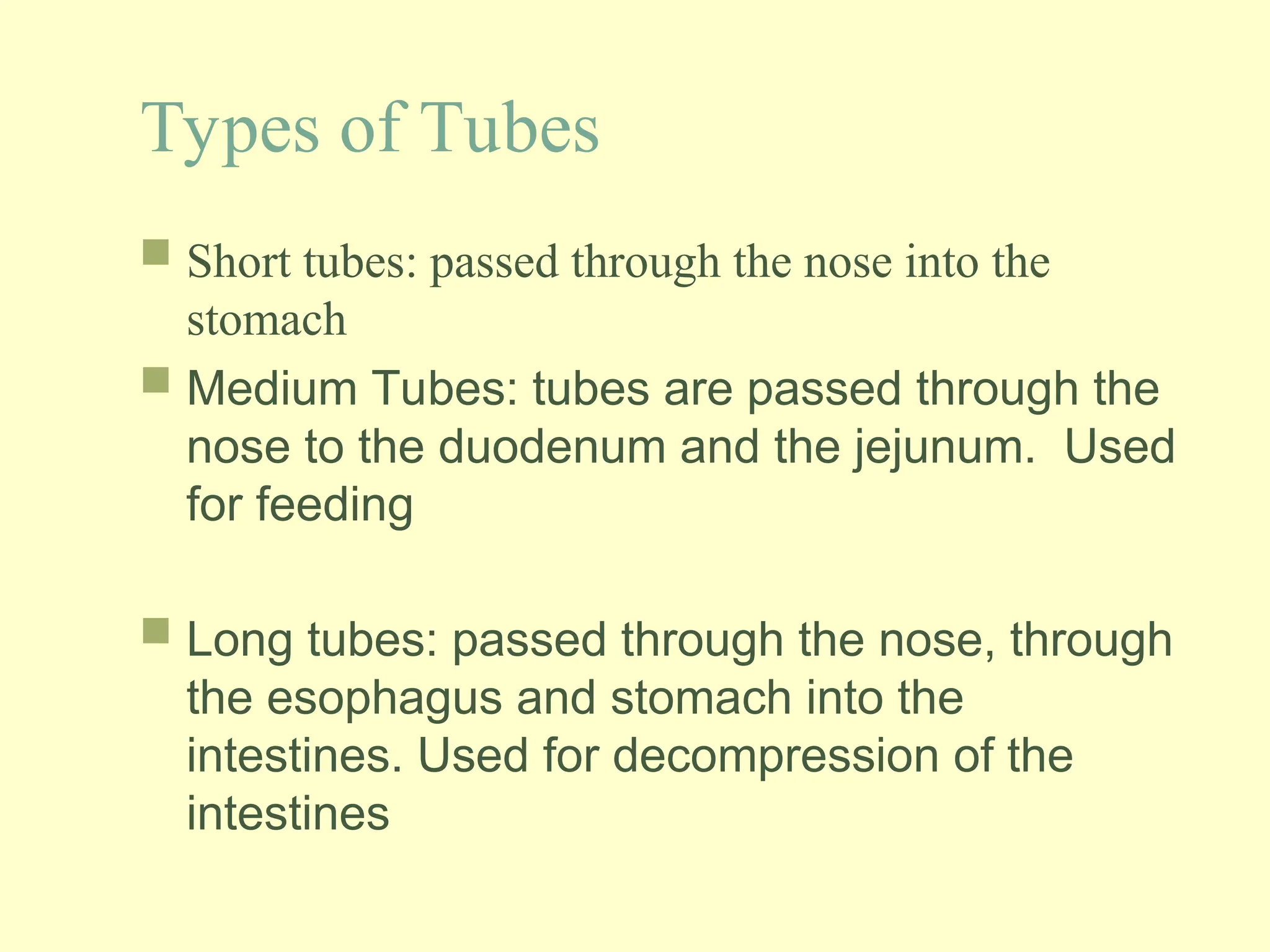
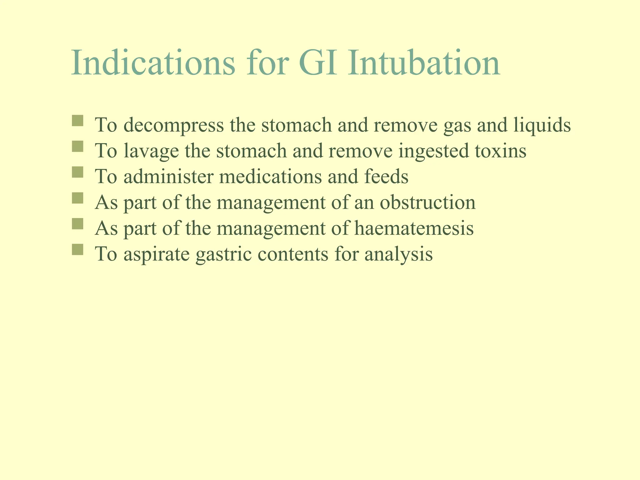
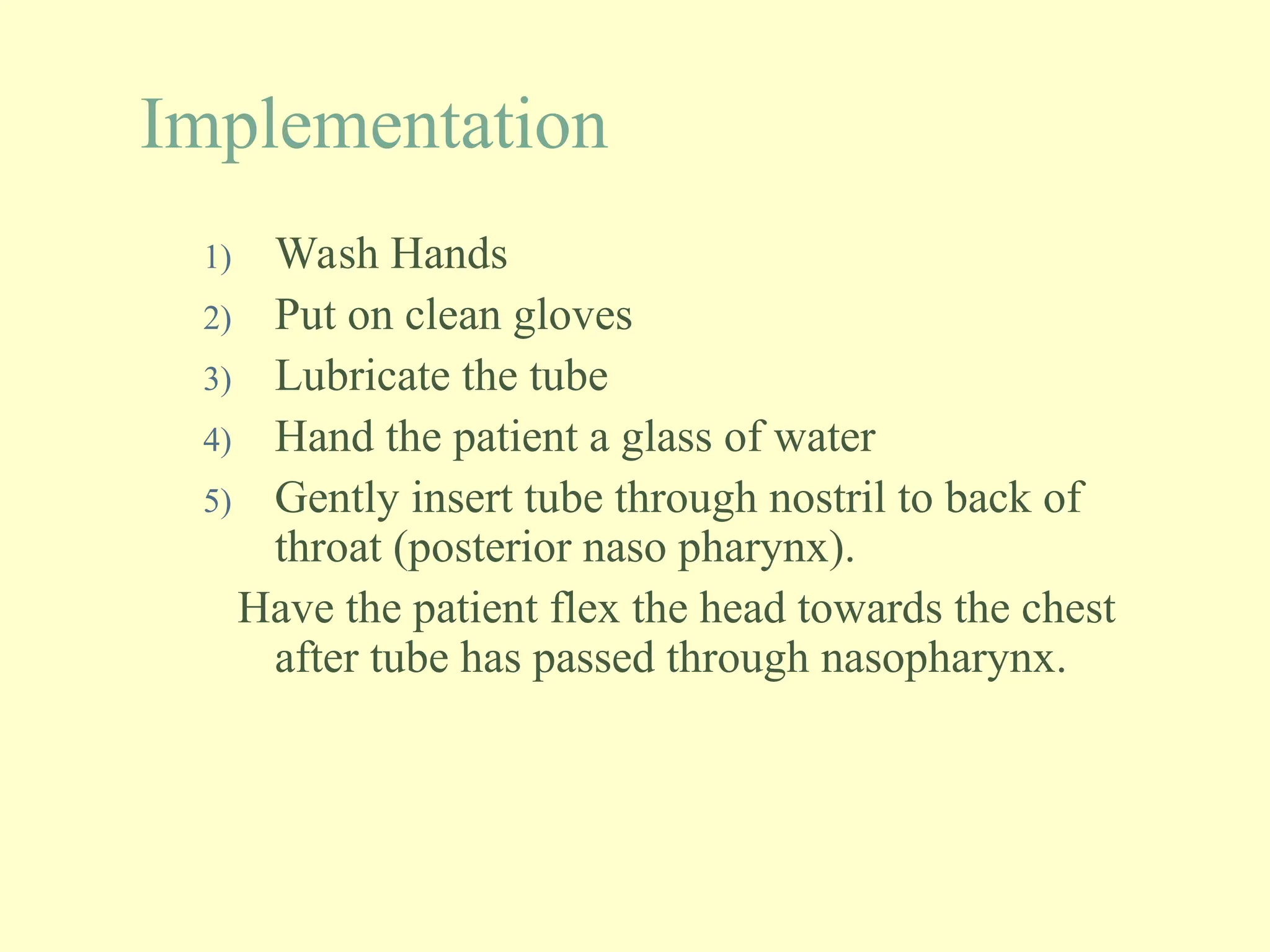
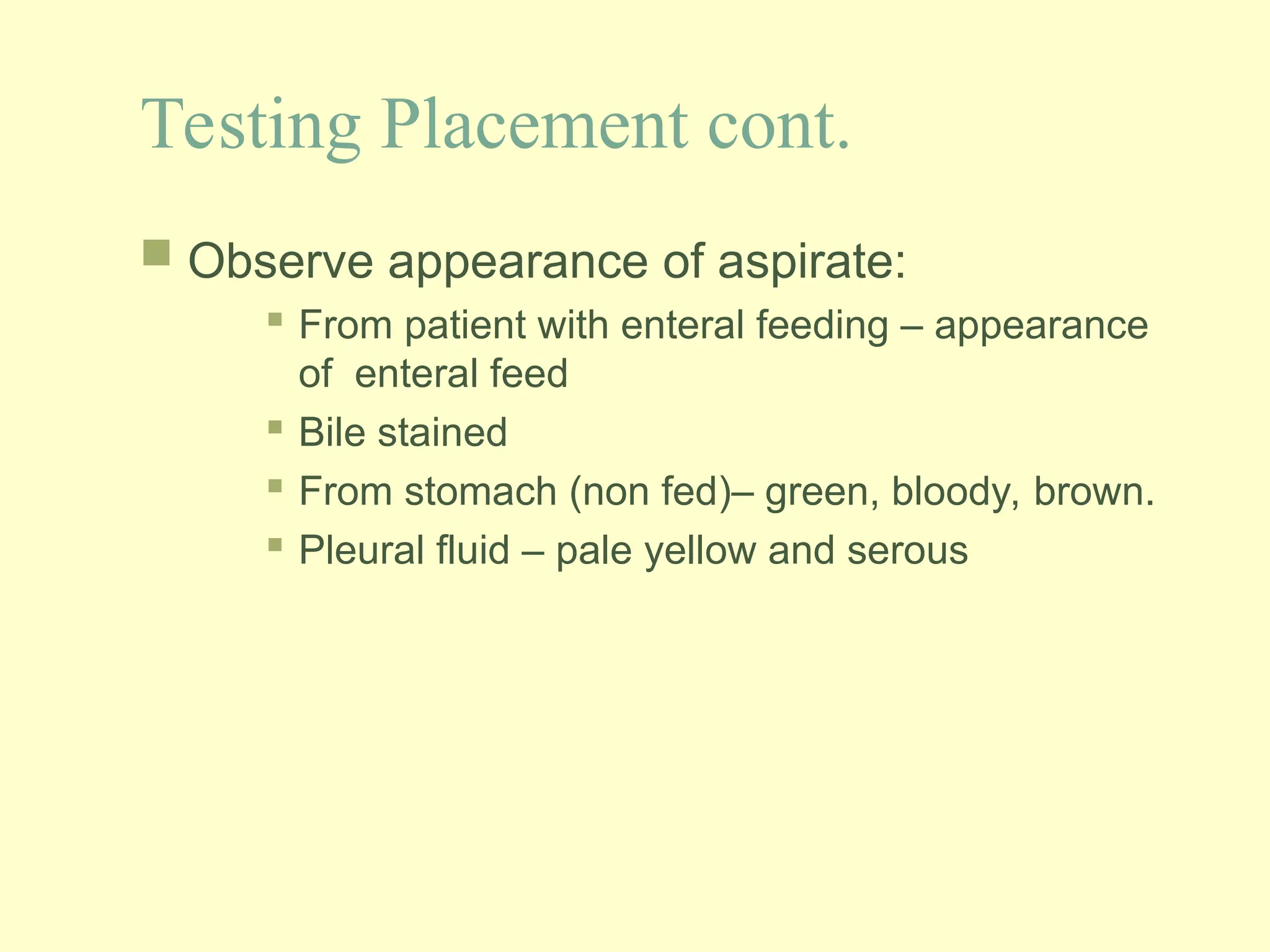
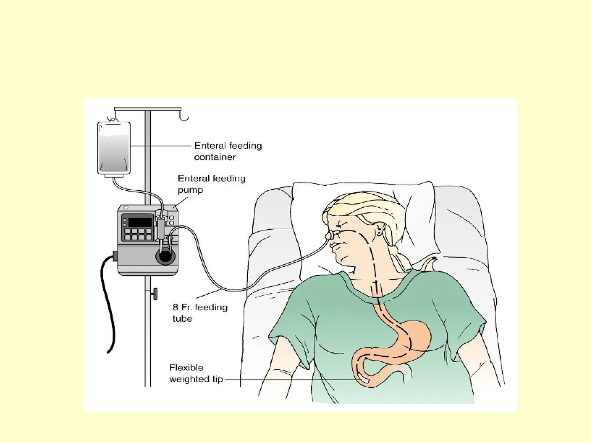
Gastrointestinal
Intubation
Nasogastric tubes
.
Nasogastric tube
 Gastrointestinal intubation deals with the inserting
of a rubber or plastic tube into the stomach,
duodenum or small intestine.
Types of Tubes
 Short tubes: passed through the nose into the
stomach
 Medium Tubes: tubes are passed through the
nose to the duodenum and the jejunum. Used
for feeding
 Long tubes: passed through the nose, through
the esophagus and stomach into the
intestines. Used for decompression of the
intestines
 Nasogastric tubes come in various sizes (8,
10, 12, 14, 16 and 18 Fr).
Indications for GI Intubation
 To decompress the stomach and remove gas and liquids
 To lavage the stomach and remove ingested toxins
 To administer medications and feeds
 As part of the management of an obstruction
 As part of the management of haematemesis
 To aspirate gastric contents for analysis
Intubating the client with an NG
tube
 Assessment:
 Who needs an NGT:
 Surgical patients
 Ventilated patients
 Neuromuscular impairment
 Patients who are unable to maintain adequate oral
intake to meet metabolic/nutritional demands
 To assess patency of the nares
Assessment cont.
 Assess patient’s medical history:
 Nose bleeds
 Nasal surgery
 Deviated nasal septum
 Anticoagulation therapy
 GI history
 Conduct a thorough physical examination.
 Assess patient’s gag reflex.
 Assess patient’s mental status.
Technique
Equipment:

14 or 16 Fr NG tube

Lubricating jelly

pH test strips

Tongue blade

Flashlight

Emesis basin

Syringes

1 inch wide tape or commercial fixation device

Suctioning available and ready

Urobag/Collection bag

Stethoscope
Technique continued…
 Explain procedure to patient and relatives
 Position the client in a sitting or high Fowler’s
position. If comatosed, semi Fowler’s.
 Examine feeding tube for flaws.
 Determine the length of tube to be inserted.
 Measure distance from the tip of the nose to the
earlobe and to the xyphoid process of the sternum.
 Prepare NG tube for insertion.
Fowler's position. Used to
promote drainage or ease
breathing. Head rest is adjusted
to desired height and bed is
raised slightly under patient's
knees
Implementation
1) Wash Hands
2) Put on clean gloves
3) Lubricate the tube
4) Hand the patient a glass of water
5) Gently insert tube through nostril to back of
throat (posterior naso pharynx).
Have the patient flex the head towards the chest
after tube has passed through nasopharynx.
Implementation Cont.
6)Emphasize the need to mouth breathe and swallow during
the procedure.
7) Swallowing facilitates the passage of the tube through the
oropharynx.
8) When the tip of the tube reaches the carina stop and listen
for air exchange from the distal end of the tube. If air is heard
remove the tube.
9) Advance tube each time client swallows until desired
length has been reached.
10) Do not force tube. If resistance is met or client starts to
cough, choke or become cyanotic stop advancing the tube and
pull back.
Implementation Cont.
11) Check placement of the tube.
X-ray confirmation
Testing pH of aspirate
12) Secure the tube with tape or commercial device.
Nasogastric Tube Position
Evaluation
 Observe the patient to determine response to procedure.
 ALERT! Persistent gagging – prolonged intubation and
stimulation of the gag reflex can result in vomiting and
aspiration.
 Coughing may indicate presence of tube in the airway.
Evaluation Cont.
 Note the location of external site marking on
the tube
 Documentation
 Size of tube, which nostril and patient’s
response.
 Record length of tube from the nostril to end of
tube.
 Record aspirate pH and characteristics
Testing Placement
 Wash hands and put on clean gloves
 Draw up 30cc of air into the syringe and attach to end
of the NG tube. Flush tube with 30cc of air prior to
attempting to aspirate fluid. Draw back on the
syringe to obtain 5 to 10 cc of gastric aspirate.
 If unable to aspirate:
 Advance tube – may be in air space above aspirate
level
 If intestinal placement suspected, withdraw tube 5 to
10 cm
 Have the patient lie on his/her left side wait 10-15 mins
and attempt aspiration again.
Testing Placement cont.
 Observe appearance of aspirate:
 From patient with enteral feeding – appearance
of enteral feed
 Bile stained
 From stomach (non fed)– green, bloody, brown.
 Pleural fluid – pale yellow and serous
Testing Placement Cont.
 If after repeated attempts, it is not possible to
aspirate fluid from a tube that was originally
established by x-ray examination to be in the desired
position and there are NO risk factors for dislocation,
tube has remained in original position and the client
is NOT experiencing any difficulty the nurse may
assume the tube is correctly placed.
Enteral Nutrition
 What is it:
 The administration of nutrients directly into the
GI tract. The most desirable and appropriate
method of providing nutrition is the oral route,
but this is not always possible.
 Nasogastric feeding is the most common route
 Nurses are the main healthcare professional
responsible for intubation
Administering Enteral Feeds
 Indications:
 Clients who are unable to maintain adequate
oral intake to meet metabolic demands
 Surgical cases
 Ventilated patients
 Neuromuscular impairment
 Generally these clients have been referred to
the Dietician.
Administering Enteral Feeds
 Contraindications:
 Clients with diffuse peritonitis.
 Severe pancreatitis
 Intestinal obstruction
 Paralytic ileus.
Complications
 Clogged/Blocked Tube- most common
 Dumping Syndrome: solution with high osmolality-
water moves into stomach and intestines from the
fluid surrounding the organs and vascular system
causing dehydration, hypotension and tachycardia
 Aspiration : ensure head of bed is elevated at least
30 degrees while feeds are being administered
Complications Cont.
 Dehydration- diarrhoea is a common problem.
 Electrolyte imbalance: hyperkalaemia and
hypernatraemia
 Oral mucosal breakdown
 Nasal irritation
nasogastrictubeinsertion-160715030455.ppt

nasogastrictubeinsertion-160715030455.ppt

  • 1.
  • 2.
    Nasogastric tube  Gastrointestinalintubation deals with the inserting of a rubber or plastic tube into the stomach, duodenum or small intestine.
  • 3.
    Types of Tubes Short tubes: passed through the nose into the stomach  Medium Tubes: tubes are passed through the nose to the duodenum and the jejunum. Used for feeding  Long tubes: passed through the nose, through the esophagus and stomach into the intestines. Used for decompression of the intestines
  • 5.
     Nasogastric tubescome in various sizes (8, 10, 12, 14, 16 and 18 Fr).
  • 6.
    Indications for GIIntubation  To decompress the stomach and remove gas and liquids  To lavage the stomach and remove ingested toxins  To administer medications and feeds  As part of the management of an obstruction  As part of the management of haematemesis  To aspirate gastric contents for analysis
  • 7.
    Intubating the clientwith an NG tube  Assessment:  Who needs an NGT:  Surgical patients  Ventilated patients  Neuromuscular impairment  Patients who are unable to maintain adequate oral intake to meet metabolic/nutritional demands  To assess patency of the nares
  • 8.
    Assessment cont.  Assesspatient’s medical history:  Nose bleeds  Nasal surgery  Deviated nasal septum  Anticoagulation therapy  GI history  Conduct a thorough physical examination.  Assess patient’s gag reflex.  Assess patient’s mental status.
  • 9.
    Technique Equipment:  14 or 16Fr NG tube  Lubricating jelly  pH test strips  Tongue blade  Flashlight  Emesis basin  Syringes  1 inch wide tape or commercial fixation device  Suctioning available and ready  Urobag/Collection bag  Stethoscope
  • 10.
    Technique continued…  Explainprocedure to patient and relatives  Position the client in a sitting or high Fowler’s position. If comatosed, semi Fowler’s.  Examine feeding tube for flaws.  Determine the length of tube to be inserted.  Measure distance from the tip of the nose to the earlobe and to the xyphoid process of the sternum.  Prepare NG tube for insertion.
  • 11.
    Fowler's position. Usedto promote drainage or ease breathing. Head rest is adjusted to desired height and bed is raised slightly under patient's knees
  • 13.
    Implementation 1) Wash Hands 2)Put on clean gloves 3) Lubricate the tube 4) Hand the patient a glass of water 5) Gently insert tube through nostril to back of throat (posterior naso pharynx). Have the patient flex the head towards the chest after tube has passed through nasopharynx.
  • 15.
    Implementation Cont. 6)Emphasize theneed to mouth breathe and swallow during the procedure. 7) Swallowing facilitates the passage of the tube through the oropharynx. 8) When the tip of the tube reaches the carina stop and listen for air exchange from the distal end of the tube. If air is heard remove the tube. 9) Advance tube each time client swallows until desired length has been reached. 10) Do not force tube. If resistance is met or client starts to cough, choke or become cyanotic stop advancing the tube and pull back.
  • 16.
    Implementation Cont. 11) Checkplacement of the tube. X-ray confirmation Testing pH of aspirate 12) Secure the tube with tape or commercial device.
  • 18.
  • 19.
    Evaluation  Observe thepatient to determine response to procedure.  ALERT! Persistent gagging – prolonged intubation and stimulation of the gag reflex can result in vomiting and aspiration.  Coughing may indicate presence of tube in the airway.
  • 20.
    Evaluation Cont.  Notethe location of external site marking on the tube  Documentation  Size of tube, which nostril and patient’s response.  Record length of tube from the nostril to end of tube.  Record aspirate pH and characteristics
  • 21.
    Testing Placement  Washhands and put on clean gloves  Draw up 30cc of air into the syringe and attach to end of the NG tube. Flush tube with 30cc of air prior to attempting to aspirate fluid. Draw back on the syringe to obtain 5 to 10 cc of gastric aspirate.  If unable to aspirate:  Advance tube – may be in air space above aspirate level  If intestinal placement suspected, withdraw tube 5 to 10 cm  Have the patient lie on his/her left side wait 10-15 mins and attempt aspiration again.
  • 22.
    Testing Placement cont. Observe appearance of aspirate:  From patient with enteral feeding – appearance of enteral feed  Bile stained  From stomach (non fed)– green, bloody, brown.  Pleural fluid – pale yellow and serous
  • 23.
    Testing Placement Cont. If after repeated attempts, it is not possible to aspirate fluid from a tube that was originally established by x-ray examination to be in the desired position and there are NO risk factors for dislocation, tube has remained in original position and the client is NOT experiencing any difficulty the nurse may assume the tube is correctly placed.
  • 24.
    Enteral Nutrition  Whatis it:  The administration of nutrients directly into the GI tract. The most desirable and appropriate method of providing nutrition is the oral route, but this is not always possible.  Nasogastric feeding is the most common route  Nurses are the main healthcare professional responsible for intubation
  • 25.
    Administering Enteral Feeds Indications:  Clients who are unable to maintain adequate oral intake to meet metabolic demands  Surgical cases  Ventilated patients  Neuromuscular impairment  Generally these clients have been referred to the Dietician.
  • 26.
    Administering Enteral Feeds Contraindications:  Clients with diffuse peritonitis.  Severe pancreatitis  Intestinal obstruction  Paralytic ileus.
  • 27.
    Complications  Clogged/Blocked Tube-most common  Dumping Syndrome: solution with high osmolality- water moves into stomach and intestines from the fluid surrounding the organs and vascular system causing dehydration, hypotension and tachycardia  Aspiration : ensure head of bed is elevated at least 30 degrees while feeds are being administered
  • 28.
    Complications Cont.  Dehydration-diarrhoea is a common problem.  Electrolyte imbalance: hyperkalaemia and hypernatraemia  Oral mucosal breakdown  Nasal irritation

Editor's Notes

  • #3 Salem can protect gastric suture lines b/c it maintains the force of suction a the drainage opening or outlets at less than 25mm hg the small vent tube (blue pig tail) controls this action.
  • #13 Flexing the head closes off glottis and reduces risk of tube entering trachea
  • #15 Carina about 25 cm in an adult
  • #27 Notes for diarrhoea adjust strength of feeds change the product antidiarrhea meds ensure clean technique research has shown many diarrhea incidents related to bacterial contamination. Dumping syndrome client will feel full,nausea,and diarrhea need to be aware of the conc. of tube feedings and observe for these effects
  • #28 hNotes for diarrhoea adjust strength of feeds change the product antidiarrheak meds ensure clean technique research has shown many diarrhea incidents related to bacterial contamination. as shown many diarrhea incidents related to bacterial contamination.