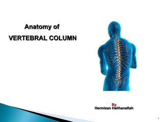
Anatomy of vertebral column
•Download as PPT, PDF•
327 likes•115,405 views

The vertebral column, or spine, is composed of 33 vertebrae in early development that fuse together into 26 vertebrae in adulthood. The vertebrae are organized into 7 cervical, 12 thoracic, 5 lumbar, 1 sacrum, and 1 coccyx vertebrae. Each vertebra has a body, vertebral arch, and 7 processes. Between the vertebrae are intervertebral discs that act as shock absorbers and allow movement. The spine has four normal curves that develop during childhood to maintain balance and absorb impacts during walking. The vertebrae permit flexion, extension, lateral flexion, and rotation movements.

Recommended
More Related Content
What's hot
What's hot (20)
Similar to Anatomy of vertebral column
Similar to Anatomy of vertebral column (20)
More from Sado Anatomist
More from Sado Anatomist (20)
Recently uploaded
Recently uploaded (20)
Anatomy of vertebral column
- 1. 1 Anatomy ofAnatomy of VERTEBRAL COLUMNVERTEBRAL COLUMN ByBy Hermizan HalihanafiahHermizan Halihanafiah
- 2. The vertebral column, also called the spine, spinal column or backbone. Composed of a series of bones called vertebrae (singular is vertebra). About 71 cm (28in): adult male. About 61 cm (24in): adult female. 2
- 3. Total number of vertebrae during early development is 33. As a child grows, several vertebrae in the sacral and coccygeal regions fuse. Adults have 26 vertebrae. *Sacrum and coccyx bones become fused.
- 4. 7 cervical vertebrae (C1 - C7 ) 12 thoracic vertebrae (T1 –T12) 5 lumbar vertebrae (L1 – L5) 1 sacrum ( 5 fused ) 1 coccyx ( 4 fused ) *The sacrum and coccyx do not have number.
- 5. The cervical, thoracic and lumbar vertebrae movable Sacrum and coccyx immovable Between adjacent vertebrae from the second cervical vertebra to the sacrum are intervertebral disc (inter = between). 5
- 6. 6
- 7. General Structure of VertebraeGeneral Structure of Vertebrae 7 1. Cervical vertebrae (C1-C7) • Formed framework of neck region • Support skull • Small in size • Presense foramen in each transverse process 2. Thoracic vertebrae (T1-T12) • Formed posterior part of thoracic cage • Articulates with associated ribs
- 8. 8 3. Lumbar Vertebrae (L1-L5) • Formed skeletal support for posterior abdominal wall • Characterized by large in size 4. Sacrum Vertebrae • Fusion of 5 sacral bones • Immovable (synostosis) • Articulates with L5 at lumbosacral joint • Articulates laterally with pelvic bone at sacroiliac joint. • Formed posterior wall of lower abdominal and pelvic cavity 5. Coccyx • Fusion of 4 coccyx bones • Immovable (synostosis) • Formed part of pelvic cavity
- 9. 1. Supports the head. 2. Help maintain balance in the upright position. 3. Enclose and protect the spinal cord. 4. Permits movement (move forward, backward, sideways, and rotate). 5. Absorbs shocks during walking. 6. Serve as a point of attachment for the ribs, pelvic girdle and muscles of the back and upper limbs. 9
- 10. In the fetus, there is a single concave curve. At 3 months after birth when infant lifts head as it begins to crawl the cervical curve develops. When child sits up, stands and walks the lumbar curve develops. 10
- 11. In adult, it shows four slight bends called normal curve: Cervical and lumbar curve are convex (bulging out) Thoracic and sacral curve are concave (cupping in)
- 12. 12
- 13. 13
- 14. The thoracic and sacral curves are called primary curves because they form first during fetal development. The cervical and lumbar curves are called secondary curves because they form later, several months after birth. All curve fully developed by age 10. However, secondary curves may be progressively lost in old age. 14
- 15. Newborn Spinal Curvature : C-shaped curve Known as Primary Curve Single curve Adult Spinal Curvature: S-shaped vertebral column Four curve (cervical, thoracic, lumbar amd sacral curve) Secondary curvatures develop after birth 15 Differences newborn and adult spinal curvature
- 16. 16
- 17. Consist of four slight bends (cervical, thoracic, lumbar, sacral) Cervical and lumbar curve are convex (bulging out). The thoracic and sacral curves are concave (cupping in). 17
- 18. 18
- 19. Increases its strength Help maintain balance in the upright position Absorb shocks during walking Help protect the vertebrae from fracture 19
- 20. Scoliosis: lateral bending of the vertebral column, usually in the thoracic region. Kyphosis: Incerase in the thoracic curve of the vertebral column. Lordosis (Hollow back) Increase in the lumbar and cervical curve of the vertebral column.
- 24. 24 Kyphosis
- 25. Typical vertebrae consists of: ◦ A body ◦ A vertebral arch (pedicles and lamina) ◦ Seven processes: two transverse processes, one spinous process, four articular processes 25
- 26. Largest part of vertebra, thick. Disc-shaped anterior portion Weight bearing portion – size increases inferiorly Its inferior and superior surfaces are roughened and give attachment to the intervertebral disc. Anterior and lateral surfaces contain nutrient foramina – pathway for blood vessels. 26
- 27. Extend backwards from the body of the vertebra. Consists of a pair of pedicles and a pair of laminae. The pedicle project backward from the body to unite with the laminae. 27
- 28. Pedicle: two short, thick processes, which project backward. the concavities above and below the pedicles are named the vertebral notches –formed IV foramina Laminae: two broad & flat plates directed backward and medialward from the pedicles. the laminae end in a single sharp, slender projection called a spinous process.
- 29. 7 processes arise from the vertebral arch: TWO TRANSVERSE PROCESS ONE SPINOUS PROCESS FOUR ARTICULAR 29
- 30. TRANSVERSE PROCESS: Extends posterolaterally for the junction between pedicle and laminae on each side (left and right) ONE SPINOUS PROCESS: A single spinous process projects posteriorly from the junction of the laminae. These 3 processes serve as points of attachment for muscles.
- 31. ARTICULAR PROCESSES (Zygapophyses): At the junction between pedicles and lamina meet, also projecting superior and inferior articular process. At the end of these processes – concave surface (facet) IAP of vertebrae above articulates with SAP of vertebrae below – zygapophysial joints (Facet Joints). 31
- 32. Between the bodies of the adjacent vertebrae C2 to the sacrum. Each disc forms a cartilaginous joint to allow slight movement of the vertebrae, and acts as a ligament to hold the vertebrae together. 32 INTERVERTEBRAL DISCS
- 33. Each vertebral discs consist of: an outer fibrous ring consisting of fibrocartilage called called annulus fibrosus (annalus = ringlike). Inner soft, pulpy, highly elastic substance called the nucleus pulposus (pulposus = pulplike), which is acts as a shock absorber, absorbing the impact of the body's daily activities and keeping the two vertebrae separated 33
- 34. Nucleus pulposus hardens and less elastic with age. Narrowing of discs and compression of the vertebrae results in a decrease in the height with age. A tear can occur within the annulus fibrosus (ring) and cause the nucleus pulposus may track into the vertebral canal or intervertebral foramen to impinge on neural structures – herniation IV discs. (prolapsed/slipped disc) 34
- 35. 1. Binds the vertebrae and forms a strong joint 2. Permits various movements of the vertebral column 3. Absorbs vertical shock and avoid friction during intervertebral joints movements. 35
- 36. Vertebral foramen contains : spinal cord and its roots, spinal meninges, ASA and PSA, Venous Plexus, fat The vertebral foramina of all vertebrae form the vertebral (spinal) canal.
- 37. Spina bifida (congenital) Two sides of vertebral arch fail to fuse during development, resulting in an open vertebral canal (cleft) Absence of spinous process Ususally in lumbosacral region Protrusion of spinal meninges (out pouch) and may contain CSF – meningocele Protrusion of part of spinal cord and meninges -myelomeningocele Abnormalities of the Vertebral canalAbnormalities of the Vertebral canal 37
- 38. 38
- 39. Between every pair of vertebrae are two apertures, the intervertebral foramen (formed by inferior and superior vertebral notches). Also called neural foramen. Passageway for nerve roots. 39 INTERVERTEBRAL FORAMEN
- 40. C1-C7 (formed framework of the neck) C1, C2 and C7 – atypical (looks weird!!!) C3-C6 – have similarities – typical ◦ Smaller bodies, short ◦ Larger vertebral arch – hence larger vertebral foramen (cervical enlargement) ◦ Transverse process consist transverse foramina @ foramen transversarium (vertebral artery) ◦ Spinous process short and bifid Cervical VertebraeCervical Vertebrae 40
- 41. C1 – Atlas Ring shaped Lack of body (fused with body of C2) Composed by lateral masses interconnected by an anterior and posterior arch Each lateral masses consist SAFacet which artculates with occipital condyle – AOJ Also consist IAF which articulates with SAF of C2 – AAJ (C1/C2) anterior arch of C1 articulates with dens of C2 – support via transverse ligament 41
- 42. C2 (Axis) Structure similar with typical cervical vertebrae but Have peg like processes called dens or odontoid process Dens projecting upwards from body of C2 toward vertebral foramen of C1 which articulates with anterior arch of C1 (AAJ). 42
- 43. Typical thoracic vertebrae ◦ Fairly large size of body ◦ Long spinous process and pointed and angled downward ◦ Vertebral foramen generally circular ◦ Transverse process projecting posterolaterally. ◦ Have 3 pairs of facets – attachment for the ribs (2 pairs of demifacets at the body and 1 pair of facet at the transverse process. Thoracic Vertebrae (T1-T12)Thoracic Vertebrae (T1-T12) 43
- 44. T1 has a superior facet and inferior demifacets for head of ribs. T2-T8 have superior and inferior demifacets for head of ribs. T9 has a superior demifacets T10-T12 have superior facet. Movements of the thoracic vertebrae are limited by thin intervetebral disc and by attachment of the ribs to the sternum (sternocostal jnt). 44
- 45. The largest and strongest – weight bearing Thickest body Spinous process is thickest and broad and project posteriorly – attachment for the large back muscles Superior articular process facing medially, inferior articular process facing laterally. Lumbar Vertebrae (L1-L5)Lumbar Vertebrae (L1-L5) 45
- 46. 46
- 47. Inverted triangular in-shaped Fusion of 5 sacral vertebrae – fusion started 16-18 y’old and completed at 30. Sacrum serve strong foundation for the pelvic girdle attachment (sacroiliac joint) Formed posterior wall of the lower abd cavity and pelvic cavity. At the superior (base), consist SAP which articulates with IAP of L5 – LSJ (L5/S1) Sacrum (S1-S5)Sacrum (S1-S5) 47
- 48. anterior surface, consist transverse ridge, mark the joining of bodies Lateral surface – smooth surface called ala Anterior ridge of sacral body which projecting forward – promontory Consist 4 pairs of anterior sacral foramina – routes for anterior rami of sacral spinal nerve 48
- 49. At the posterior surface consist posterior sacral foramina – routes for posterior rami of sacaral spinal nerves. Sacral canal- continuation of vertebral canal from L5 (routes for roots of sacral and coccygeal spinal nerves), and terminates as a opening called sacral hiatus (routes for S5 and co1 spinal nerve). On either side of the sacral hiatus, consist sacral cornua. Median sacral crest – fusion of spinous process Lateral sacral crest – fusion of transverse process 49
- 50. 50
- 51. Inverted Triangular in shaped Fusion of 4 coccyx vertebrae Dorsal surface, 2 projection called coccygeal cornua – attachment for sacrococcygeal ligament and also attachment for pelvic floor muscles (levator ani) Coccyx vertebrae (co1-co4)Coccyx vertebrae (co1-co4) 51
- 52. Flexion (to bend) Extension (to stretch out) Hyperextension Lateral flexion Rotation – right and left rotate 52
- 54. Flexion: C1 (atlas): allows for forward and backward motion of the head. Extension: Straightening the joint, moving the spine back Lateral Flexion (Abduction): moving the spine to the side (left or right); the neck moves toward the shoulder. Rotation: C2 (axis) for rotation making a "no" motion. Turning the spine to the side (right or left); the neck turns toward the shoulder. 54
- 55. 55 Flexion and extension Lateral flexion Rotation
- 56. Flexion: moving the spine forward, the thorax moves toward the pelvis. Extension / Hyperextension: Straightening the joint by moving the spine back, the thorax moves away from the pelvis. Lateral Flexion (Abduction): moving the spine to the side (left or right), the thorax moves to the side toward the pelvis. Rotation: turning the spine to the side (right or left); the thorax rotates to one side. 56
- 59. Hyperextension is a straightening movement that goes beyond the normal, healthy boundaries of the joint 59